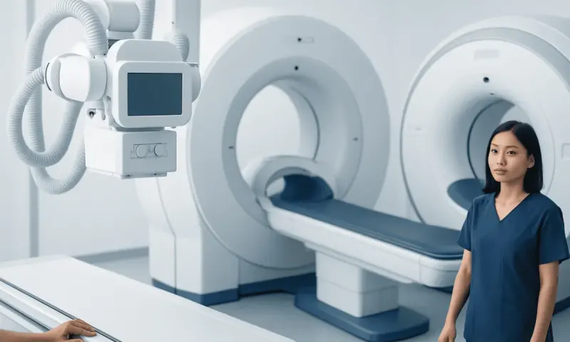
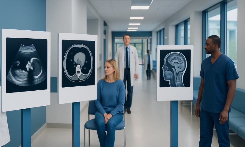
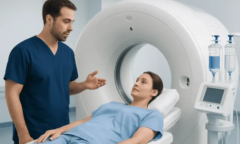
CT Scan vs MRI vs Ultrasound: Which Scan Fits?
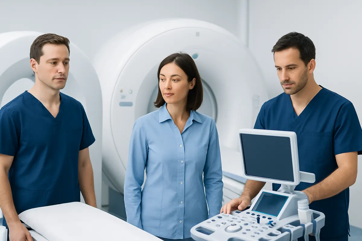
Cat scan vs MRI vs ultrasound describes three imaging methods that answer different clinical questions. CT is often chosen for speed and detail, MRI for soft tissues and the nervous system, and ultrasound for real-time views of many organs, blood flow and pregnancy without ionizing radiation.
Cat Scan vs MRI vs Ultrasound: a side-by-side comparison
A CAT scan is another name for a CT (computed tomography) scan. CT, MRI and ultrasound are not interchangeable tests: each creates images in a different way and is particularly useful for particular tissues and clinical situations. The clinician’s aim is to select the test that can answer the medical question safely and efficiently.
The table below offers a practical comparison. In some situations, more than one scan may be needed because each test can provide complementary information.
- CT/CAT scan: Uses X-rays and computer processing to create cross-sectional images. It is fast and commonly used for trauma, suspected internal bleeding, lung conditions, many abdominal problems, bone injuries and urgent assessments.
- MRI: Uses magnetic fields and radio waves rather than X-rays. It provides very detailed images of soft tissues, including the brain, spinal cord, nerves, muscles, ligaments, joints and many pelvic organs.
- Ultrasound: Uses sound waves transmitted through a handheld probe. It is useful for examining abdominal and pelvic organs, pregnancy, thyroid conditions, superficial lumps, the heart and blood flow in vessels.
- Radiation: CT involves ionizing radiation. MRI and ultrasound do not use ionizing radiation.
- Time and experience: Ultrasound and CT are often relatively quick. MRI usually takes longer and requires lying still in a scanner, which can feel confined for some people.
Contrast material may be used with CT or MRI to make blood vessels, inflammation, tumors or organs easier to see. Ultrasound may also use Doppler techniques to assess blood flow. The imaging team reviews allergies, kidney function, pregnancy and implanted devices before recommending contrast or selecting a scan.
How clinicians tell the scans apart
Clinicians begin with the suspected problem rather than choosing a scan based on a general ranking of accuracy. They consider where symptoms are located, how quickly answers are needed, whether the concern involves bone, air-filled lungs, soft tissue, blood vessels or fluid, and whether the person can safely have contrast material or enter an MRI scanner.
CT is particularly effective at showing bone, calcification, many chest findings and acute bleeding. It can image a large area quickly, which is important after significant injury or when a serious emergency is being considered. Modern CT protocols are designed to use the lowest radiation exposure that can still provide clinically useful images.
MRI creates strong contrast between different soft tissues. This helps doctors assess structures that may look similar on CT, such as brain tissue, spinal discs, ligaments, tendons and some tumors. MRI may not be suitable for everyone, including some people with certain implanted medical devices or metal fragments; the MRI safety team checks this carefully.
Ultrasound is portable and dynamic. The clinician or sonographer can often apply gentle pressure, ask the patient to point to the most tender area, observe an organ moving and evaluate blood flow at the same appointment. Image quality can be affected by bowel gas, body shape and the depth of the structure being examined.
What can an ultrasound show that a CT scan cannot?
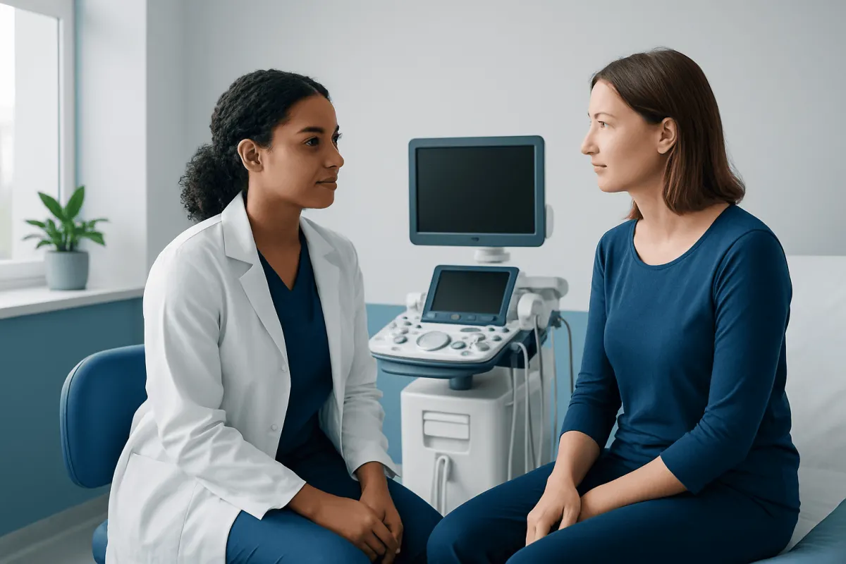
Ultrasound can show movement in real time. For example, it can observe a fetus moving, evaluate heart valve motion during echocardiography, assess whether a lump moves with swallowing, or help examine a painful area while the patient identifies the exact site of discomfort. CT provides a detailed snapshot but does not offer the same bedside, live assessment.
Doppler ultrasound can measure the direction and speed of blood flow. This is especially useful when evaluating blood flow in arteries and veins, assessing possible venous clots, checking circulation in the neck or legs, or monitoring blood supply to organs and transplanted tissues. CT angiography can also assess vessels, but it uses radiation and usually requires iodinated contrast material.
Ultrasound can guide procedures in real time, including some biopsies, drainage procedures and injections. It can help the clinician avoid nearby vessels and direct a needle toward a visible target. It is also commonly the first imaging method for pregnancy because it does not use ionizing radiation.
However, ultrasound cannot see through bone or air-filled bowel and lungs in the same way as CT. Therefore, it may not adequately assess deeper structures, complex injuries or areas obscured by gas. A clinician may order CT or MRI when ultrasound findings are unclear or when a broader view is needed.
Why do doctors choose CT over MRI?
Doctors may choose CT over MRI when speed is essential. CT is commonly available in emergency settings and can rapidly evaluate the head, chest, abdomen and pelvis. This can be valuable after trauma, when looking for internal bleeding, evaluating certain strokes, or assessing severe abdominal symptoms that may require urgent treatment.
CT is often better than MRI for seeing fractures, small bone details, calcifications and many conditions affecting the lungs. Because air is naturally present in the lungs, CT is a central test for investigating many chest symptoms and abnormalities. It can also be used when a patient cannot lie still for the longer duration of an MRI examination.
MRI may be impractical or unsafe for some people because of certain implanted devices, retained metal fragments, severe claustrophobia or an inability to remain still. CT may be the safer or more feasible option in these circumstances. In other cases, a clinician may use strategies such as an open MRI option, additional support or carefully supervised medication to help a person complete an MRI if it is the most informative test.
The trade-off is that CT uses ionizing radiation. The benefit of obtaining timely, useful information generally outweighs this risk when CT is medically indicated, but unnecessary repeat scans should be avoided. Patients can tell their healthcare team about recent imaging so prior examinations may be reviewed when appropriate.
What can a MRI show that a CT scan cannot?
MRI can show subtle differences among soft tissues that CT may not distinguish as clearly. It is especially useful for evaluating the brain and spinal cord, including inflammation, some types of stroke, multiple sclerosis-related changes, tumors, nerve compression and disorders of the pituitary region. For certain neurological concerns, neurological conditions may require MRI as part of a wider clinical assessment.
In the musculoskeletal system, MRI can show ligaments, tendons, cartilage, menisci, muscle injuries, bone marrow changes and early stress injuries. CT can show a fracture very well, but MRI may reveal soft-tissue damage or bone marrow swelling when a fracture is not obvious on X-ray or CT.
MRI can also provide detailed assessment of many pelvic, liver, bile duct and vascular conditions. Depending on the clinical question, it may characterize a finding seen on ultrasound or CT without using ionizing radiation. Specialized MRI sequences can show fluid, fat, blood products and tissue composition in ways that guide diagnosis and treatment planning.
MRI is not automatically the best test for every problem. It takes longer than CT, may be more sensitive to movement, and is less useful for some lung and acute bone assessments. A radiologist and requesting clinician decide whether MRI findings are likely to change care before recommending it.
What is more accurate, an ultrasound or CT scan?
Neither ultrasound nor CT is universally more accurate. Accuracy depends on what is being examined, the quality of the study, the person’s anatomy and the question being asked. Ultrasound can be highly accurate for certain gallbladder, pelvic, thyroid, testicular, vascular and pregnancy-related concerns, particularly when performed by an experienced professional.
CT often provides a more comprehensive view of deep structures and can be more reliable when bowel gas, body size or depth limits ultrasound images. It is frequently preferred for many injuries, complex abdominal conditions, kidney stones, lung disorders and suspected internal bleeding. CT may also reveal conditions outside the area that initially seemed responsible for symptoms.
For some concerns, ultrasound is the appropriate first test because it is radiation-free, readily available and can provide a focused answer. If it is inconclusive or suggests a problem requiring a broader assessment, the clinician may recommend CT, MRI or another test. The sequence is chosen to balance diagnostic value, safety and urgency.
Imaging results are not diagnoses on their own. A scan may identify an incidental finding that is unrelated to symptoms, while some conditions may need blood tests, physical examination, follow-up imaging or specialist review for a clear diagnosis.
Preparing for results and knowing what to do next
Preparation depends on the examination. Some CT, MRI and abdominal ultrasound studies require fasting for several hours, while others do not. Patients should follow the instructions given by the imaging department and report pregnancy or possible pregnancy, kidney disease, previous reactions to contrast material, allergies, asthma and all medicines and supplements.
Before MRI, patients should report any pacemaker, implanted pump, cochlear implant, clips from prior surgery, metal fragments, joint replacements or other implanted materials. They may need to remove jewelry, hearing aids, removable dental devices and clothing with metallic parts. The imaging staff can confirm whether an implant is MRI-compatible or whether another test is preferable.
After the scan, a radiologist interprets the images and sends a report to the requesting clinician. The clinician explains what the findings mean in the context of the person’s symptoms and medical history. If further assessment is needed, this may involve follow-up imaging, laboratory testing, a biopsy or referral to another specialist.
Acıbadem Health Point’s multidisciplinary specialists and JCI-accredited hospitals support international patients who need diagnostic imaging and follow-up care, with investigations selected according to individual clinical needs.
When to seek medical care
Medical advice is appropriate for persistent, worsening or unexplained symptoms, especially pain, a new lump, ongoing severe headaches, changes in vision, unexplained weakness, prolonged fever, blood in urine or stool, or changes in bowel or bladder habits. A healthcare professional can determine whether imaging is needed and which test is most suitable.
Urgent assessment is important for symptoms that may indicate an emergency, such as sudden weakness or difficulty speaking, a sudden severe headache, chest pain, severe shortness of breath, fainting, significant injury, uncontrolled bleeding, severe abdominal pain, or new confusion. Emergency services or the nearest emergency department should be contacted rather than waiting for an outpatient scan.
People who have already received an imaging report should contact the requesting clinician if they do not understand the findings, symptoms change, or they have questions about recommended follow-up. They should not delay urgent care because a previous scan was normal, as new or evolving symptoms can require reassessment.
Frequently asked questions
01Is a CAT scan the same as a CT scan?
Yes. CAT scan stands for computed axial tomography, while CT stands for computed tomography. Both terms describe an imaging test that uses X-rays and computer processing to produce detailed cross-sectional images.
02Does MRI use radiation?
MRI does not use ionizing radiation. It uses a strong magnetic field and radio waves to create images. MRI safety screening remains important because the magnetic field can affect certain implants or metal objects.
03Which scan is best for abdominal pain?
The best scan depends on the location, severity and likely cause of the pain. Ultrasound is often used first for concerns involving the gallbladder, pelvis or pregnancy, while CT may be preferred for many urgent or complex abdominal conditions. A clinician chooses based on the symptoms and examination findings.
04Can ultrasound detect cancer?
Ultrasound can identify a mass or an area that looks different from nearby tissue, but it usually cannot confirm cancer by itself. Further imaging, follow-up monitoring or a biopsy may be needed to determine the cause of an abnormal finding.
05Is CT or MRI better for a head injury?
CT is commonly the first scan after an acute head injury because it is fast and can detect bleeding and skull fractures. MRI may be used later when more detailed assessment of brain tissue, nerves or certain subtle injuries is needed. The decision depends on the injury and clinical examination.
06Can a person have CT, MRI and ultrasound on the same day?
Sometimes more than one imaging test is needed, and the timing depends on the clinical situation. The medical team considers urgency, contrast use, preparation requirements and whether each test will add useful information. Many people only need one appropriately selected examination.
This article is for general information only and is not a substitute for professional medical advice. Please consult a qualified doctor about your individual situation.
Joint and spine care in Turkey — expert assessment & treatment
JCI-accredited · board-certified surgeons · reply within 24h
Add us as a Preferred Source to see more of our trusted health content across Google Search, AI Overviews and Discover.