Optic Neuropathy: Vision Loss Patterns and Neuro-Ophthalmology Evaluation

Optic neuropathy refers to damage or dysfunction of the optic nerve, the cable that carries visual information from the eye to the brain. Recognizing the pattern of vision loss and arranging a neuro-ophthalmology evaluation can help identify the cause and guide timely, appropriate care.
Overview
Optic neuropathy is a medical term for injury, inflammation, or impaired function of the optic nerve. The optic nerve is made of more than a million nerve fibers that transmit signals from the retina at the back of the eye to the brain, where sight is interpreted. When these fibers are affected, the person may notice blurred vision, dim vision, missing areas in the visual field, reduced color perception, or changes that are first detected during an eye examination.
The condition is not a single disease. It is a group of disorders with different causes, patterns, and levels of urgency. Some forms develop suddenly over hours or days, while others progress slowly over weeks, months, or years. Optic neuropathy may involve one eye or both eyes, and it may be associated with eye pain, headache, neurologic symptoms, or no discomfort at all.
Because the optic nerve is part of the central nervous system, evaluation often requires a careful connection between eye findings and neurologic health. Neuro-ophthalmology focuses on this connection. A neuro-ophthalmologist assesses visual function, the optic nerve, eye movements, and related brain or systemic conditions to determine why the optic nerve is not working normally.
Vision Loss Patterns and Symptoms
The pattern of vision loss provides important clues. Some people experience central vision loss, making reading, face recognition, or detailed work difficult. Others notice a dark or dim area above, below, or to one side of their vision. In certain optic nerve disorders, vision may seem washed out, colors may look faded, or red objects may appear less bright in one eye compared with the other.
Optic neuropathy often causes reduced contrast sensitivity, meaning that objects may be visible but less clear in low light or against a similar background. A person may also notice that vision is worse after exercise or heat exposure, particularly in some inflammatory or demyelinating conditions. In mild cases, the person may not be aware of a problem until one eye is covered or a visual field test is performed.
Symptoms that may accompany optic neuropathy include pain with eye movement, headache, tenderness around the temples, double vision, eye pressure, or neurologic symptoms such as numbness, weakness, or imbalance. These symptoms do not point to one diagnosis by themselves, but they help clinicians decide which tests are needed and how urgently care should be arranged.
- Central blur or a blind spot can occur with optic neuritis, hereditary optic neuropathy, toxic or nutritional causes, and compressive lesions.
- Altitudinal field loss, where the upper or lower half of vision is affected, may suggest reduced blood flow to the optic nerve.
- Gradual side vision loss can be seen with glaucoma or long-standing compression.
- Reduced color vision is common in many optic nerve disorders and may be more noticeable than loss of sharpness at first.
Causes and Risk Factors

Inflammatory optic neuropathy, often called optic neuritis, occurs when the optic nerve becomes inflamed. It may be associated with demyelinating conditions such as multiple sclerosis or related disorders, but it can also occur after infections or with autoimmune conditions. It commonly affects younger adults, although optic nerve inflammation can occur at any age.
Ischemic optic neuropathy results from reduced blood flow to the optic nerve. Non-arteritic anterior ischemic optic neuropathy is more common in older adults and is associated with vascular risk factors such as high blood pressure, diabetes, sleep apnea, and small, crowded optic nerve anatomy. Arteritic ischemic optic neuropathy, most often related to giant cell arteritis, requires urgent medical assessment because it is an inflammatory blood vessel condition that can threaten vision.
Compressive optic neuropathy occurs when a mass, swelling, aneurysm, or other structural problem presses on the optic nerve or optic chiasm. This can cause gradual vision loss, visual field changes, and sometimes changes in eye movement or hormone-related symptoms if the area near the pituitary gland is involved. Toxic and nutritional optic neuropathies may develop with certain medications, alcohol misuse, tobacco exposure, heavy metals, or deficiencies such as vitamin B12 or folate deficiency.
Other causes include inherited optic nerve disorders, traumatic injury, radiation-related damage, infection, and glaucoma. Risk factors vary by cause, so a complete medical history is essential. The clinician may ask about medications, family history, vascular health, autoimmune disease, infections, weight changes, cancer history, occupational exposures, diet, and recent neurologic symptoms.
Neuro-Ophthalmology Evaluation and Diagnosis
A neuro-ophthalmology evaluation begins with a detailed history of how the vision changed. The timing is especially important: sudden, painful, gradual, fluctuating, or progressive vision loss can suggest different causes. The doctor also reviews general health, medications, recent infections, headaches, neurologic symptoms, and family history.
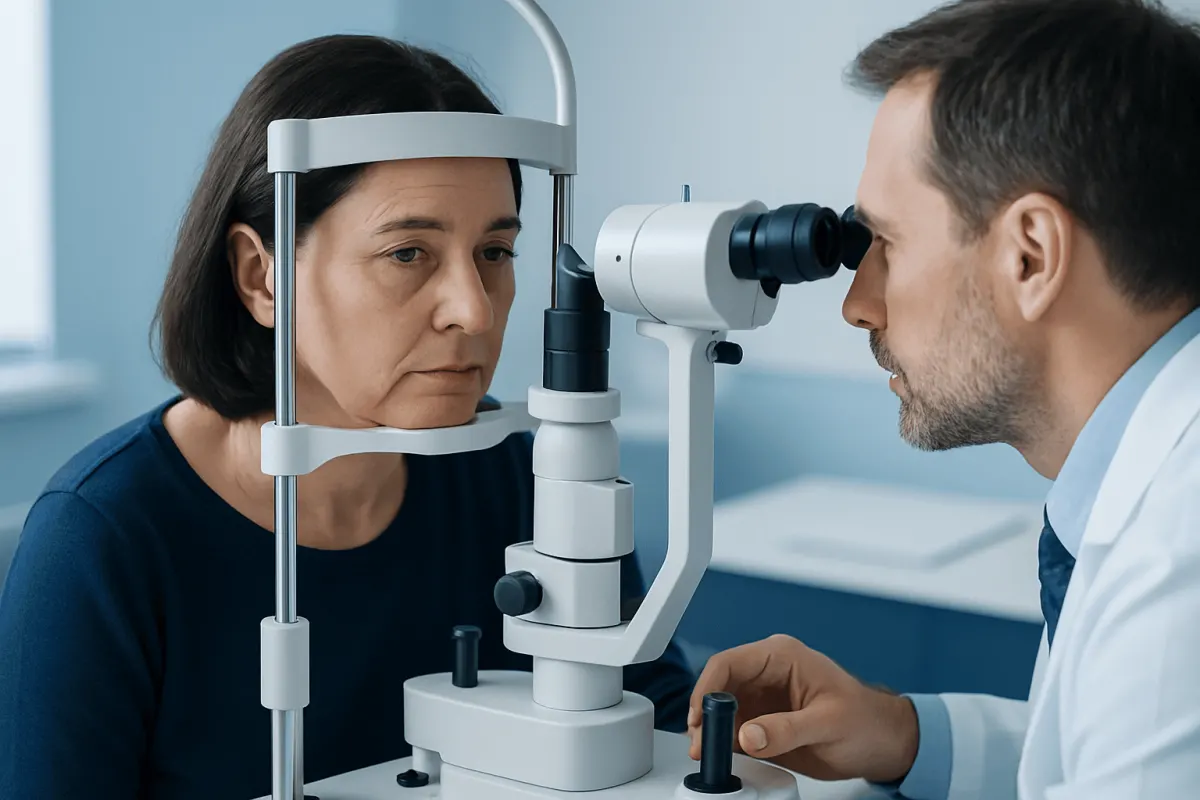
The examination usually includes visual acuity testing, color vision testing, pupil examination, eye pressure measurement, and a dilated examination of the optic nerve and retina. A key sign is a relative afferent pupillary defect, sometimes called an RAPD, which shows that one optic nerve is sending a weaker signal to the brain. The optic nerve may look swollen, pale, cupped, or normal, depending on the type and stage of disease.
Visual field testing maps areas of missing or reduced vision and is one of the most useful tools for understanding optic neuropathy. Optical coherence tomography, or OCT, measures the thickness of the retinal nerve fiber layer and ganglion cell layer, helping clinicians estimate structural damage and monitor change over time. Fundus photography may document the appearance of the optic disc for comparison at future visits.
Additional tests are selected based on the suspected cause. MRI of the brain and orbits with contrast may be used to evaluate inflammation, compression, demyelination, or other neurologic conditions. Blood tests may assess inflammation markers, infections, autoimmune disease, vitamin levels, and metabolic health. In selected cases, genetic testing, lumbar puncture, vascular imaging, or referral to neurology, rheumatology, endocrinology, or oncology may be appropriate.
Treatment Options
Treatment for optic neuropathy depends on the underlying cause. This is why careful diagnosis is so important. In some cases, the aim is to reduce inflammation, in others to protect remaining nerve function, treat a systemic illness, remove pressure on the nerve, or prevent further damage. Recovery varies widely and depends on the cause, severity, and timing of treatment.
Inflammatory optic neuritis may be monitored or treated with corticosteroids in selected situations, particularly when faster visual recovery is desired or when the pattern suggests a higher-risk inflammatory disorder. Some autoimmune optic neuropathies require additional immune-directed therapy under specialist care. If infection is suspected, treatment is guided by the specific organism and the patient’s overall condition.
Ischemic optic neuropathy management focuses on identifying and treating vascular and inflammatory risks. If giant cell arteritis is suspected, doctors typically begin urgent treatment while confirming the diagnosis, because early treatment can help protect the other eye and reduce systemic complications. For non-arteritic ischemic optic neuropathy, care often includes evaluation of blood pressure, diabetes, cholesterol, sleep apnea, and medication-related factors, although no treatment reliably reverses established damage in every patient.
Compressive optic neuropathy may require neurosurgical, endocrine, oncologic, or radiation oncology evaluation, depending on the cause. Toxic or nutritional optic neuropathy is managed by stopping the harmful exposure when possible and correcting deficiencies under medical supervision. Glaucoma-related optic neuropathy is treated by lowering eye pressure and monitoring the optic nerve and visual field over time.
Prevention, Monitoring, and Self-Care
Not all optic neuropathies can be prevented, especially inherited, inflammatory, or sudden ischemic forms. However, some risk factors can be reduced. Managing diabetes, blood pressure, cholesterol, and sleep apnea supports overall vascular health, including the small blood vessels that supply the optic nerve. Avoiding tobacco and excessive alcohol and maintaining a balanced diet also help reduce the risk of toxic and nutritional optic nerve injury.
Patients should take medications only as prescribed and should inform their doctors about all prescription medicines, over-the-counter products, and supplements. Some drugs can rarely affect the optic nerve, particularly when taken long term or in higher-risk patients. Regular eye examinations are important for people with glaucoma, diabetes, a strong family history of eye disease, or previous optic nerve problems.
Monitoring often includes repeat visual fields and OCT scans. These tests help determine whether the condition is stable, improving, or progressing. Patients may find it helpful to note changes in reading ability, color perception, night vision, and side vision, but home observation cannot replace formal testing. If one eye is affected, covering each eye separately from time to time may make new changes easier to notice.
When to See a Doctor
Any new or unexplained vision loss should be evaluated by an eye care professional. Prompt assessment is especially important when vision changes occur suddenly, affect one eye more than the other, cause a new blind spot, or are associated with pain on eye movement. New double vision, severe headache, neurologic symptoms, or loss of vision in part of the visual field also deserves timely medical attention.
People over 50 should seek urgent care if vision loss occurs with scalp tenderness, jaw pain while chewing, fever, unexplained weight loss, shoulder or hip aching, or new temple headache. These symptoms can occur with giant cell arteritis, a condition that requires rapid treatment. A clinician may order blood tests and begin treatment before all results are available if the suspicion is high.
Patients with known optic neuropathy should keep scheduled follow-up appointments even if vision feels stable. Some optic nerve conditions progress slowly and may be detected earlier on OCT or visual field testing than by symptoms alone. At Acıbadem Health Point, multidisciplinary specialists in JCI-accredited hospitals evaluate and treat optic nerve and neuro-ophthalmic conditions for international patients, coordinating care when eye, neurologic, vascular, or systemic factors overlap.
Frequently asked questions
01Is optic neuropathy the same as optic neuritis?
No. Optic neuropathy is the broader term for optic nerve damage or dysfunction from many possible causes. Optic neuritis is one type of optic neuropathy caused by inflammation of the optic nerve.
02Can optic neuropathy cause permanent vision loss?
It can, depending on the cause and severity. Some forms, such as many cases of typical optic neuritis, may improve significantly, while others may leave lasting visual field or color vision changes. Early diagnosis helps identify treatable causes and reduce the risk of further damage.
03Why is color vision tested in optic nerve disease?
The optic nerve is important for transmitting color and contrast information. In many optic neuropathies, colors may look faded or less bright even when the eye chart score is only mildly reduced. Comparing color perception between the two eyes can reveal subtle optic nerve dysfunction.
04Will an MRI always be needed for optic neuropathy?
Not always. MRI is commonly used when inflammation, compression, demyelinating disease, tumor, or an unexplained pattern of vision loss is suspected. In other cases, the diagnosis may rely more on the eye examination, OCT, visual field testing, blood tests, and medical history.
05Can glasses or cataract surgery correct optic neuropathy vision loss?
Glasses can improve focusing problems, and cataract surgery can improve vision when cataract is a major cause of blur. However, optic neuropathy affects the nerve signal itself, so optical correction cannot fully restore vision that is limited by optic nerve damage. An eye examination can help distinguish these causes.
06What should a patient bring to a neuro-ophthalmology appointment?
It is helpful to bring previous eye records, visual field tests, OCT scans, MRI or CT reports and images if available, a medication list, and information about medical conditions. Patients should also be ready to describe when symptoms began, whether vision changed suddenly or gradually, and whether pain, headache, or neurologic symptoms occurred.
This article is for general information only and is not a substitute for professional medical advice. Please consult a qualified doctor about your individual situation.
Explore treatments in Turkey — costs, top hospitals & a free quote
JCI-accredited · board-certified surgeons · reply within 24h
Add us as a Preferred Source to see more of our trusted health content across Google Search, AI Overviews and Discover.