Visual Field Testing in Neuro-Ophthalmology: What Abnormal Results Can Mean
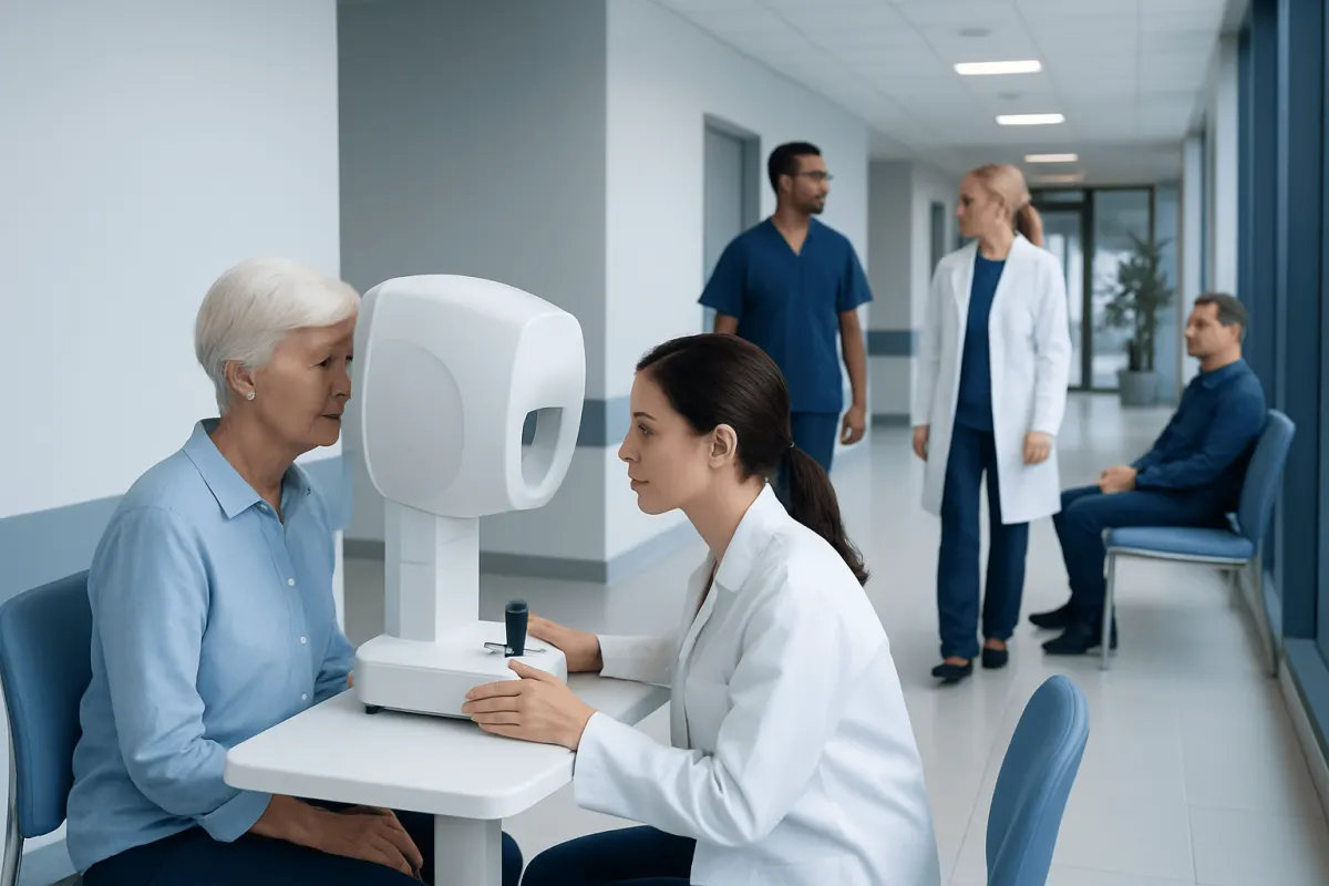
Visual field testing is a key neuro-ophthalmology examination that measures side vision, central vision, and blind spots that may not be obvious in daily life. Abnormal results can help doctors locate whether a problem may involve the eye, optic nerve, visual pathways, or brain.
Overview
Visual field testing, also called perimetry, measures the full area a person can see while looking straight ahead. Standard eye charts measure sharp central vision, but they do not show how well the side vision, upper and lower field, or small blind spots are working. In neuro-ophthalmology, this test is especially useful because the visual pathways extend from the eye through the optic nerve, optic chiasm, optic tract, brainstem connections, and visual cortex at the back of the brain.
During a visual field test, the person usually looks at a central target and presses a button when small lights appear in different locations. The machine records which lights were seen, which were missed, and how sensitive each area of the visual field is. Results are displayed as maps that help the doctor identify patterns. These patterns can be more informative than a simple pass or fail result.
Abnormal visual field results can point toward eye conditions, such as glaucoma or retinal disease, but they can also suggest optic nerve disorders, pituitary region problems, stroke, migraine-related changes, inflammation, or other neurological conditions. The result is interpreted together with the medical history, eye examination, pupil reactions, color vision, optic nerve appearance, and when needed, imaging tests such as MRI.
Why Neuro-Ophthalmologists Use Visual Field Testing
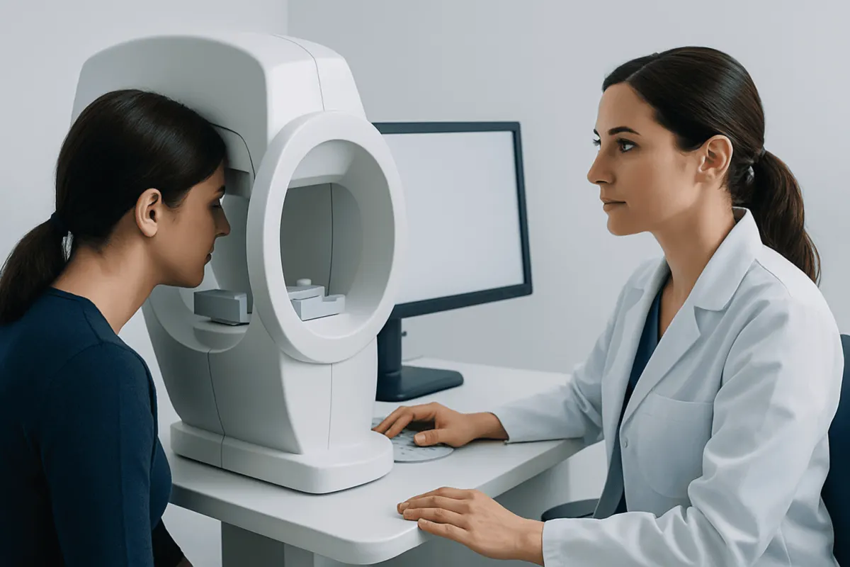
Neuro-ophthalmology focuses on conditions that affect both vision and the nervous system. Some of these conditions are subtle at first. A person may read well and have normal central vision, yet still miss objects to one side, bump into door frames, have difficulty driving, or notice that part of a page seems absent. Visual field testing can detect and measure these changes in a structured way.
The test also helps doctors identify where along the visual pathway a problem may be located. For example, damage in one eye or optic nerve often causes field loss in that same eye, while a problem behind the optic chiasm in the brain may cause matching field loss in the same side of both eyes. This localization is one reason visual field testing is so valuable in neuro-ophthalmic diagnosis.
Visual field testing is also used to monitor change over time. In conditions such as glaucoma, optic neuritis, compressive optic neuropathy, or recovery after a stroke, repeated tests can show whether the visual field is stable, improving, or worsening. Doctors often compare several tests rather than relying on a single measurement, because learning effect, tiredness, and attention can influence the result.
What Happens During the Test
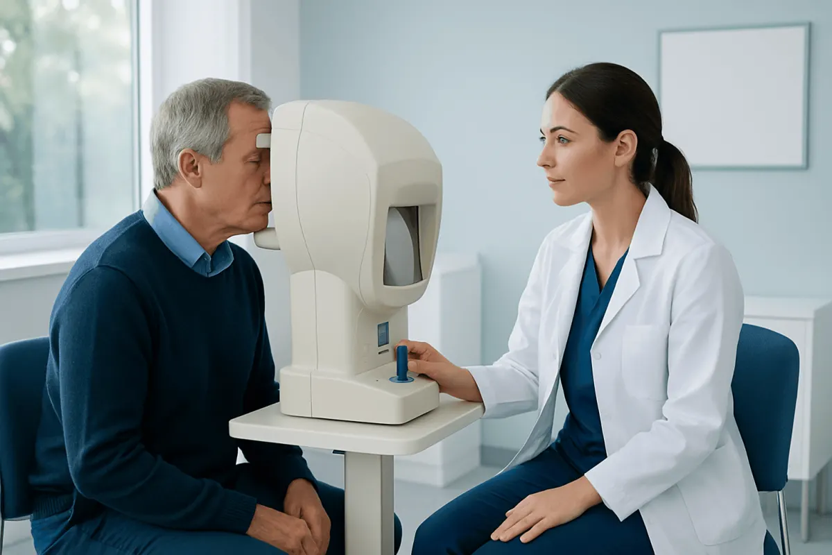
The most common method is automated static perimetry. The person sits at a bowl-shaped instrument, covers one eye, and looks at a central fixation light. Small points of light appear in different locations and at different brightness levels. The person presses a button each time a light is seen. Each eye is tested separately, and the test usually takes several minutes per eye, depending on the strategy used.
Good preparation improves accuracy. The person should be comfortably positioned, use the correct corrective lens if provided by the clinic, and try to blink normally. It is not necessary to see every light; some are intentionally dim and may be missed even by healthy eyes. The goal is to respond honestly when a light is seen, while keeping the gaze on the central target rather than searching around the bowl.
Other forms of visual field assessment may also be used. Confrontation visual field testing is a quick bedside or clinic screening method. Goldmann kinetic perimetry, in which moving light targets are used, may be helpful for certain neurological or low-vision situations. In children, older adults, or people who find automated testing difficult, the doctor may choose a method that best fits the patient’s attention, vision level, and clinical question.
Common Abnormal Visual Field Patterns
Visual field reports often include terms such as scotoma, hemianopia, quadrantanopia, generalized depression, or arcuate defect. These words describe the shape and location of areas where sensitivity is reduced or absent. The pattern matters because different diseases tend to affect different parts of the visual pathway.
Some important patterns include:
- Central scotoma: A blind or dim spot near the center of vision. This may occur with optic nerve inflammation, optic neuropathy, macular disease, or toxic and nutritional optic nerve problems.
- Arcuate defect: A curved area of field loss often associated with glaucoma, although other optic nerve conditions can sometimes resemble it.
- Altitudinal defect: Loss of the upper or lower half of vision in one eye, which may suggest certain optic nerve or retinal blood flow problems.
- Bitemporal hemianopia: Loss of the outer visual field in both eyes. This pattern can occur when the optic chiasm is compressed, for example by a pituitary region mass.
- Homonymous hemianopia: Loss of the same side of the visual field in both eyes. This usually points to a problem behind the optic chiasm, such as in the optic tract, optic radiation, or visual cortex.
- Quadrantanopia: Loss of one quarter of the visual field, which may help localize neurological disease within the brain’s visual pathways.
These patterns are clues, not final diagnoses. For example, a drooping eyelid can mimic an upper field defect, an incorrect lens can blur the test, and poor fixation can create misleading results. A neuro-ophthalmologist checks the reliability indices on the report and compares the field map with the physical examination before deciding what the abnormal result means.
Possible Causes of Abnormal Results
Abnormal visual field testing can be caused by conditions affecting the eye itself. Glaucoma is a common reason for progressive peripheral field loss. Retinal disorders, macular disease, retinal vascular problems, or previous eye injuries may also create characteristic defects. Cataract or other media opacities can cause more generalized reduction in sensitivity rather than a sharply localized neurological pattern.
Optic nerve conditions are another major group. Optic neuritis, ischemic optic neuropathy, compressive optic neuropathy, hereditary optic neuropathies, and toxic or nutritional optic neuropathies can affect central vision, color vision, pupil reactions, and the visual field. The doctor may look closely at the optic disc for swelling, pallor, cupping, or other changes that match the field result.
Neurological causes are particularly important in neuro-ophthalmology. Strokes, tumors, aneurysms, demyelinating disease, traumatic brain injury, inflammatory disorders, and raised intracranial pressure can affect the visual pathways. Pituitary and parasellar lesions may cause chiasmal field loss, while occipital lobe problems may cause homonymous visual field defects. Because the causes vary widely, the next steps depend on the pattern, timing, severity, and associated symptoms.
How Doctors Interpret and Confirm the Findings
Interpreting visual field testing begins with checking whether the test is reliable. The report may show fixation losses, false-positive responses, false-negative responses, and gaze tracking information. High error rates do not mean the person did anything wrong; the test is demanding, and fatigue, anxiety, dry eyes, or misunderstanding the instructions can affect performance. Sometimes the best next step is simply to repeat the test under better conditions.
If the result appears reliable, the doctor compares it with the clinical examination. This may include visual acuity, color vision, pupils, eye movements, eyelid position, intraocular pressure, retinal examination, and optic nerve assessment. Optical coherence tomography may be used to measure retinal nerve fiber layer and macular ganglion cell thickness, which can support or clarify suspected optic nerve disease.
When a neurological cause is possible, imaging or laboratory evaluation may be recommended. MRI of the brain and orbits is often used to evaluate the optic nerves, chiasm, and visual pathways. In selected cases, blood tests, vascular assessment, inflammatory markers, infectious disease testing, or consultation with neurology, endocrinology, rheumatology, or neurosurgery may be appropriate. The purpose is to connect the visual field pattern with the person’s overall health picture.
Treatment Options and Follow-Up
Treatment depends on the underlying cause rather than on the visual field test alone. For glaucoma, treatment may involve eye drops, laser procedures, or surgery to reduce eye pressure and protect remaining vision. For optic neuritis or inflammatory optic nerve disease, management depends on the suspected cause and may include observation, anti-inflammatory treatment, or investigation for related neurological conditions. For compressive lesions near the optic chiasm, care may involve endocrinology, neurosurgery, radiation oncology, or other specialties depending on the diagnosis.
Some visual field defects improve when the cause is treated, especially if the condition is identified early and the nerve fibers have not been permanently damaged. Other defects may remain stable or only partially improve. In such cases, follow-up testing helps determine whether the condition is controlled and whether daily-life adaptations may help. Low-vision rehabilitation, occupational therapy, scanning strategies, and safety modifications can support independence for people with persistent field loss.
Doctors usually schedule repeat visual field testing at intervals based on the diagnosis and level of risk. The same testing strategy and machine are often preferred for comparison. Patients can help by bringing previous test records, reporting any new symptoms, and taking regular medications as prescribed. At Acıbadem Health Point, multidisciplinary specialists in JCI-accredited hospitals evaluate and treat neuro-ophthalmic conditions for international patients when coordinated eye and neurological care is needed.
When to See a Doctor
A person should seek prompt medical attention if visual field loss appears suddenly, worsens quickly, or is accompanied by neurological symptoms. Warning signs include new weakness or numbness on one side, difficulty speaking, severe sudden headache, new double vision, confusion, imbalance, or loss of consciousness. These symptoms require urgent assessment because they may involve the brain, blood vessels, or optic nerve.
Non-urgent but timely evaluation is also important for gradual changes. Examples include bumping into objects on one side, difficulty reading because words disappear, trouble driving, new blind spots, worsening night navigation, or a visual field result that the patient does not understand. People with glaucoma, pituitary disease, multiple sclerosis, previous stroke, optic nerve disease, or unexplained headaches may need periodic visual field testing as part of ongoing care.
Patients should not try to interpret a visual field printout in isolation. The maps, grayscale images, probability plots, and numerical indices can be confusing without clinical context. A qualified ophthalmologist, neuro-ophthalmologist, or neurologist can explain whether the abnormality is likely real, whether repeat testing is needed, and what further evaluation or treatment is appropriate.
Frequently asked questions
01Is visual field testing painful?
No. Visual field testing is non-invasive and does not involve pain. Some people find it tiring because it requires concentration, but the test can usually be paused if the person needs a short break.
02Does an abnormal visual field test always mean a brain problem?
No. Abnormal results can come from many causes, including glaucoma, retinal disease, cataract, eyelid drooping, optic nerve conditions, or test reliability issues. The pattern of the defect and the rest of the examination help determine whether a neurological cause is likely.
03Can a visual field test be wrong?
Yes, a single test can be misleading. Fatigue, poor fixation, misunderstanding the instructions, dry eyes, or an incorrect lens can affect the result. Doctors often repeat the test or compare it with other findings before making a diagnosis.
04What is hemianopia?
Hemianopia means loss of half of the visual field. When the same side is missing in both eyes, it is called homonymous hemianopia and often points to a problem behind the optic chiasm in the brain’s visual pathways. The cause may require neurological assessment and imaging.
05Can lost visual field come back?
Sometimes it can improve, depending on the cause and how quickly treatment begins. Inflammation, compression, or some vascular conditions may show partial recovery, while other damage may be permanent. Follow-up testing helps doctors understand the direction of change.
06How should a patient prepare for visual field testing?
Patients should bring their glasses or contact lens information, use prescribed eye drops as usual unless told otherwise, and tell the technician if they are tired or uncomfortable. During the test, it helps to keep looking at the central target, blink normally, and press the button only when a light is seen.
This article is for general information only and is not a substitute for professional medical advice. Please consult a qualified doctor about your individual situation.
Explore treatments in Turkey — costs, top hospitals & a free quote
JCI-accredited · board-certified surgeons · reply within 24h
Add us as a Preferred Source to see more of our trusted health content across Google Search, AI Overviews and Discover.