MRI vs Biopsy for Prostate Cancer: When Each Is Used
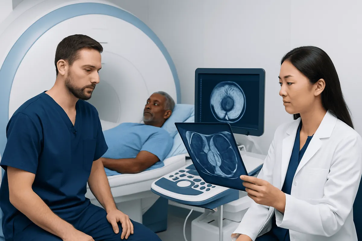
MRI and biopsy have different but complementary roles in prostate cancer assessment. MRI can show areas that may need closer evaluation, while a biopsy is usually required to confirm cancer and determine its grade.
MRI vs Biopsy for Prostate Cancer: Side-by-Side Comparison
For MRI vs biopsy for prostate cancer, the key difference is that MRI looks at the prostate, while biopsy examines prostate tissue under a microscope. A multiparametric MRI (mpMRI) can identify areas that appear more or less suspicious for clinically significant cancer. A biopsy can confirm whether cancer cells are present and provide information about the cancer grade.
| Feature | Prostate MRI | Prostate biopsy |
|---|---|---|
| Main purpose | Find and assess suspicious areas within the prostate | Confirm or rule out cancer by examining tissue |
| What it involves | Magnetic imaging, usually without radiation | Needle samples taken from the prostate, commonly with ultrasound guidance |
| What the result shows | Level of suspicion, location and possible local spread | Cancer presence, type and grade when cancer is found |
| Can it diagnose cancer definitively? | No; it estimates likelihood | Yes, when cancer cells are identified in the sample |
| Common role in care | Often performed before biopsy to help decide and plan sampling | Performed when confirmation is needed or risk remains concerning |
| Main limitations | May miss small or low-grade cancers and can produce uncertain findings | Can miss a cancer if the sampled tissue does not include it; carries small procedure-related risks |
Neither test automatically replaces the other. Clinicians interpret MRI, prostate-specific antigen (PSA) results, PSA density, digital rectal examination findings, family history, age, ethnicity, prior biopsy results and overall health together. The appropriate sequence is individualized rather than based on one test alone.
How Clinicians Put MRI and Biopsy Results Together
A prostate MRI is usually reported using the PI-RADS scoring system. Scores range from 1 to 5, with higher scores indicating a greater likelihood of clinically significant prostate cancer. The report also describes the size and location of any lesion, which can help a urologist direct biopsy needles more precisely if sampling is recommended.
Biopsy results answer a different question: what does the tissue show? If cancer is present, a pathologist evaluates its pattern and assigns a Grade Group, often based on the Gleason grading system. This information helps clinicians discuss whether active surveillance, surgery, radiation therapy or another approach may be appropriate.
A suspicious MRI does not always mean cancer. Benign prostate enlargement, inflammation and previous changes in the gland can sometimes resemble a tumor on imaging. Conversely, a negative MRI reduces the likelihood of significant cancer but cannot eliminate it completely, particularly when PSA levels, examination findings or inherited risk factors remain concerning.
If prostate cancer is confirmed, the care team may use imaging and pathology together to understand the condition more fully. Patients can learn more about prostate cancer and the tests commonly used to assess it.
Is MRI More Accurate Than Biopsy for Prostate Cancer?

MRI and biopsy are accurate in different ways, so it is not quite correct to say that one is simply more accurate than the other. MRI is helpful for detecting and locating areas that may contain clinically significant cancer, especially cancers that could be missed by untargeted sampling. It can also help avoid unnecessary biopsies for some people with a low-risk clinical picture and a reassuring scan.
However, biopsy remains the standard way to confirm a prostate cancer diagnosis because it directly examines cells. A conventional systematic biopsy takes samples from predetermined areas of the prostate. A targeted biopsy directs samples toward a lesion seen on MRI, and many clinicians combine targeted and systematic samples to reduce the chance of missing important disease.
Both tests have limitations. MRI quality and interpretation matter, and small, low-volume or less visible cancers may not be seen. A biopsy examines only selected tissue, so it may miss a tumor or underestimate its grade. For this reason, an apparently negative result may still need follow-up when clinical suspicion remains high.
Is It Better to Get a Prostate MRI or Biopsy First?
For many people who have not had a biopsy before and have an elevated PSA level or another reason for concern, guidelines support considering multiparametric MRI before biopsy. This approach can help identify people who are more likely to benefit from a biopsy and can guide targeted sampling when a suspicious area is found.
There are exceptions. A clinician may recommend proceeding directly to biopsy if the risk of significant cancer appears high based on PSA, examination findings or other factors, if MRI is unavailable or unsuitable, or if a previous MRI does not explain persistently concerning results. MRI may also be less suitable for some people with certain implanted devices, severe claustrophobia or other practical considerations, although many devices are MRI-compatible.
When MRI shows no suspicious lesion, the next step depends on the overall risk. Some people can be monitored with repeat PSA testing and clinical review. Others may still need biopsy because a normal-looking scan does not fully exclude cancer. A urologist can explain the balance of benefits and limitations for the individual situation.
Can You Tell If a Prostate Tumor Is Cancerous From an MRI?
No. MRI can show that an area has imaging features associated with cancer, but it cannot prove that a prostate tumor is cancerous. Radiologists use MRI findings to estimate suspicion and assign a PI-RADS score, not to make a tissue diagnosis.
Inflammation, infection, scarring and benign nodules can sometimes look suspicious on MRI. In addition, MRI cannot always determine the exact grade of a tumor. When the result would change care, a biopsy is generally needed to identify cancer cells and determine their grade.
MRI remains highly valuable because it can show where a lesion is located, whether there are features suggesting extension beyond the prostate, and which area should be sampled. This supports more informed biopsy planning and can contribute to treatment decisions after diagnosis.
How Long Should I Wait Between a Prostate MRI and a Biopsy?
There is no single required waiting period between a prostate MRI and biopsy. If MRI identifies a concerning lesion and biopsy is recommended, it is commonly arranged within weeks, based on the level of concern, symptoms, local scheduling and the person’s health. The clinician will advise on the appropriate timing.
When possible, MRI is often performed before biopsy because bleeding and inflammation caused by biopsy can affect image interpretation for a period afterward. If a person has already had a biopsy, the care team may recommend waiting before MRI so post-biopsy changes are less likely to obscure the scan. The ideal interval varies with the clinical question and the imaging protocol.
Before a biopsy, patients should tell the clinical team about blood-thinning medicines, allergies, prior infections, urinary symptoms and any implanted medical devices. The team will provide individualized instructions about medicines, preparation and signs of infection or other complications that require prompt contact after the procedure.
What to Do for Common Result Combinations
A raised PSA with a suspicious MRI finding commonly leads to an MRI-targeted biopsy, often combined with systematic sampling. If biopsy confirms cancer, the next decisions are based on Grade Group, PSA, imaging, the amount of tissue involved and personal preferences. Some low-risk cancers can be monitored through active surveillance, while others require treatment.
A raised PSA with a negative MRI may lead to monitoring or biopsy depending on PSA density, PSA trend, examination findings and risk factors. Persistent concern after a negative biopsy may lead to repeat MRI, repeat biopsy or a different sampling approach. A negative test is reassuring, but it should be interpreted within an ongoing follow-up plan rather than in isolation.
If cancer treatment is needed, options may include prostatectomy, radiation-based treatment, hormone therapy or other therapies depending on the stage and biology of the disease. The best option depends on the individual, and patients should have time to discuss possible benefits, side effects and alternatives with an experienced multidisciplinary team.
Acıbadem Health Point’s multidisciplinary specialists at JCI-accredited hospitals evaluate and treat prostate conditions for international patients, with care plans based on imaging, pathology and each patient’s clinical needs.
When to Seek Medical Care
A person should arrange medical review if they have a persistently elevated or rising PSA level, an abnormal digital rectal examination, blood in the urine or semen, recurrent urinary infections, or a strong family history of prostate or related cancers. Urinary symptoms such as weak flow, frequent urination or waking at night are common with benign prostate enlargement and do not by themselves diagnose cancer, but they still merit assessment if they are new, persistent or bothersome.
Urgent medical advice is appropriate for inability to pass urine, fever or chills with urinary symptoms, severe pelvic pain, or feeling unwell after a prostate biopsy. These symptoms can have several causes, including infection, and prompt evaluation is important.
People considering MRI or biopsy should discuss their personal risk and concerns with a urologist. Shared decision-making can help clarify whether monitoring, MRI, biopsy or a combination of approaches is the most suitable next step.
Frequently asked questions
01Does a high PSA mean that I have prostate cancer?
No. PSA can rise for several reasons, including benign prostate enlargement, inflammation, infection, recent ejaculation or urinary procedures. A PSA result is interpreted alongside age, PSA trend, prostate size, examination findings, MRI and, when needed, biopsy.
02What is an MRI-targeted prostate biopsy?
An MRI-targeted biopsy uses MRI findings to guide sampling of a suspicious area in the prostate. It may be performed with MRI-ultrasound fusion technology, cognitive targeting or, less commonly, directly in an MRI scanner. It is often combined with systematic samples from other prostate areas.
03Can a negative prostate MRI rule out cancer?
A negative MRI lowers the likelihood of clinically significant prostate cancer, but it does not rule it out completely. Whether biopsy can be deferred depends on the PSA level, PSA density, examination findings, family history and other individual risk factors.
04Is a prostate biopsy painful?
A biopsy is usually performed with local anesthetic, and sedation may be offered in some settings. People may feel pressure or brief discomfort, but the experience varies. The clinical team can explain the technique, pain-control options and expected recovery before the procedure.
05What are the risks of a prostate biopsy?
Temporary blood in the urine, semen or stool and short-term discomfort can occur after biopsy. Infection and urinary retention are less common but important risks, which is why preparation and aftercare instructions should be followed closely. Patients should contact their care team promptly if they develop fever, chills, worsening pain or difficulty urinating.
06If biopsy finds low-risk prostate cancer, is treatment always needed?
Not always. Some low-risk prostate cancers grow slowly and can be managed with active surveillance, which includes regular PSA tests, examinations, imaging and sometimes repeat biopsy. Treatment is considered if monitoring shows signs that the cancer is becoming more likely to cause harm or if the patient prefers treatment after informed discussion.
This article is for general information only and is not a substitute for professional medical advice. Please consult a qualified doctor about your individual situation.
Oncology care in Turkey — second opinion and treatment plan
JCI-accredited · board-certified surgeons · reply within 24h
Add us as a Preferred Source to see more of our trusted health content across Google Search, AI Overviews and Discover.