Breast Cancer Diagnosis: Exams, Imaging and Biopsy
Breast Prostate Cancer Diagnosis: Tests and Next Steps" class="ahp-ilk">cancer diagnosis usually begins with screening or a breast change noticed by a patient or clinician, then progresses through imaging and, when needed, a biopsy. A biopsy is the only test that can confirm whether cells are cancerous and provides information that helps plan treatment.
Overview: How Breast Cancer Is Diagnosed
Breast cancer diagnosis combines several steps rather than relying on one test alone. A clinician considers a person’s symptoms, personal and family history, breast examination findings and imaging results. If an area looks suspicious, a sample of tissue is taken and examined in a laboratory.
Screening aims to find breast cancer before it causes symptoms. Diagnostic testing, by contrast, investigates a particular concern, such as a lump, nipple discharge, skin change or an unexpected finding on a screening mammogram. Screening results that require further assessment are common and do not by themselves mean that cancer is present.
When cancer is confirmed, laboratory testing of the biopsy sample helps identify the type and characteristics of the cancer. These results, together with imaging and overall health information, support an individualized care plan. For an overview of the condition and its treatment pathways, readers may consult breast cancer care.
Screening Tests and Who May Benefit
Breast screening is intended for people without breast symptoms. Mammography uses low-dose X-rays to look for small masses, distortions in breast tissue or tiny calcium deposits that can sometimes be early signs of cancer. Screening recommendations vary by country, age, sex assigned at birth, breast density, family history and other risk factors, so an individual should discuss the right schedule with a doctor.
People with a higher-than-average risk may need earlier or more frequent screening. This can include people with certain inherited gene changes, a strong family history of breast or ovarian cancer, previous chest radiation at a young age, or some prior breast biopsy findings. In selected higher-risk individuals, breast MRI may be used alongside mammography.
Breast self-awareness is also helpful. This means knowing what is usual for the breasts and seeking assessment for a new or persistent change, rather than relying on self-examination alone as a replacement for screening. A routine health assessment can help review personal risk and preventive care needs; health check-up packages may include appropriate consultations and tests based on individual circumstances.
Symptoms That Can Lead to Diagnostic Testing

Many breast changes are caused by non-cancerous conditions, including cysts, hormonal changes, infections or benign growths. Even so, it is important not to delay assessment of a change that is new, persistent or worsening. Breast pain alone is common and is often not caused by cancer, but pain that is focal or continues should be discussed with a clinician.
Changes that may lead to diagnostic testing include:
- a new lump, thickened area or swelling in the breast or underarm;
- a change in breast shape, size or contour that is not expected for the individual;
- skin dimpling, redness, scaling or an orange-peel-like texture;
- a nipple that newly turns inward, develops a rash or produces bloody or clear discharge; and
- a sore on the breast or nipple that does not heal.
Clinical assessment begins with a focused history and breast examination. The clinician may assess both breasts and the lymph nodes around the underarm and collarbone. Imaging is usually the next step when there is a concerning symptom or examination finding.
Imaging: Mammogram, Ultrasound and MRI
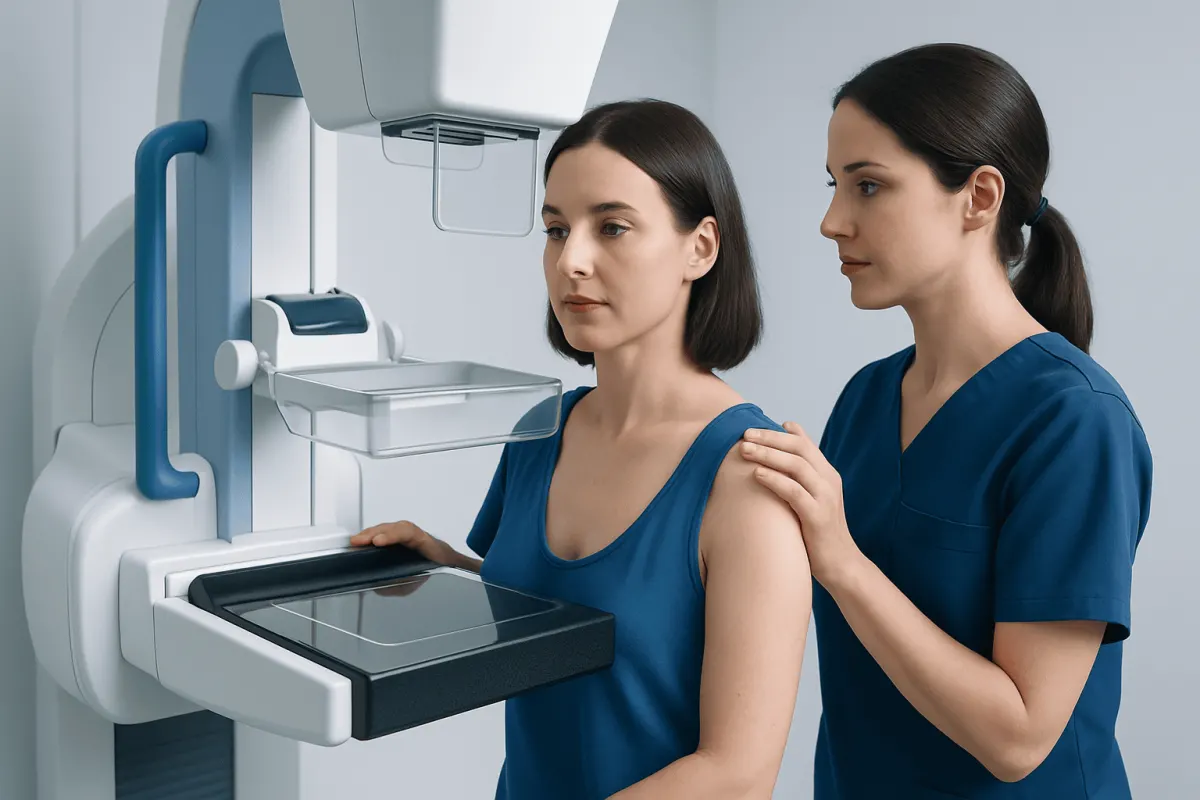
Diagnostic mammography takes targeted images of a specific breast area and may include additional views beyond those used for screening. During the examination, each breast is briefly compressed between plates to keep it still and produce clear images. Compression can be uncomfortable for a short time but should not cause lasting harm.
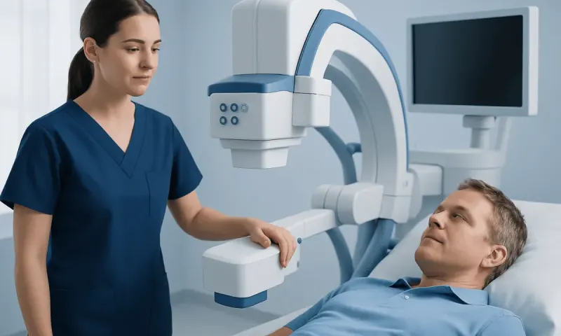
Breast ultrasound uses sound waves and does not involve radiation. It is often used to assess a lump or an area seen on mammography, and it can help distinguish a fluid-filled cyst from a solid mass. Ultrasound may also guide a needle during a biopsy.
Breast MRI uses magnetic fields and contrast material to create detailed images. It may be appropriate for some people at increased risk, for evaluating the extent of a known cancer, or when other imaging does not answer an important clinical question. MRI is sensitive, meaning it can detect many changes, but it can also identify findings that need further testing and are ultimately benign.
Imaging reports may use a standardized assessment category to communicate how likely a finding is to be benign and what follow-up is recommended. A clinician should explain the result in context, including whether routine screening, short-interval imaging or biopsy is advised.
Breast Biopsy: How It Works and What to Expect
A breast biopsy is the procedure used to confirm or rule out cancer. It removes cells or small tissue samples from an abnormal area so that a pathologist can examine them under a microscope. Most suspicious imaging findings do not turn out to be cancer, but imaging alone cannot always provide a definite answer.
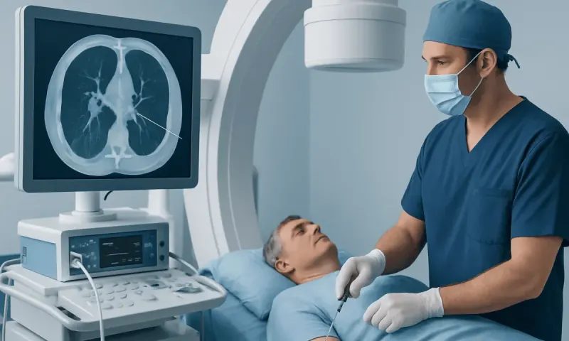
Needle biopsy is the most common approach. Depending on the location and appearance of the finding, the radiologist may use ultrasound, mammography or MRI guidance to place the needle accurately. A fine-needle aspiration collects cells, while a core needle biopsy removes small cylinders of tissue and is commonly used because it provides more information. Surgical biopsy is less common and may be considered if needle biopsy is not suitable or does not provide a clear result.
Before a biopsy, the care team reviews medications, allergies, bleeding risks and pregnancy status where relevant. Blood-thinning medication may need special planning, but it should never be stopped without advice from the prescribing clinician. The procedure is usually performed with local anesthetic; the person remains awake, and pressure rather than sharp pain is generally felt once the area is numb.
After samples are collected, a small marker clip may be placed at the site. This is safe for most people and can help identify the area on future imaging. The samples are sent for pathology testing, and the care team explains when and how results will be provided. For information about coordinated diagnostic and treatment planning, see breast cancer cancer Treatment Options: Surgery, Chemo, Radiation & Beyond" class="ahp-ilk">treatment options.
Results, Staging and Further Tests
If a biopsy shows no cancer, the radiologist and pathologist should confirm that the pathology result matches the imaging finding. This is called radiology-pathology concordance. When the findings do not match, additional sampling or surgical assessment may be recommended to ensure the area has been adequately evaluated.
If cancer is found, the pathology report identifies the cancer type and may describe its grade. Tests are usually performed to determine hormone receptor status, including estrogen and progesterone receptors, and HER2 status. These features do not describe a person’s worth or prognosis on their own; they help the specialist team choose treatments that are most likely to be appropriate.
Further tests may be needed to establish the stage, which describes the size and location of the cancer and whether it has spread to nearby lymph nodes or elsewhere. Depending on the clinical situation, this may involve further breast imaging, lymph node evaluation or scans. Not every person needs the same tests, and extensive body scans are not routinely required for every early breast cancer diagnosis.
Receiving results can be emotionally difficult. It can help to bring a trusted person to appointments, write down questions and ask for copies or explanations of reports. A multidisciplinary team may include breast surgeons, medical oncologists, radiation oncologists, radiologists, pathologists, nurses and supportive care professionals.
Benefits, Limitations, Risks and Recovery
The main benefit of screening and timely diagnostic evaluation is the opportunity to detect cancer at an earlier stage or clarify that a change is benign. However, all screening tests have limitations. Mammograms can miss some cancers, especially in dense breasts, and they can detect changes that require extra testing but are not cancer.
Imaging may lead to false-positive findings, meaning a test appears abnormal although cancer is not present. Less commonly, screening may identify a slow-growing cancer that might never have caused symptoms. These possibilities are part of informed screening decisions and can be discussed with a doctor in relation to personal risk.
After a needle biopsy, mild bruising, tenderness or swelling is common and generally settles over several days to one or two weeks. Most people can return to gentle usual activities the same day or the next day, although strenuous exercise and heavy lifting may be limited briefly. The team may recommend a supportive bra, a cold pack and suitable non-prescription pain relief if medically appropriate.
Complications from needle biopsy are uncommon but can include bleeding, infection or a larger collection of blood under the skin. A person should contact the clinical team if there is increasing redness, warmth, drainage, fever, severe pain or swelling that is getting worse. A biopsy does not cause breast cancer to spread.
When to Seek Medical Care
A person should arrange a medical appointment promptly for a new breast or underarm lump, bloody nipple discharge, a persistent localized breast change, or skin or nipple changes that do not resolve. It is also important to follow up on a recall after a screening mammogram, even though many recalls lead to benign results.
Urgent medical advice is appropriate if a breast becomes rapidly red, hot and swollen, particularly with fever or feeling unwell, as infection may need prompt treatment. People who are pregnant, breastfeeding or have breast implants should still seek assessment for concerning changes; the choice of imaging can be adapted to the situation.
People with a strong family history of breast, ovarian, pancreatic or prostate cancer may benefit from a discussion about risk assessment and genetic counselling. Acıbadem Health Point’s multidisciplinary specialists and JCI-accredited hospitals diagnose and treat breast conditions for international patients, with care planned according to clinical needs.
Frequently asked questions
01Can a mammogram diagnose breast cancer on its own?
A mammogram can identify an abnormal area that may need further evaluation, but it cannot confirm cancer on its own. Confirmation generally requires a biopsy, in which tissue is examined by a pathologist.
02Does being called back after a mammogram mean cancer is likely?
No. A callback means that additional images or testing are needed to clarify a finding. Many callbacks are due to normal tissue overlap, cysts or other benign changes.
03What happens during a core needle breast biopsy?
The breast is numbed with local anesthetic, and imaging guidance is used to position a hollow needle at the area of concern. Several small tissue samples are collected, usually during an outpatient visit, and sent to a laboratory.
04How long does it take to receive breast biopsy results?
Timing varies according to the laboratory and whether additional specialized testing is needed. The care team should explain the expected timeframe and arrange a discussion of the results.
05Can breast ultrasound replace a mammogram?
Ultrasound and mammography provide different information, so one does not routinely replace the other. Ultrasound is often used as an additional test to investigate a specific finding or to guide biopsy.
06Do all breast lumps need a biopsy?
Not all lumps need a biopsy. A clinician may determine through examination and imaging that a finding is clearly benign or can be safely monitored, while suspicious or unclear findings usually need tissue sampling.
This article is for general information only and is not a substitute for professional medical advice. Please consult a qualified doctor about your individual situation.
Oncology care in Turkey — second opinion and treatment plan
JCI-accredited · board-certified surgeons · reply within 24h
Add us as a Preferred Source to see more of our trusted health content across Google Search, AI Overviews and Discover.