Melanoma and Nonmelanoma Skin Cancer: Diagnosis and Outlook
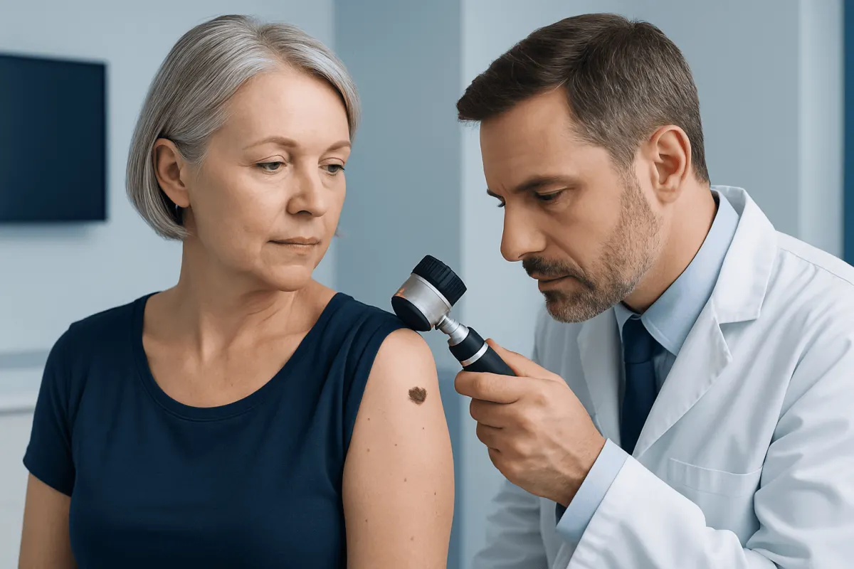
Melanoma and nonmelanoma skin cancer are different forms of cancer that begin in skin cells. Early assessment of a new, changing or non-healing skin lesion helps doctors confirm the diagnosis, choose appropriate treatment and discuss the likely outlook.
Understanding melanoma and nonmelanoma skin cancer
Melanoma and nonmelanoma skin cancer are cancers that start in the skin, but they arise from different types of cells and can behave differently. Melanoma develops in melanocytes, the cells that make skin pigment. Although it is less common than nonmelanoma skin cancer, melanoma is more likely to grow into deeper layers of skin and spread to other parts of the body if it is not diagnosed early.
Nonmelanoma skin cancer is a broad term that most often refers to basal cell carcinoma and squamous cell carcinoma. Basal cell carcinoma usually grows slowly and rarely spreads, while squamous cell carcinoma can be more likely to spread when it is large, deep, recurrent or located in certain areas. Both are often treatable, particularly when found at an early stage.
Skin cancer can occur anywhere on the body, including places that receive little sun exposure. It may appear on the face, scalp, ears, neck, hands, trunk, legs, soles of the feet, under the nails or in genital areas. A diagnosis is not possible from appearance alone, so a suspicious lesion should be assessed by a qualified clinician.
Skin changes that may need assessment
Melanoma may begin in an existing mole or appear as a new dark or unusual mark. One useful way to notice potentially concerning changes is the ABCDE approach: asymmetry, uneven borders, varied colour, a diameter that is increasing, and evolution or change over time. A spot that looks distinctly different from a person’s other moles, sometimes called an “ugly duckling,” also deserves attention.
Basal cell carcinoma may look like a pearly or shiny bump, a pink patch, a sore that repeatedly crusts or bleeds, or a scar-like area without a clear injury. Squamous cell carcinoma may appear as a rough, scaly patch, a firm red bump, a wart-like growth or a sore that does not heal. These descriptions are not definitive because benign conditions can look similar.
- A mole or spot that changes in size, shape, colour or sensation
- A lesion that bleeds, crusts, ulcerates or does not heal
- A new dark streak beneath a nail or a changing nail pigment band
- A persistent scaly, tender or raised patch of skin
- New swelling in nearby lymph nodes, particularly after a known skin Prostate Cancer Diagnosis: Tests and Next Steps" class="ahp-ilk">cancer diagnosis
Many skin marks are harmless. However, monitoring a changing lesion rather than having it examined may delay diagnosis, so it is sensible to arrange a skin review when there is uncertainty.
Causes and factors that increase risk
Ultraviolet, or UV, radiation is a major preventable cause of many skin cancers. UV exposure comes mainly from sunlight and artificial tanning devices. Repeated sun exposure over many years is strongly associated with basal cell and squamous cell carcinoma, while intermittent intense exposure and sunburns can contribute to melanoma risk.
Risk is also influenced by personal characteristics and medical history. People with fair skin, light eyes or hair, a tendency to burn, numerous moles, atypical moles, or a family history of melanoma may have a higher risk. Skin cancer can still occur in people of every skin tone, and lesions may be diagnosed later in darker skin if changes are less readily recognised.
Other factors include older age, previous skin cancer, a weakened immune system, previous radiation treatment, chronic wounds or inflammation, and certain inherited conditions. Having a risk factor does not mean a person will develop cancer, and having no recognised risk factors does not eliminate the possibility. The most useful response is ongoing awareness of the skin and appropriate UV protection.
How doctors diagnose and stage skin cancer
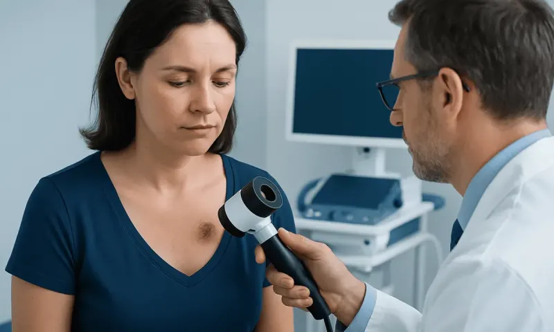
Diagnosis usually starts with a medical history and examination of the skin. The clinician may ask when the lesion appeared, whether it has changed, whether it bleeds or itches, and about sun exposure, previous skin cancers and family history. A dermatoscope, a handheld device that magnifies and illuminates the skin, may help the clinician evaluate a lesion more closely.
A biopsy is the only way to confirm whether a suspicious area is cancerous. During a biopsy, the clinician removes all or part of the lesion and sends it to a laboratory for examination. The pathology report identifies the cancer type and can provide details that help guide management. For melanoma, these may include tumour thickness, ulceration and other microscopic features.
If melanoma or a higher-risk squamous cell carcinoma is diagnosed, further assessment may be recommended to determine whether it has spread beyond the original site. This can include examination of nearby lymph nodes, imaging in selected situations, and sometimes sentinel lymph node biopsy. Staging describes the extent of cancer and helps the care team discuss treatment and outlook.
Modern treatment approaches
For many skin cancers, surgery is the main treatment. A clinician removes the cancer along with a margin of surrounding healthy-looking tissue. The method depends on the cancer type, size, location, depth and whether it has returned after previous treatment. Some lesions can be treated with procedures such as curettage and cautery, cryotherapy, topical medicines or light-based treatments when clinically appropriate.
Mohs micrographic surgery may be considered for selected basal cell or squamous cell carcinomas, especially on areas where preserving healthy tissue is important, such as the face, ears, hands or genital region. During this technique, tissue is removed in stages and examined until the edges are clear. Radiation therapy can be an alternative or an additional treatment for some patients when surgery is not suitable or when there is a meaningful risk of recurrence.
Melanoma is generally treated with surgical excision. Depending on the stage and pathology findings, treatment may also include sentinel lymph node biopsy, immunotherapy, targeted therapy or, less commonly, radiation therapy. Advanced melanoma and advanced squamous cell carcinoma are increasingly managed with systemic medicines selected according to the individual cancer and the person’s overall health.
The care plan should be developed with a dermatologist, surgeon, oncologist and other specialists as needed. Melanoma and other skin cancers require follow-up because a person who has had one skin cancer can develop another in the future.
Outlook and follow-up after treatment
The outlook for skin cancer depends on the specific diagnosis, how deeply it has grown, whether it has spread, the location of the tumour, its biological features and the person’s general health. Basal cell carcinoma is very often managed successfully with local treatment. Squamous cell carcinoma also commonly has a favourable outlook when treated early, though certain high-risk tumours need closer monitoring.
For melanoma, earlier-stage disease is generally associated with a more favourable outlook than melanoma that has reached lymph nodes or distant organs. Pathology and staging results allow the doctor to give information that is relevant to the individual rather than relying on broad comparisons. Newer systemic treatments have expanded options for many people with higher-risk or advanced melanoma.
Follow-up appointments are important after treatment. They may include examination of the treated area, a full-skin check and, for selected cancers, lymph node examinations or imaging. The schedule varies according to the type and stage of cancer. Between appointments, patients can become familiar with their own skin and report new or changing lesions promptly.
Lowering UV exposure and caring for the skin
UV protection is an important part of preventing skin damage and lowering the risk of future skin cancers. It is helpful to seek shade when UV levels are high, wear a broad-brimmed hat, sunglasses and protective clothing, and use a broad-spectrum sunscreen with an SPF of 30 or higher on exposed skin. Sunscreen should be applied generously and reapplied according to the product instructions, especially after swimming or sweating.
Artificial tanning devices should be avoided because they expose the skin to harmful UV radiation. Sun protection is needed throughout the year, not only on hot or sunny days, because UV radiation can still reach the skin through clouds and reflect from surfaces such as water, sand and snow.
A regular self-check can support, but never replace, professional skin examinations. A person may use a mirror or ask a trusted family member to check areas that are difficult to see, such as the scalp, back and backs of the legs. Photographing a stable mole can sometimes help identify change, but a changing or suspicious lesion should be reviewed rather than monitored at home alone.
When to seek medical care
A person should arrange a medical appointment promptly for a mole that is changing, an unexplained new dark mark, a skin sore that does not heal, or a lesion that repeatedly bleeds, crusts or becomes painful. Assessment is also appropriate for a new growth that enlarges over several weeks or months, even if it is not painful.
People with a personal history of skin cancer, many atypical moles, a strong family history of melanoma or immune suppression may benefit from planned professional skin checks. The clinician can advise how often these should take place based on individual risk.
Acıbadem Health Point’s multidisciplinary specialists and JCI-accredited hospitals support international patients who need assessment and treatment for skin cancer. Care decisions should always be based on pathology results, stage, personal health needs and discussion with the treating team.
Frequently asked questions
01What is the main difference between melanoma and nonmelanoma skin cancer?
Melanoma starts in pigment-producing melanocytes, whereas nonmelanoma skin cancer usually refers to basal cell carcinoma and squamous cell carcinoma. Melanoma is less common but has a greater tendency to spread if it is not detected and treated early. All suspicious skin lesions should be assessed by a clinician.
02Can skin cancer develop in areas that are not exposed to the sun?
Yes. Although UV exposure is a major risk factor, skin cancer can develop anywhere on the body, including the soles of the feet, under nails, the scalp and genital areas. Regular awareness of the whole skin surface is useful for everyone.
03Does an itchy mole mean melanoma?
Itching alone does not mean that a mole is melanoma, as many harmless skin conditions can itch. However, itching together with a mole that changes in shape, size, colour or surface should be checked. A clinician may recommend a biopsy if the lesion appears suspicious.
04How is skin cancer confirmed?
Skin cancer is confirmed with a biopsy. A doctor removes all or part of the abnormal area and a pathologist examines the tissue under a microscope. This identifies the cancer type and provides information needed to plan treatment.
05Is skin cancer always treated with surgery?
Surgery is commonly used and is often the main treatment for early skin cancers. Depending on the diagnosis and location, other options may include topical treatments, cryotherapy, radiation therapy, immunotherapy or targeted therapy. The most suitable approach depends on the individual cancer and the patient’s health.
06Can skin cancer come back after treatment?
Some skin cancers can recur at the original site, and people who have had skin cancer have an increased chance of developing a new skin cancer elsewhere. Follow-up visits, self-awareness and sun protection are therefore important after treatment. The care team can recommend a follow-up schedule based on the cancer type and risk level.
This article is for general information only and is not a substitute for professional medical advice. Please consult a qualified doctor about your individual situation.
Hair transplant in Turkey — costs, graft counts & a free assessment
JCI-accredited · board-certified surgeons · reply within 24h
Add us as a Preferred Source to see more of our trusted health content across Google Search, AI Overviews and Discover.