How Varicose veins Is Diagnosed: Tests, Imaging & What to Expect
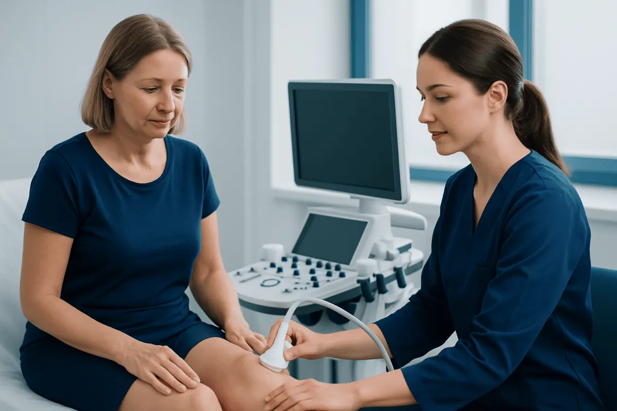
Varicose veins diagnosis usually begins with a clinical assessment of the legs, followed by duplex ultrasound when symptoms, visible veins or treatment planning warrant a closer look. The test is painless and helps clinicians identify faulty vein valves, map blood flow and rule out important complications.
Overview: how varicose veins are diagnosed
Varicose veins diagnosis is based on a person’s symptoms, medical history, a physical examination and, when needed, an ultrasound scan of the leg veins. The aim is not only to identify visible enlarged veins, but also to understand whether vein valves are allowing blood to flow backward, a problem called venous reflux.
Healthy leg veins carry blood back to the heart. Small one-way valves inside the veins help prevent blood from falling backward when a person stands. When these valves weaken, blood can pool in superficial veins near the skin, causing twisting or bulging veins and sometimes aching, heaviness, swelling, itching or skin changes.
Not every visible vein requires extensive testing. However, evaluation is particularly useful when symptoms affect daily activities, swelling is persistent, skin changes are present, a vein has bled, or a person is considering treatment. The findings allow the clinician to develop an individual plan rather than basing decisions on appearance alone.
What happens at the first appointment
The appointment usually starts with questions about the person’s concerns. A clinician may ask when the veins appeared, whether symptoms worsen after standing, whether there is ankle swelling or night cramping, and whether there has been bleeding, a leg ulcer, a blood clot or previous vein treatment.
Medical and family history also matters. Pregnancy, jobs involving prolonged standing or sitting, reduced mobility, excess body weight, prior leg injury, hormone use and a family history of varicose veins can all be relevant. The clinician will also review medications and conditions that may influence circulation or the safety of a future procedure.
During the examination, the legs are usually assessed while the person is standing because veins are easier to see when filled with blood. The clinician looks for enlarged veins, swelling, tenderness, discoloration, eczema-like skin irritation, hardened skin and slow-healing wounds around the ankle. Pulses in the feet may also be checked to assess arterial circulation before compression is recommended.
- Visible bulging or branching surface veins
- Ankle or lower-leg swelling
- Brownish skin staining, itching or inflammation
- Areas of tenderness, warmth or firmness along a vein
- Signs of a current or previous venous leg ulcer
Duplex ultrasound: the main imaging test
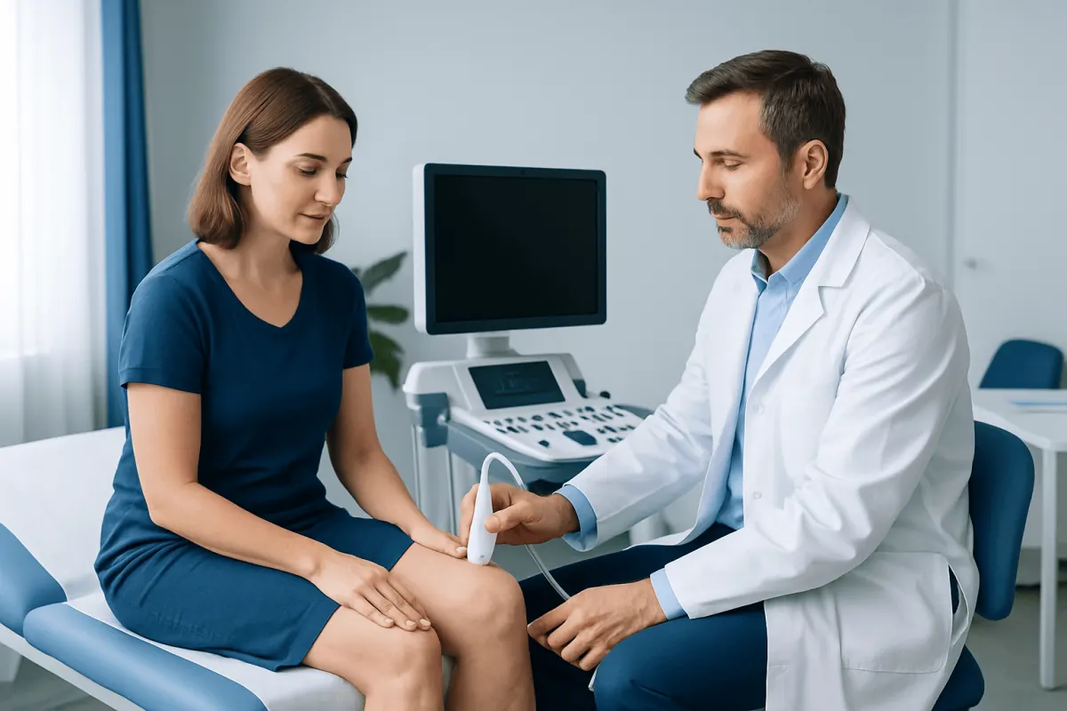
Duplex ultrasound is the principal test used in varicose veins diagnosis. It combines standard ultrasound, which creates images of veins and surrounding tissues, with Doppler ultrasound, which shows the direction and speed of blood flow. It is noninvasive, uses no ionizing radiation and usually does not require injections, fasting or special preparation.
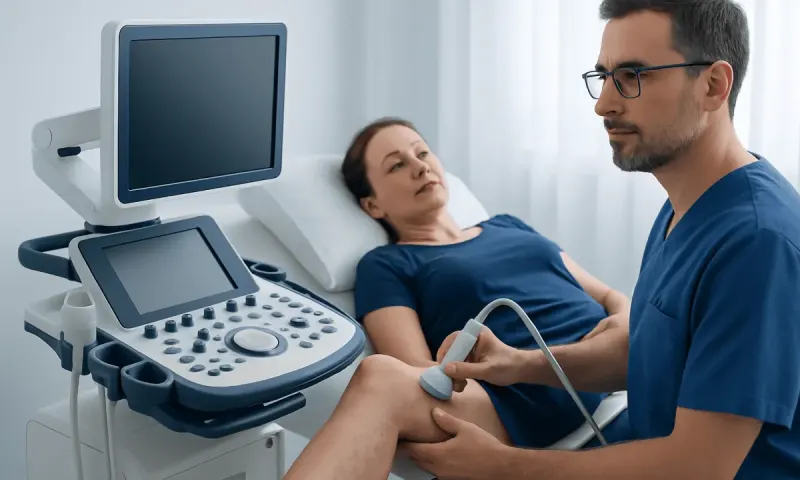
A trained sonographer or clinician applies gel to the skin and moves a handheld probe over the leg. The examination may be performed with the person lying down and standing. Gentle pressure and brief squeezing of the calf can help demonstrate how blood moves through the veins and whether valves close effectively.
The scan maps the superficial veins that commonly cause varicose veins, the deep veins within the leg and the connecting veins between them. It can show reflux, obstruction, old clot-related changes and the location of veins that may be suitable for treatment. Results are used to guide decisions about conservative care or varicose veins treatment.
Many people receive preliminary information after the scan, although the full interpretation and care plan may be discussed at a follow-up consultation. If an ultrasound is not immediately needed, a clinician can explain why observation or self-care is reasonable and what changes should prompt reassessment.
Other tests and when they may be needed
Most people do not need MRI, CT scans or invasive vein imaging for uncomplicated varicose veins. Duplex ultrasound usually provides the information needed to diagnose reflux and plan treatment. This focused approach avoids unnecessary tests while still giving a detailed picture of leg vein circulation.
Additional imaging may occasionally be considered if symptoms suggest a blockage higher in the pelvis or abdomen, if there is a complex history of deep vein thrombosis, or if ultrasound findings do not fully explain the problem. Depending on the clinical question, the specialist may consider CT venography, MR venography or other targeted tests. These are not routine screening tests for visible leg veins.
Blood tests are also not typically used to diagnose varicose veins. They may be ordered for a separate reason, such as assessing a suspected clotting disorder, infection, anemia after significant bleeding or readiness for a planned procedure. A D-dimer blood test may sometimes contribute to assessment of suspected deep vein thrombosis, but it does not diagnose varicose veins.
If arterial disease is a concern, clinicians may use an ankle-brachial index, which compares blood pressure at the ankle and arm. This can be important before strong compression therapy is advised, because compression is not appropriate for everyone.
Understanding results and planning next steps
Results are considered alongside symptoms and examination findings. Some people have obvious varicose veins with little or no reflux, while others have significant reflux that causes swelling, skin changes or recurrent discomfort. A scan may also identify chronic venous insufficiency, meaning long-term problems with vein function that require ongoing attention.
The report may describe which veins show reflux, such as the great or small saphenous vein, and whether the deep veins are open and functioning. It may also note tributary veins, perforator veins or signs of a previous clot. These details help the specialist select the safest and most appropriate approach.
When symptoms are mild, initial management often includes regular movement, avoiding long periods of stillness when possible, elevating the legs at rest and discussing professionally fitted compression garments. Compression can reduce symptoms for some people, but it does not remove faulty valves or eliminate all varicose veins.
If symptoms continue, complications develop or the person wishes to discuss definitive management, ultrasound mapping supports procedural planning. The available options and their suitability depend on the vein pattern, general health, prior treatments and personal goals.
Procedures after diagnosis: candidacy, process and recovery
Diagnosis does not automatically mean a procedure is necessary. A clinician may discuss treatment when varicose veins cause persistent pain, heaviness, swelling, skin changes, bleeding, recurrent inflammation, ulcers or substantial impact on quality of life. Treatment may also be considered for cosmetic concerns after an appropriate vascular assessment.
Modern procedures often close or remove the affected superficial vein while blood naturally reroutes through healthier veins. Depending on the ultrasound findings, options may include thermal ablation using heat, ultrasound-guided foam sclerotherapy, medical adhesive closure or removal of smaller surface veins. The specialist will explain which option fits the anatomy and whether any medicines or health conditions need attention beforehand.
Many vein procedures are performed on an outpatient basis with local anesthesia, sometimes with additional sedation. The typical process includes ultrasound marking of the target vein, skin cleaning, treatment through a small needle or tiny incision, and a brief check of blood flow afterward. The exact steps differ by procedure, so patients should ask what their planned treatment involves.
Recovery is often relatively quick, with walking encouraged soon after treatment. Temporary bruising, tenderness, tightness or mild pulling sensations can occur. Follow-up ultrasound may be arranged to confirm closure of the treated vein and check for uncommon complications. Expected benefits include reduced symptoms and improvement in the treated veins, while risks can include skin staining, inflammation, nerve irritation, recurrence, clotting complications or an incomplete response.
When to seek medical care
A routine medical appointment is appropriate for persistent leg aching, heaviness, swelling, itching, visible enlarging veins, skin changes near the ankles or a wound that is slow to heal. Early assessment can clarify whether symptoms are related to vein disease or another condition affecting the legs.
Urgent assessment is important for sudden swelling of one leg, new significant calf pain, warmth or redness, especially after surgery, travel, immobility or a previous clot. These symptoms can have several causes, including deep vein thrombosis, which needs prompt evaluation.
Emergency care is needed if leg symptoms occur with chest pain, shortness of breath, coughing blood, fainting, or severe dizziness. A varicose vein that starts bleeding should be managed by lying down, raising the leg and applying firm direct pressure with a clean cloth or bandage while urgent medical help is arranged if bleeding does not stop promptly.
Acıbadem Health Point’s multidisciplinary specialists and JCI-accredited hospitals assess and treat varicose vein conditions for international patients, including ultrasound-based evaluation and individualized treatment planning.
Frequently asked questions
01Can varicose veins be diagnosed without an ultrasound?
A clinician can often identify varicose veins through medical history and examination. However, duplex ultrasound is commonly used when symptoms are present, complications are suspected or treatment is being considered because it shows blood-flow direction and valve function.
02Is duplex ultrasound for varicose veins painful?
Duplex ultrasound is generally painless and noninvasive. The probe may apply light pressure to the leg, and the clinician may briefly squeeze the calf or ask the person to change position to assess blood flow.
03Do I need to prepare for a varicose vein ultrasound?
Usually, no special preparation is needed. Comfortable clothing that allows access to the legs is helpful, and patients should bring information about medications, prior blood clots, previous vein procedures and relevant medical history.
04Can an ultrasound detect a blood clot as well as varicose veins?
Yes, duplex ultrasound can often evaluate the deep and superficial veins for signs of a current clot, obstruction or changes from an older clot. If a clot is suspected based on symptoms, the timing and urgency of testing should be determined by a clinician.
05Why are my legs examined while standing?
Standing allows leg veins to fill and makes visible varicose veins easier to assess. It may also help ultrasound testing reveal reflux that is less apparent when a person is lying down.
06Will I need treatment if the scan shows venous reflux?
Not necessarily. Treatment decisions depend on symptoms, skin changes, complications, the extent of reflux and personal preferences. Some people benefit from lifestyle measures and compression, while others may be candidates for a procedure.
This article is for general information only and is not a substitute for professional medical advice. Please consult a qualified doctor about your individual situation.
Persistent digestive symptoms? Get evaluated in Turkey
JCI-accredited · board-certified surgeons · reply within 24h
Add us as a Preferred Source to see more of our trusted health content across Google Search, AI Overviews and Discover.