Liver Lesions on Imaging: Which Findings Usually Need More Tests Before Treatment

Key Takeaways
- Many liver lesions are benign and only need observation, not immediate treatment.
- Imaging details such as size, shape, contrast behavior, and background liver health help guide next steps.
- MRI, contrast-enhanced CT, ultrasound, and blood tests may be used to clarify uncertain findings.
- A lesion that is new, changing, or seen in a higher-risk patient usually deserves a more careful workup.
- Treatment should not be decided from a single image alone when the diagnosis is not secure.
Liver lesions are common imaging findings, and many are harmless. The key question is not only what the scan shows, but whether the appearance, size, and clinical context suggest that more tests are needed before any treatment is planned.
Overview
A liver lesion is a spot or area seen on an imaging study that differs from the surrounding liver tissue. These findings are often discovered by chance during an ultrasound, CT scan, or MRI done for another reason. For many people, the first reaction is worry, but the reality is more reassuring: a large share of liver lesions are benign and never cause trouble.
The important clinical question is not simply whether a lesion exists, but whether its appearance fits a common harmless pattern or whether it remains too uncertain to label confidently. In that second situation, doctors usually recommend further evaluation before discussing any treatment. This stepwise approach helps avoid unnecessary procedures while still identifying lesions that deserve closer attention.
For international patients, this often means comparing the original scan report with dedicated liver imaging, reviewing prior studies if they exist, and making sure the interpretation is tied to the person’s medical history rather than the image alone. The most useful plan is usually the one that explains the finding clearly and reduces uncertainty without rushing to intervention.
Symptoms
Most liver lesions do not cause symptoms and are found incidentally. That is why imaging findings can feel surprising; a person may feel well and still be told that a “spot” was seen on the liver. In many cases, the lesion is unrelated to the reason the scan was ordered.
When symptoms do occur, they are usually nonspecific. Some people notice right upper abdominal discomfort, fullness after meals, nausea, or a sense of pressure. These symptoms are not enough on their own to identify the lesion type, but they may prompt a doctor to look more closely, especially if they are persistent or getting worse.
It is also important to separate symptoms caused by the lesion itself from signs of underlying liver disease. Jaundice, unexplained weight loss, fever, easy bruising, or swelling in the abdomen may point to a broader medical issue that needs prompt assessment. A careful history helps determine whether the imaging finding is truly incidental or part of a larger picture.
Causes & Risk Factors

Liver lesions can arise from many different processes. Some are benign growths such as hemangiomas, focal nodular hyperplasia, and certain cysts. Others may reflect inflammation, infection, fatty liver-related changes, or, less commonly, a primary liver cancer or a metastasis that spread from another organ.
Risk factors matter because the same imaging appearance can mean different things in different people. A lesion in a person with a healthy liver and no cancer history is often approached differently from a similar lesion in someone with cirrhosis, chronic hepatitis B or C, or a known cancer elsewhere in the body. The background liver, medications, alcohol use, and previous infections all influence the level of concern.
Lesions are more likely to require additional testing when the images are not classic for a benign diagnosis, when the lesion is larger than expected for a common incidental finding, or when there is evidence of growth on prior scans. Doctors also pay close attention to enhancement patterns after contrast is given, because these patterns can strongly suggest one diagnosis over another. When the pattern is not clear, the safest course is to continue evaluating rather than guessing.
Diagnosis
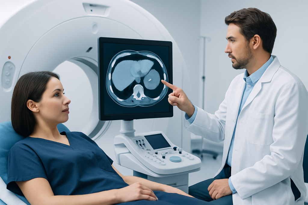
Diagnosis usually begins with reviewing the original imaging study in detail. Radiologists look at the lesion’s size, shape, borders, density or signal characteristics, and the way it takes up contrast. They also consider whether the scan was performed with a liver-specific protocol or whether the lesion was seen only on a study done for another reason.
When the appearance is not definitive, doctors may recommend a dedicated liver MRI or a multiphase contrast CT scan. These studies can show blood flow and tissue characteristics more clearly than a routine scan. Ultrasound, including contrast-enhanced ultrasound in some settings, may also help clarify whether the lesion behaves like a benign mass or a lesion that needs closer attention.
Blood tests may support the evaluation, especially when underlying liver disease is suspected. Liver function tests, hepatitis testing, and tumor markers may be ordered in selected cases, though no blood test alone can identify every lesion type. If the diagnosis is still uncertain after imaging and lab work, a biopsy may be discussed, but only when the result is likely to change management and the procedure is considered appropriate for that person.
For patients traveling for care, one helpful step is to bring the actual image files, not only the written report. A second review by a specialist can sometimes prevent unnecessary worry by confirming a benign pattern, or it can speed up the next stage if the lesion truly needs more investigation.
Treatment Options
Treatment depends entirely on what the lesion is. That is why clarifying the diagnosis comes first. A cyst, hemangioma, or focal nodular hyperplasia may need no treatment at all unless it causes symptoms or remains atypical on imaging. In contrast, lesions that represent cancer or have a significant risk of becoming cancerous are managed very differently.
When the diagnosis is uncertain, the “treatment” may simply be more information. This can include a short period of imaging follow-up to check stability, a specialized scan, or consultation with a hepatologist, radiologist, surgeon, or oncologist. If the lesion is confirmed to be benign, observation is often enough. If it is suspicious or proven malignant, the plan may involve surgery, ablation, systemic therapy, or another targeted approach based on the type and stage of disease.
People are sometimes told about a lesion before any decision has been made. In that setting, it is reasonable to slow down and ask whether the current images are truly diagnostic. A measured approach protects patients from both overtreatment and delay. At Acibadem Health Point, multidisciplinary specialists and JCI-accredited hospitals work together to diagnose and treat liver lesions for international patients when coordinated care is needed.
Prevention & Self-care
Not every liver lesion can be prevented, especially benign incidental findings. Still, overall liver health matters because a healthier liver is easier to interpret and monitor. Limiting alcohol, maintaining a healthy weight, managing diabetes, and reducing the risk of viral hepatitis all support long-term liver well-being.
After a lesion is found, self-care is mostly about follow-through. Keeping copies of reports and images, attending scheduled scans, and asking for a clear explanation of the next step are practical ways to stay involved in care. If a doctor recommends a repeat scan, the timing matters; delayed follow-up can make it harder to determine whether a lesion is stable.
People who live in another country or plan to travel for evaluation should also think ahead about continuity. It helps to know which specialist will review the images, how follow-up will be shared with the home physician, and what symptoms should trigger earlier contact. When a lesion is judged likely benign, this organized approach can replace anxiety with a concrete plan.
When to See a Doctor
Medical review is appropriate whenever a liver lesion is newly discovered and the report does not clearly state that it has a classic benign appearance. A person should also seek further assessment if the lesion is growing, if imaging is inconsistent across reports, or if there is known liver disease or a history of cancer. In those settings, more testing before treatment is a standard and sensible step.
Prompt assessment is especially important if there are warning signs such as jaundice, unexplained weight loss, persistent fever, abdominal swelling, vomiting, or new pain that does not settle. These symptoms do not automatically mean something serious, but they do warrant a clinician’s review. The goal is to match the urgency of the workup to the real level of concern.
Even when the finding is likely benign, patients are entitled to a clear explanation. Asking whether the lesion is typical, indeterminate, or suspicious can help frame the discussion. If the answer is uncertain, a specialist review is often the right next move rather than immediate treatment.
FAQ
Frequently asked questions about liver lesions often center on whether every spot is dangerous and whether treatment is always necessary. The short answer is no. The meaning of a liver lesion depends on the imaging pattern, the person’s medical background, and whether the finding is stable over time.
Below are practical answers to the questions doctors hear most often when a liver lesion is first reported. They are meant to help patients understand why additional testing is sometimes recommended before any treatment is considered.
FAQ 1
Are all liver lesions cancer? No. Many liver lesions are benign and are found incidentally during scans done for unrelated reasons. Doctors use the imaging appearance and the patient’s history to decide whether the lesion looks harmless or needs more workup.
FAQ 2
Why would a doctor order more tests after a scan? Extra tests are recommended when the lesion does not have a classic benign pattern or when the background liver history raises concern. A dedicated MRI, multiphase CT, ultrasound, or follow-up scan can often clarify the diagnosis without jumping to treatment.
FAQ 3
Can a blood test tell what kind of liver lesion it is? Blood tests can provide clues about liver health or inflammation, but they usually cannot identify the exact type of lesion on their own. Imaging remains central, and biopsy is sometimes needed if the diagnosis is still unclear after scans and lab work.
FAQ 4
What does “indeterminate” mean on an imaging report? It means the scan did not show a pattern that is clearly benign or clearly suspicious. In that situation, the next step is often a more specialized study or a short interval follow-up, not immediate treatment.
FAQ 5
Should someone seek a second opinion for a liver lesion? A second opinion can be very helpful when the report is uncertain, when treatment is being considered, or when images were done outside a specialist liver setting. Bringing the original image files and reports allows another expert to review the finding in context.
FAQ 6
When is a liver lesion usually treated right away? Immediate treatment is not the norm for most incidental liver lesions. Rapid treatment is more likely when imaging strongly suggests a cancerous lesion, when symptoms are significant, or when a specialist confirms that the diagnosis is clear and action is needed.
References
- American College of Radiology
- Radiological Society of North America
- American Association for the Study of Liver Diseases
- National Cancer Institute
- Mayo Clinic
This article is for general information only and is not a substitute for professional medical advice. Please consult a qualified doctor about your individual situation.