Lingula Of Lung

Key Takeaways
- The lingula is part of the left upper lobe of the lung and sits near the heart.
- It can be affected by infections, inflammation, mucus plugging, and less commonly structural lung disease.
- Symptoms are often similar to other lung problems, such as cough, shortness of breath, or chest discomfort.
- Chest imaging is often used to evaluate lingular changes, especially when symptoms persist.
- Treatment depends on the cause and may include medication, airway clearance, or monitoring.
Medically reviewed by the Acıbadem clinical team — July 13, 2026
The lingula of the lung is a small, tongue-like part of the left upper lobe that helps with normal breathing and can be involved in infections or other lung conditions. Understanding its anatomy and common problems can make lung scan results and treatment plans easier to discuss with a doctor.
Overview
The lingula of the lung is a small, tongue-shaped portion of the left upper lobe. Its name comes from the Latin word for “little tongue,” which is a helpful way to picture its shape and position. Although it is only one part of the lung, it can become clinically important when infection, inflammation, or airway blockage affects it.
For many people, the lingula is first mentioned after a chest X-ray or CT scan. That can sound technical, but the finding usually simply describes an anatomic location rather than a disease on its own. In day-to-day lung care, doctors think about the lingula the same way they think about other lung segments: what it looks like on imaging, whether symptoms are present, and whether the airways are open and working well.
Because the lingula lies close to the heart and the front of the chest, changes in this area may be noticed during evaluation for cough, fever, breathlessness, or an abnormal scan. For international patients arranging care from another country, this type of report is often one piece of a larger picture, and it is best interpreted alongside the person’s symptoms, medical history, and current medications.
Symptoms
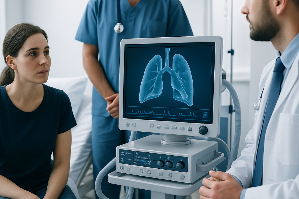
The lingula itself does not cause symptoms when it is healthy. When it becomes involved in a lung condition, the symptoms usually reflect the underlying problem rather than the anatomy of the lingula alone. A person may notice a lingering cough, mucus production, chest tightness, mild wheezing, or a feeling that breathing is not as easy as usual.
Infections affecting the lingula can lead to fever, fatigue, and pain or discomfort when breathing deeply. If mucus is not clearing well, the same area may repeatedly seem “stuck” on imaging, which can happen even when symptoms are mild. Some people have little to no discomfort and learn about lingular involvement only after a scan done for another reason.
Symptoms that deserve medical attention include cough that lasts more than a few weeks, coughing up blood, unexplained shortness of breath, recurrent fevers, or chest pain that is new or worsening. These symptoms do not point to one specific diagnosis, but they do help a doctor decide whether the lingula or another part of the lungs needs closer evaluation.
Causes & Risk Factors

Several different issues can involve the lingula. The most common are respiratory infections, especially when inflammation causes the small airways to swell and produce mucus. In some cases, a plugged airway can lead to partial collapse of the lingular tissue, a pattern sometimes discussed in radiology reports.
Other causes may include bronchiectasis, which is a long-term widening of the airways that can make it harder to clear secretions, and inflammatory conditions that affect the lungs more broadly. Less commonly, airway obstruction from a foreign body, scarring, or a growth may be responsible. A doctor considers these possibilities based on age, symptoms, past lung illness, smoking history, immune status, and imaging findings.
Risk is higher in people who have frequent chest infections, chronic cough, asthma, chronic obstructive pulmonary disease, difficulty clearing mucus, or immune suppression. Prior tuberculosis, aspiration problems, and certain structural lung conditions can also make recurrent lingular problems more likely. Not everyone with these risk factors develops a lingular problem, but they can shape how closely the lungs are monitored.
Diagnosis
Diagnosis usually begins with a conversation about symptoms and a physical examination. A doctor may listen for crackles, wheezing, or reduced breath sounds and ask how long the cough has been present, whether fever has occurred, and whether infections keep returning. This history helps distinguish a short-lived respiratory illness from a more persistent process.
Imaging is often the next step. A chest X-ray may show an area of opacity, volume loss, or recurrent changes in the left upper lobe region, while a CT scan offers a more detailed view of the lingula and nearby airways. Depending on the situation, tests may also include blood work, sputum analysis, breathing tests, or bronchoscopy if a doctor needs to look directly inside the airways.
For people traveling internationally for evaluation, it is helpful to bring prior images and reports, not only written summaries. Comparing older and newer scans can show whether a lingular finding is stable, improving, or changing over time. That comparison often matters more than the name of the anatomical segment itself.
Treatment Options
Treatment depends entirely on the cause. If an infection is present, a doctor may recommend medication and supportive care. If mucus clearance is part of the problem, therapy may focus on helping the airways open and drain better so the lingula can re-expand or recover more fully.
Common approaches may include inhaled therapies when airway inflammation is present, physiotherapy or breathing techniques to loosen secretions, and hydration and rest as appropriate. When bronchiectasis is involved, care often centers on long-term secretion management and treating flare-ups early. If there is concern about obstruction, further evaluation may be needed to identify and address the cause.
In selected cases, bronchoscopy or more specialized procedures may be considered, especially if imaging suggests a blockage or if symptoms do not improve as expected. Surgery is not a routine treatment for lingular findings, but it may be discussed in uncommon situations such as persistent localized disease that does not respond to other care. The best plan is usually individualized and based on the full lung picture rather than the lingula alone.
Prevention & Self-care
There is no specific way to “protect” the lingula on its own, but healthy lung habits can reduce the chance of repeated problems. Avoiding smoking and secondhand smoke, staying up to date on recommended vaccines, and treating respiratory infections promptly all support better airway health. People with chronic lung disease may also benefit from a tailored routine to keep mucus from building up.
Simple self-care can make a meaningful difference, especially after a recent infection. Drinking enough fluids, following the prescribed inhaler or airway-clearance plan, walking or moving gently as tolerated, and using breathing exercises if advised can support recovery. If a person is recovering while abroad, clear written instructions and a follow-up plan are especially important so treatment continues smoothly after travel.
Because the lingula is sometimes mentioned on a report without an obvious cause, it is wise not to self-interpret the scan wording. A doctor can explain whether the finding is a normal variation, a temporary change after infection, or something that needs monitoring. That conversation is often reassuring and prevents unnecessary worry.
When to See a Doctor
A medical evaluation is appropriate if cough, wheezing, chest discomfort, or breathlessness lasts longer than expected or keeps returning. It is also sensible to seek care after recurrent chest infections, especially if imaging repeatedly points to the same area of the left lung. Persistent symptoms deserve attention even when they seem mild, because they can reveal a treatable airway issue.
Urgent assessment is important for coughing up blood, significant shortness of breath, blue lips, severe chest pain, high fever with weakness, or confusion. These symptoms may signal a serious infection or another condition that needs prompt treatment. If someone is already under care, worsening symptoms should be reported rather than waiting for a scheduled visit.
For international patients, follow-up planning matters as much as the initial diagnosis. A specialist may recommend repeat imaging, medication review, or a handoff to a local physician after travel. Acibadem Health Point’s multidisciplinary specialists and JCI-accredited hospitals can diagnose and treat lingular and other lung conditions for international patients, with coordinated care that helps bridge evaluation and recovery across borders.
Living With a Lingular Finding
When a scan mentions the lingula, the phrase can sound more serious than it actually is. In many cases, it is simply a description of where a change is located. The most useful question is not “What is the lingula?” but “What is happening there, and does it need treatment or follow-up?”
That distinction matters because the same location can be involved in very different situations, from a short-term infection to a chronic airway condition. Clear communication with the care team helps people understand whether the finding is expected to resolve, should be monitored, or needs additional testing. Keeping copies of reports and images can make that discussion easier over time.
With the right diagnosis and a practical plan, most lingular problems can be managed effectively. The key is to treat the cause, support the airways, and watch for changes rather than focusing on the anatomical term alone.
Frequently asked questions
Is the lingula of the lung a separate organ?
No. The lingula is a small part of the left upper lobe of the lung, not a separate organ. It is simply a named region that doctors use when describing anatomy or imaging findings.
Why would a scan mention the lingula?
A scan may mention the lingula because there is inflammation, infection, mucus plugging, scarring, or another change in that specific area. Sometimes the report is simply describing a location rather than naming a disease.
Can lingular problems cause a cough?
Yes, but the cough is usually caused by the underlying lung issue affecting the lingula, such as infection or airway irritation. A cough that lasts a long time or keeps returning should be evaluated.
Is treatment always needed if the lingula looks abnormal on imaging?
Not always. Some findings are temporary or mild and may only need observation, while others require medication or further testing. A doctor decides based on symptoms, scan details, and medical history.
How is a lingular problem different from pneumonia in general?
Pneumonia is an infection of the lungs, while the lingula is just one part of the left lung where pneumonia can occur. The location helps doctors describe exactly where the infection or other change is seen.
What should an international patient bring to a lung appointment?
It helps to bring previous scans, written radiology reports, medication lists, and discharge summaries in English if possible. These documents make it easier for the specialist to compare findings and plan the next step.
References
- Mayo Clinic
- Cleveland Clinic
- National Heart, Lung, and Blood Institute
- Radiological Society of North America
- Merck Manual Professional Edition
This article is for general information only and is not a substitute for professional medical advice. Please consult a qualified doctor about your individual situation.