What Is Foot Xray? A Complete Medical Guide

Key Takeaways
- A foot xray uses a small amount of radiation to create images of the bones and joints in the foot.
- It is often used after injuries, for ongoing pain, or when arthritis, deformity, or infection is suspected.
- The test is usually quick and painless, with no special recovery needed afterward.
- Xrays are helpful for bone problems, but soft tissue injuries may need ultrasound, MRI, or other tests.
- A doctor should interpret the results together with symptoms, physical examination, and medical history.
A foot xray is a common imaging test that helps doctors look for fractures, joint problems, arthritis, deformities, and some infections in the bones of the foot. It is quick, painless, and often one of the first tests used when a foot injury or persistent foot pain needs clearer evaluation.
Overview
A foot xray is a standard medical imaging test that shows the bones, joints, and overall alignment of the foot. For many people, it is the first step in understanding why the foot hurts, swells, looks misshapen, or does not bear weight normally.
The test works by sending a small amount of X-ray energy through the foot. Dense structures such as bone appear clearly on the image, which helps doctors look for fractures, arthritis, dislocations, bone spurs, and changes in the shape of the foot. It is especially useful after a twist, fall, impact, or sports injury, but it can also help evaluate pain that develops more slowly.
For international patients who are planning care abroad, a foot xray is often part of the first orthopedic assessment. It can help clinicians decide whether urgent treatment is needed, whether more advanced imaging is necessary, or whether a conservative treatment plan is appropriate.
Symptoms and Reasons a Foot Xray May Be Needed
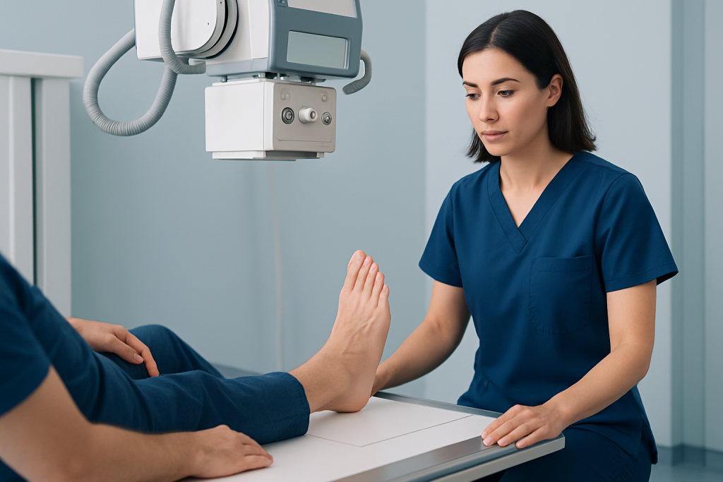
A foot xray is not ordered for every ache or strain. Doctors usually recommend it when symptoms suggest a bone, joint, or alignment problem that cannot be understood well enough by examination alone. The goal is to match the imaging test to the symptom pattern, so the evaluation stays focused and useful.
Common reasons include pain after an injury, swelling that does not improve, bruising over the foot, difficulty walking, tenderness over a bone, or a visible change in foot shape. It may also be used when a person has long-lasting stiffness, limited movement, or pain related to standing and walking.
- Suspected fracture or stress fracture
- Dislocation or bone misalignment
- Arthritis in the foot joints
- Deformities such as bunions or flatfoot changes
- Foreign body in the foot, such as after a puncture injury
- Bone infection or other unusual bone changes
Sometimes the xray is normal even though the foot still hurts. That does not mean the symptom is unreal; it may mean the problem involves ligaments, tendons, cartilage, or nerves, which may need a different kind of imaging.
Causes and Risk Factors for Foot Problems That Show on Xray

The reasons behind foot problems vary widely, but several patterns commonly lead to xray evaluation. Sudden trauma, such as a fall, sports injury, or twisting the foot, can cause fractures or joint displacement. Repetitive stress from running, jumping, or long periods of standing may lead to stress fractures that become visible on imaging over time.
Chronic conditions can also change what the xray shows. Arthritis may narrow joint spaces or create bone spurs. Bunions can alter the shape and alignment of the front part of the foot. In some people, diabetes, circulation problems, or inflammatory disease can increase the chance of foot complications that require imaging.
Risk factors depend on the condition being considered, but doctors often pay special attention to people with osteoporosis, prior foot injuries, high-impact activity, poorly fitting footwear, and jobs or sports that place repeated strain on the feet. A foot xray helps separate one possible cause from another, which can guide the next step in care.
How Diagnosis Is Made
The xray itself is only one part of diagnosis. Before ordering it, a clinician usually asks how the pain began, where it is located, whether the person can bear weight, and whether there has been trauma, fever, numbness, or a wound. A physical examination then helps identify tenderness, swelling, range of motion, and any visible deformity.
A foot xray may be taken from more than one angle, because different views can reveal different details. This helps the doctor assess the bones and joints in a more complete way. In some cases, special weight-bearing views are used to understand alignment while the person is standing.
If the xray does not fully explain the symptoms, other tests may be recommended. Ultrasound can help evaluate tendons or fluid collections, MRI can show soft tissue and bone marrow details, and CT can offer a more detailed look at complex fractures or bone structure. The best test depends on the clinical question, not just the symptom itself.
Treatment Options After the Results
Treatment depends on what the foot xray shows. A minor fracture may need rest, protection, and follow-up imaging, while a more serious break may require immobilization, reduction, or surgery. If the xray shows arthritis or a deformity, treatment may focus on footwear changes, activity adjustment, anti-inflammatory measures, or specialist referral.
When no fracture is seen, the plan may still include careful management of pain and movement. This can involve temporary support, ice, elevation, gradual return to activity, or physiotherapy if the problem is related to mechanics or overuse. The xray helps doctors avoid treating blindly and instead choose a plan that fits the actual structure of the foot.
For patients traveling from another country, treatment may also include coordination of records, review of previous imaging, and follow-up planning before returning home. When surgery or prolonged care is needed, it helps to have a clear explanation of the findings and a practical recovery plan that can be continued locally after travel.
Prevention and Self-care
Not every foot problem can be prevented, but sensible habits can reduce the risk of injury and strain. Supportive shoes, gradual increases in exercise, and attention to pain signals all matter. People who run, train, or stand for long periods often benefit from pacing activity and replacing worn-out footwear in time.
If a foot injury occurs, early self-care should be gentle and safe. Resting the foot, limiting weight-bearing when needed, and elevating it can help while waiting for medical advice. It is important not to assume that a severe bruise or swelling is “just a sprain,” because some fractures can look similar at first.
- Wear shoes that fit well and support the arch
- Increase activity levels gradually
- Use protective gear in sports and work settings when appropriate
- Do not ignore persistent pain after an injury
- Follow treatment instructions carefully if a fracture or stress injury is diagnosed
People with diabetes or reduced sensation in the feet should inspect their feet regularly and seek care early for wounds, swelling, or new pain. In these situations, timely evaluation can prevent complications.
When to See a Doctor
Medical assessment is important when foot pain is severe, does not improve, or follows an injury. A doctor should also be consulted if the foot looks crooked, the person cannot take steps, or swelling and bruising develop quickly after trauma. These signs may indicate a fracture, dislocation, or another problem that should not be left to guesswork.
Immediate attention is especially important if there is an open wound, fever, numbness, color change, or increasing pain that feels out of proportion to the injury. People with diabetes, poor circulation, or immune suppression should seek advice early, even for injuries that seem small.
When care is sought promptly, a foot xray can help speed up the right diagnosis and avoid unnecessary delay. That is often the difference between simple recovery guidance and a more complex treatment plan, so it is worth getting checked when symptoms do not fit a minor strain.
At Acibadem Health Point, multidisciplinary specialists and JCI-accredited hospitals diagnose and treat foot conditions for international patients, with imaging and follow-up organized to support safe, coordinated care.
What to Expect During the Test
A foot xray is usually simple and quick. The person may be asked to remove shoes, socks, and any metal objects from the area. A radiologic technologist positions the foot on the xray table or plate and may ask for several views so the doctor can see the bones from different angles.
The test does not usually hurt. Some discomfort can happen if the foot is already injured and needs to be moved into position, but the imaging itself is brief. In many cases, the person can leave immediately afterward and resume normal activities unless another injury-specific instruction has been given.
Pregnancy should always be mentioned before any xray, even though foot imaging uses a limited body area and protective measures may be considered. The care team will decide whether the test is appropriate and whether extra precautions are needed.
Limits of Foot Xray and What Results Can Mean
Although a foot xray is very useful, it cannot show everything. Ligaments, tendons, and many soft tissue injuries may appear normal even when they are causing pain. Early stress fractures can also be difficult to see at first, which is why symptoms and follow-up matter.
Results may show a fracture, arthritis, bone alignment changes, infection-related bone damage, or a foreign body. In some people, the image simply helps confirm that no major bone injury is present. Either result can be clinically valuable because it narrows the possibilities and helps guide the next decision.
For the most accurate interpretation, the xray should be read in context. A normal image does not automatically end the evaluation if the foot remains swollen, unstable, or painful. Doctors may repeat imaging or choose another modality if symptoms continue.
Frequently asked questions
01What is a foot xray used for?
A foot xray is used to look at the bones and joints of the foot. Doctors commonly order it to check for fractures, dislocations, arthritis, deformities, foreign bodies, or other bone-related problems.
02Does a foot xray show a sprain or tendon injury?
Usually not in detail. Sprains and tendon injuries involve soft tissues, so an xray may look normal even when those structures are injured. If symptoms point to soft tissue damage, a doctor may suggest ultrasound or MRI instead.
03How long does a foot xray take?
The imaging itself is usually very quick, often only a few minutes. Extra time may be needed for registration, position changes, or if several views are required.
04Do I need to prepare for a foot xray?
Most people do not need special preparation. It is helpful to wear clothing and footwear that are easy to remove, and to tell the care team about pregnancy, recent surgery, implants, or prior injuries.
05Is a foot xray safe?
A foot xray uses a small amount of radiation and is generally considered safe when medically needed. The care team uses the lowest reasonable dose and may add shielding or other precautions depending on the situation.
06What if my foot xray is normal but pain continues?
A normal xray does not rule out all causes of foot pain. The problem may involve ligaments, tendons, nerves, or an early bone injury that is not yet visible, so follow-up with a doctor is still important.
This article is for general information only and is not a substitute for professional medical advice. Please consult a qualified doctor about your individual situation.
Joint and spine care in Turkey — expert assessment & treatment
JCI-accredited · board-certified surgeons · reply within 24h
Add us as a Preferred Source to see more of our trusted health content across Google Search, AI Overviews and Discover.