Varus Deformity: Causes, Symptoms, and Treatment
Varus deformity describes an inward angulation of a bone or joint, meaning the segment farther from the body turns toward the midline. It is often discussed at the knee as bow-legged alignment, but it can also affect the foot, ankle, elbow, hip, or other parts of the skeleton.
Overview: What Is Varus Deformity?
Varus deformity is a medical term for an inward tilt or angulation of a limb segment relative to the body’s midline. In the lower limb, the part below the affected joint points inward. At the knee, this usually creates a bow-legged appearance: when the ankles are together, the knees may remain apart. The clinical term for this pattern at the knee is genu varum.
Varus is the opposite of valgus, in which the limb segment angles outward. These words describe alignment rather than the exact reason for the alignment. A varus position may arise during normal growth, after an injury, because of bone or joint disease, or as a consequence of uneven joint degeneration.
The meaning of varus depends on the location. For example, knee varus may change how body weight passes through the knee, while hindfoot or ankle varus may affect foot contact and balance. A healthcare professional considers the whole limb, joint movement, symptoms, age, and imaging findings rather than judging alignment from appearance alone.
How Varus Alignment Can Affect the Body
Alignment helps distribute forces across bones, cartilage, ligaments, muscles, and joints. With knee varus, the line of weight-bearing may shift toward the medial, or inner, compartment of the knee. Over time, this can increase stress in that area, particularly when cartilage has already been damaged or lost.
Not every person with varus alignment has discomfort or functional limitations. Mild, stable alignment differences may cause no symptoms and require only observation. Problems are more likely when the deformity is substantial, progresses over time, occurs after trauma, or is combined with pain, instability, stiffness, or reduced mobility.
In adults, varus knee alignment may occur alongside medial compartment osteoarthritis. The two can influence one another: joint wear can contribute to a change in alignment, and altered alignment can increase loading in the affected compartment. However, an X-ray finding alone does not determine how a person feels or which treatment is appropriate.
Symptoms and Signs
The most visible sign is a limb that appears to curve outward overall, with the lower segment angled inward at the joint. In knee varus, a person may notice a gap between the knees when standing with the feet or ankles close together. The degree of visible bowing does not always match the degree of symptoms.
When symptoms are present, they can include pain on the inner side of the knee, stiffness after rest, reduced ability to walk long distances, swelling after activity, or a sense that the knee is less steady. A person may also develop changes in walking pattern, such as favoring one leg. In foot or ankle varus, recurrent ankle sprains, uneven shoe wear, pressure areas, or difficulty finding comfortable footwear can occur.
Children with physiologic bowing are usually otherwise well, active, and free from pain. Concerning features include a deformity that is clearly more pronounced on one side, worsens rather than improves, starts after early childhood, or occurs with limping, pain, short stature, or other signs of illness. These features do not confirm a serious cause, but they warrant medical assessment.
Causes and Risk Factors
In babies and young children, bowing of both legs may be a normal part of development. Many infants have some genu varum, partly related to their position before birth. As children begin standing and walking, alignment changes gradually; the legs commonly become less bowed and later may temporarily look mildly knock-kneed. A clinician can help distinguish expected developmental variation from a condition needing follow-up.
Other causes include previous fractures that healed in an altered position, growth plate injuries, congenital skeletal differences, and disorders that affect bone strength or growth. Nutritional vitamin D deficiency and rickets are examples of conditions that can affect bone mineralization. Blount disease is a growth disorder involving the upper shinbone that can produce progressive varus alignment, usually at the knee.
In adults, common contributors include osteoarthritis affecting one side of the knee, prior ligament or meniscus injury, past fractures, and long-standing altered loading. Excess body weight can add to joint load and may worsen symptoms in people with knee arthritis, although it is not the sole cause of varus alignment. Family history, certain neuromuscular conditions, and occupation or sports involving repetitive joint stress may also be relevant in some individuals.
Because the same appearance can have different causes, it is important not to assume that all bowing is developmental or that all adult varus deformity is caused by arthritis. Identifying the underlying reason helps clinicians recommend safe, proportionate care.
Assessment and Diagnosis
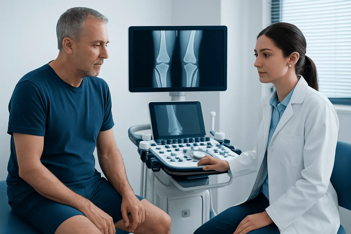
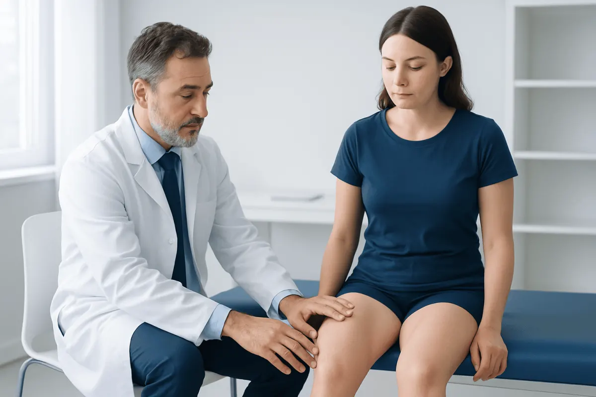
Assessment starts with a medical history and physical examination. The clinician may ask when the alignment was first noticed, whether it is changing, whether there was an injury, and whether pain limits work, sport, or daily activities. They will usually observe standing posture and walking, compare both limbs, assess joint motion and stability, and check for tenderness, swelling, muscle weakness, or differences in leg length.
Weight-bearing X-rays are often used when a deformity is persistent, symptomatic, progressive, or being considered for treatment. Full-length standing images can help show the mechanical axis of the leg and identify whether the angulation mainly arises in the thighbone, shinbone, knee joint, foot, or a combination of areas. Imaging may also show signs of arthritis, old injury, or changes around a growth plate.
Further tests are selected only when clinically indicated. Blood tests may be considered if a metabolic bone condition is suspected. MRI can be useful for evaluating cartilage, menisci, ligaments, or other soft tissues, while CT may help with detailed surgical planning in selected cases. In children, growth pattern and serial examinations are especially informative.
Treatment Options and Everyday Management
Treatment for varus deformity is individualized. It is based on the person’s age, the cause and location of the deformity, its severity and progression, symptoms, activity needs, and joint health. A painless developmental alignment pattern in a young child may need reassurance and planned review rather than active treatment. Parents should avoid unproven devices or exercises marketed as ways to straighten a child’s legs without medical guidance.
For symptomatic knee varus, non-surgical care may include activity adjustment, physiotherapy, strengthening of the muscles that support the hip and knee, and strategies to improve mobility and movement control. Supportive footwear or a prescribed orthotic may be useful in selected foot and ankle alignment problems. A clinician may also discuss weight management when appropriate, as gradual weight reduction can decrease knee joint loading for some people with overweight or obesity.
Medication may be considered for pain related to a specific condition, such as osteoarthritis, but it does not correct bone alignment. The choice of pain relief should take account of other medical conditions and medicines. If testing identifies a nutritional or metabolic bone disorder, treatment focuses on correcting that underlying problem under clinical supervision.
When deformity is significant, progressing, or causing substantial pain or functional difficulty despite non-surgical measures, surgery may be discussed. In growing children, guided growth procedures may sometimes help redirect growth. In selected adolescents and adults, an osteotomy can realign bone to redistribute joint load. For advanced arthritis affecting the knee, joint replacement may be considered when appropriate. These decisions require careful assessment by an orthopedic specialist.
When to Seek Medical Care
A routine medical appointment is appropriate for persistent bowing beyond the expected early-childhood period, a change in alignment over time, new adult-onset bowing, or any concern about walking and balance. Evaluation is also advisable when one leg looks more bowed than the other, a child has delayed growth or other health concerns, or a past fracture may have healed with altered alignment.
Prompt assessment is important if varus deformity is accompanied by significant pain, swelling, inability to bear weight, a new limp, repeated falls, joint locking, fever, or redness and warmth around a joint. Following a recent injury, urgent care may be needed if there is visible deformity, severe pain, numbness, or a cold or pale foot.
An orthopedic clinician can clarify the source of alignment changes and explain whether observation, rehabilitation, or another approach is suitable. Acıbadem Health Point’s multidisciplinary specialists and JCI-accredited hospitals assess and treat bone and joint alignment conditions for international patients.
Frequently asked questions
01Is varus deformity the same as bow legs?
At the knee, varus deformity commonly describes bow-legged alignment, also called genu varum. However, varus can also describe inward angulation at other joints, including the ankle, foot, or elbow. The underlying cause needs to be assessed separately.
02Can varus deformity correct itself?
Normal bowing in infants and young toddlers often improves naturally as growth and walking development continue. Whether correction is expected depends on the child’s age, whether both legs are affected similarly, and whether there are signs of an underlying condition. Persistent or progressive bowing should be reviewed by a clinician.
03Does varus deformity always cause knee pain?
No. Some people have mild varus alignment without pain, stiffness, or limitations. Symptoms are more likely when alignment changes joint loading or occurs with arthritis, injury, instability, or another joint problem.
04Can exercise straighten a varus knee?
Exercise generally cannot change a fixed bone deformity in adults. However, a tailored physiotherapy program may strengthen supporting muscles, improve movement, and reduce symptoms for some people. Exercises should be selected according to the cause of the alignment and the person’s joint condition.
05How is varus deformity measured?
Clinicians assess alignment during a physical examination and commonly use standing X-rays when more accurate measurement is needed. Full-leg weight-bearing imaging can show how the mechanical axis passes through the knee and where the angulation originates. Measurements help with monitoring and treatment planning.
06When is surgery considered for varus deformity?
Surgery may be considered when the deformity is severe or worsening, causes major pain or functional difficulty, or contributes to substantial joint damage despite appropriate non-surgical care. The procedure depends on age, growth potential, the location of the deformity, and the condition of the joint. An orthopedic specialist can explain potential benefits, limitations, and recovery considerations.
This article is for general information only and is not a substitute for professional medical advice. Please consult a qualified doctor about your individual situation.
Joint and spine care in Turkey — expert assessment & treatment
JCI-accredited · board-certified surgeons · reply within 24h
Add us as a Preferred Source to see more of our trusted health content across Google Search, AI Overviews and Discover.