Uterine Fibroids Diagnosis: Exams and Imaging
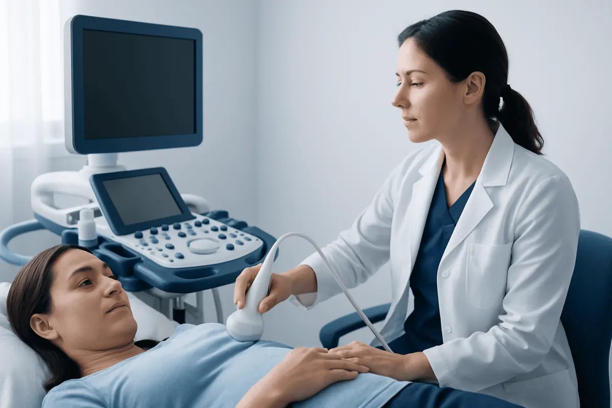
Uterine fibroids diagnosis commonly starts with a review of symptoms, medical history and a pelvic examination, followed by pelvic ultrasound. MRI, hysteroscopy or blood tests may be recommended when the diagnosis is uncertain, symptoms are significant or treatment planning requires a more detailed view.
Overview: how uterine fibroids are diagnosed
Uterine fibroids diagnosis usually begins during a gynecological consultation. A clinician asks about menstrual bleeding, pelvic pressure or pain, urinary or bowel symptoms, pregnancy history and plans for future fertility. They then perform a pelvic examination when appropriate and usually arrange a pelvic ultrasound to look at the uterus.
Fibroids, also called leiomyomas or myomas, are noncancerous growths of uterine muscle. They may develop within the uterine wall, project toward the outer surface of the uterus or grow into the uterine cavity. Their location and size can influence symptoms, fertility considerations and the most suitable management approach.
Some fibroids are found incidentally during a routine pelvic examination, pregnancy scan or imaging performed for another reason. Others are investigated because of heavy or prolonged periods, anemia, pelvic fullness, frequent urination, constipation or difficulty conceiving. A diagnosis does not automatically mean treatment is needed; the next steps depend on symptoms and individual goals.
Symptoms and clinical assessment
Symptoms alone cannot confirm fibroids because heavy periods and pelvic discomfort can have several possible causes. However, a clear description of symptoms helps the clinician decide which tests are useful and whether there may be another condition that also needs assessment.
During the appointment, patients may be asked when symptoms began, whether periods have changed, how often sanitary products are changed, whether there is bleeding between periods, and whether pain occurs during sex or menstruation. Questions may also cover urinary frequency, constipation, tiredness, previous pelvic surgery, medications and family history.
A pelvic examination may reveal an enlarged, irregularly shaped or firm uterus, which can suggest fibroids. Small fibroids may not be felt on examination, and an examination cannot reliably show their exact number or location. Imaging is therefore usually needed to clarify the findings.
- Heavy, prolonged or painful menstrual periods
- Pelvic pressure, bloating or a feeling of fullness
- Frequent urination or difficulty fully emptying the bladder
- Constipation or rectal pressure
- Fatigue, breathlessness or dizziness that may indicate anemia
- Fertility concerns, recurrent pregnancy loss or pregnancy-related symptoms
Tests used to confirm fibroids
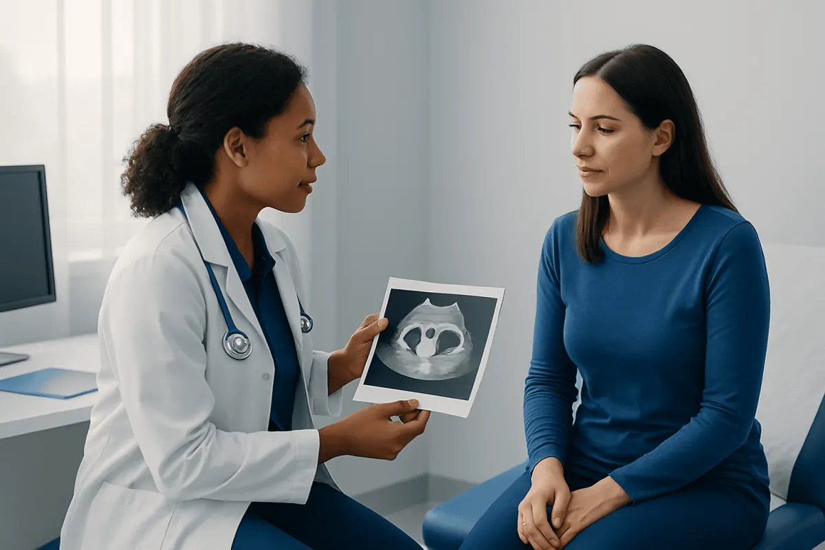
Pelvic ultrasound is the first-line imaging test for most people with suspected fibroids. It is safe, does not use radiation and can show the uterus, ovaries and many fibroids. A transabdominal ultrasound is performed over the lower abdomen, while a transvaginal ultrasound uses a slim probe placed in the vagina to provide closer images of the uterus. Sometimes both approaches are used.
MRI provides more detailed images than ultrasound and can be particularly useful when there are multiple fibroids, a large uterus, uncertain ultrasound findings or a need to plan a procedure. It can help distinguish fibroids from other uterine conditions and produce a detailed map of their size, number, location and tissue characteristics. Access to timely MRI or specialist imaging review may also support efficient treatment planning where clinically appropriate.
Blood tests may be advised if menstrual bleeding is heavy. A full blood count can identify anemia, and additional tests may be selected based on the person’s symptoms and medical history. These tests do not diagnose fibroids themselves, but they help assess the impact of bleeding and guide safe care.
If symptoms suggest a fibroid growing into the uterine cavity, a clinician may recommend saline infusion sonography or hysteroscopy. In saline infusion sonography, sterile fluid is placed in the uterus during ultrasound to outline the cavity. Hysteroscopy uses a thin camera passed through the vagina and cervix to examine the uterine cavity directly; it can also allow selected cavity fibroids to be treated during the same pathway.
What to expect during imaging and diagnostic procedures
Most diagnostic tests for fibroids are outpatient procedures. For a pelvic ultrasound, patients may be asked to arrive with a comfortably full bladder for an abdominal scan. A transvaginal scan may be performed with an empty bladder. The examination generally takes a short time, and normal activities can usually be resumed immediately.
For a pelvic MRI, the patient lies still on a table that moves into the scanner. The test is painless, although the machine makes loud sounds. Some scans use contrast material administered through a vein to improve tissue detail. The imaging team checks for factors such as kidney problems, previous contrast reactions, pregnancy or implanted devices before the examination.
Hysteroscopy may be performed in a clinic or operating setting depending on the reason for the test and whether treatment is planned. A speculum is inserted, then a narrow telescope passes through the cervix into the uterus. Local anesthesia, sedation or general anesthesia may be used in some circumstances. Mild cramping or light bleeding afterward can occur.
Although these tests are generally low risk, every procedure has considerations. Transvaginal ultrasound may cause temporary discomfort. MRI contrast reactions are uncommon but possible. Hysteroscopy has small risks including infection, bleeding, uterine perforation or fluid-related complications. The care team explains individual benefits, alternatives and risks before a procedure.
How results guide treatment planning
Test results help the clinician determine whether fibroids are likely to be responsible for symptoms and whether watchful waiting, medicines or a procedure is most appropriate. Important details include the fibroids’ number, size, rate of change when previous scans are available, and relationship to the uterine cavity, bladder and bowel.
For people hoping to become pregnant, the position of a fibroid can be especially relevant. Fibroids that distort the uterine cavity may affect fertility or pregnancy in some cases, while many fibroids do not prevent pregnancy. A fertility-focused assessment considers imaging findings alongside age, ovarian reserve where relevant, fallopian tube status and other possible factors.
When a procedure is being considered, detailed imaging supports shared decision-making. Options may include hysteroscopic removal of cavity fibroids, myomectomy to remove fibroids while preserving the uterus, uterine artery embolization, or hysterectomy for people who do not wish to preserve the uterus. The best option depends on symptoms, fibroid characteristics, overall health and reproductive preferences. Further information about evaluation and management is available through uterine fibroid treatment options.
Some imaging findings can overlap with other conditions, such as adenomyosis, ovarian cysts or less commonly other pelvic masses. If the appearance is not typical, the clinician may recommend additional imaging, follow-up scanning or referral to a gynecologist with relevant expertise rather than making assumptions from a single test.
Recovery, follow-up and self-care after testing
Recovery after a standard pelvic ultrasound or MRI is immediate for most patients. Results may be discussed on the day of the scan or at a follow-up appointment, depending on local processes and whether a specialist radiology review is needed. It can be helpful to keep a symptom and menstrual diary while awaiting results.
After hysteroscopy, mild cramping and light spotting may last for a short period. Patients are usually given specific instructions about pain relief, bathing, sexual activity, tampon use and returning to work or exercise. If sedation or general anesthesia was used, they should arrange for a responsible adult to accompany them home and follow the discharge advice provided.
Until the assessment is complete, people with heavy periods can ask their clinician about ways to manage bleeding and protect iron levels. A balanced diet containing iron-rich foods may support general health, but suspected anemia should be evaluated rather than self-treated alone. Iron supplements and medicines for menstrual bleeding should be used under professional guidance.
Follow-up scans are not required for every fibroid. They may be considered if symptoms change, a fibroid was difficult to characterize, pregnancy is planned, or conservative monitoring has been chosen. Patients should ask what findings were seen, whether the uterine cavity is affected and what changes would warrant earlier review.
When to seek medical care
Medical assessment is appropriate for persistent heavy or prolonged periods, bleeding between periods, pelvic pressure, worsening pain, unexplained fatigue or new urinary and bowel symptoms. These symptoms are often treatable, and early evaluation can identify whether fibroids or another condition is contributing.
Urgent medical advice is needed for very heavy vaginal bleeding, fainting, marked dizziness, chest discomfort, shortness of breath, severe or sudden pelvic pain, fever with pelvic pain, or pain and bleeding during pregnancy. These symptoms do not always indicate a serious problem, but they require prompt assessment.
People with fibroid symptoms who are trying to conceive, have experienced recurrent pregnancy loss or are planning fertility treatment should discuss imaging results with a gynecologist or fertility specialist. The goal is to understand whether a fibroid is relevant before deciding on observation or treatment.
At Acıbadem Health Point, multidisciplinary specialists in JCI-accredited hospitals evaluate and treat uterine fibroids for Myomectomy Recovery for International Patients: The Usual Travel Timeline" class="ahp-ilk">international patients, using appropriate gynecological assessment and imaging to support individualized care plans.
Frequently asked questions
01Can uterine fibroids be diagnosed with an ultrasound?
Yes. Pelvic ultrasound is the most common test used to identify uterine fibroids and assess their size and location. Depending on the clinical question, an abdominal ultrasound, transvaginal ultrasound or both may be used.
02Why might an MRI be needed for uterine fibroids?
MRI may be recommended when ultrasound does not provide enough detail or when a procedure is being considered. It can show the number, position and characteristics of fibroids more precisely and help plan care.
03Do blood tests show whether someone has fibroids?
Blood tests cannot confirm fibroids directly. They are often used to check for anemia or iron deficiency when heavy menstrual bleeding is present and to assess overall health before treatment.
04Is a transvaginal ultrasound painful?
A transvaginal ultrasound may feel uncomfortable or create mild pressure, but it is usually not painful. Patients can tell the sonographer if they feel pain or would like the examination paused.
05Can a pelvic exam diagnose fibroids on its own?
A pelvic examination can sometimes suggest fibroids if the uterus feels enlarged or irregular. Imaging is generally needed to confirm the diagnosis and determine the fibroids’ exact size and location.
06What happens if fibroids are found but there are no symptoms?
Many fibroids do not require treatment if they are not causing symptoms or affecting fertility plans. A clinician may recommend observation and review if symptoms develop or change.
This article is for general information only and is not a substitute for professional medical advice. Please consult a qualified doctor about your individual situation.
Persistent digestive symptoms? Get evaluated in Turkey
JCI-accredited · board-certified surgeons · reply within 24h
Add us as a Preferred Source to see more of our trusted health content across Google Search, AI Overviews and Discover.