The Talus Bone: Function, Injuries, and Care

The bone talus, also called the talus bone, is a small but essential bone in the ankle that connects the lower leg to the foot and helps transfer body weight during standing and walking. Its unique anatomy and blood supply mean that significant talus injuries need timely medical assessment and careful follow-up.
What Is the Bone Talus?
The bone talus, or talus bone, is one of the seven tarsal bones in the rear part of the foot. It sits directly beneath the tibia, or shinbone, and forms a central part of the ankle joint. The talus receives force from the leg and distributes it into the foot when a person stands, walks, runs, or jumps.
Unlike many bones, the talus has no muscles attached to it. Instead, it is held in place by surrounding ligaments and fits closely with the tibia, fibula, heel bone (calcaneus), and navicular bone. This arrangement allows the ankle and hindfoot to move smoothly while remaining stable under body weight.
The talus is not the same as the calcaneus. The calcaneus is the larger heel bone below the talus. Together, these bones form the subtalar joint, which helps the foot adapt to uneven ground by allowing inward and outward tilting movements.
Why the Talus Is Important for Movement
The upper rounded surface of the talus, called the talar dome, meets the tibia and fibula to create the main ankle joint. This joint mainly permits the foot to move upward toward the shin and downward away from it. These movements are necessary for walking, climbing stairs, squatting, and many sporting activities.
Below the talus, joint surfaces connect with the calcaneus at the subtalar joint. In front, the talar head connects with the navicular bone. These linked joints help coordinate foot position, balance, and shock absorption as the body moves over the foot.
A large proportion of the talus is covered with articular cartilage. Cartilage reduces friction where bones meet, but it also means there is relatively little surface area for blood vessels to enter the bone. This feature is clinically important because a serious injury can sometimes disrupt blood flow and slow healing.
Problems affecting the talus can therefore cause symptoms not only at the ankle, but also around the back, inner, or outer side of the hindfoot. The exact location of pain depends on the part of the talus and the nearby joint involved.
Common Talus Conditions and Injuries

A talus fracture is a break in the bone. It may involve the talar neck, body, head, or one of the smaller projections on either side. These fractures often occur after substantial force, such as a fall from height, a road traffic collision, or a forceful twisting injury. Less severe injuries may be mistaken initially for a routine ankle sprain, especially when swelling and pain are prominent.
An osteochondral lesion of the talus involves damage to cartilage and the underlying bone on the talar dome. It may develop after an ankle sprain or fracture, although not every case has a clearly remembered injury. Some people notice deep ankle pain, swelling after activity, catching, clicking, or a feeling that the ankle is not moving normally.
Another concern after a major talus injury is avascular necrosis, sometimes called osteonecrosis. This means part of the bone is damaged because its blood supply has been reduced or interrupted. It does not occur after every fracture, but the risk can be greater with displaced fractures or dislocations. A clinician may monitor healing with repeat imaging when this risk is present.
Arthritis can also affect the ankle or subtalar joint after a talus injury. This may happen when joint cartilage has been injured at the time of trauma or when the joint heals with altered alignment. Symptoms can include stiffness, pain with weight-bearing, and reduced tolerance for walking on uneven surfaces.
Symptoms That May Involve the Talus
Symptoms vary with the cause and severity of the problem. Following an acute injury, a person may have intense ankle or hindfoot pain, rapid swelling, bruising, tenderness, and difficulty or inability to put weight through the foot. The ankle may look misshapen after a fracture-dislocation, which requires urgent assessment.
More gradual talar problems may cause deep pain in the ankle, stiffness after rest, swelling after exercise, or discomfort when walking on slopes and uneven ground. Some people with cartilage-related injuries report catching, locking, or a sense of instability, although these symptoms can have several possible causes.
Not all ankle pain comes from the talus. Sprains, tendon problems, stress injuries, inflammatory arthritis, nerve irritation, and other conditions can produce similar symptoms. A medical assessment helps identify the source rather than relying on symptoms alone.
It is sensible to avoid repeatedly “testing” a painful ankle by continuing impact activity. Early protection may limit further discomfort and makes it easier for a clinician to examine the area accurately.
How Talus Problems Are Diagnosed
A clinician usually begins by asking how symptoms started, whether there was an injury, where pain is felt, and whether the person can bear weight. The examination may include checking swelling, skin condition, circulation, sensation, tenderness, range of motion, and ankle stability. The other leg may be examined for comparison.
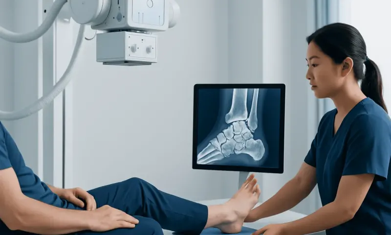
Plain X-rays are often the first imaging test after an injury. However, some small fractures, cartilage injuries, and early changes in bone blood supply may not be fully visible on initial X-rays. If clinical concern remains, a doctor may recommend computed tomography (CT) for detailed bone images or magnetic resonance imaging (MRI) to assess bone marrow, cartilage, ligaments, and soft tissues.
In selected cases, repeat X-rays or follow-up scans are used to assess alignment and bone healing over time. This follow-up is especially important after a displaced talus fracture because joint alignment and circulation to the bone can influence longer-term outcomes.
Accurate diagnosis supports appropriate treatment planning. It also helps distinguish a minor soft-tissue injury from conditions that require more protection, specialist review, or surgery.
Treatment Options and Recovery
Treatment depends on the specific diagnosis, the location of any fracture, whether bone fragments have moved out of position, the stability of nearby joints, skin condition, and the person’s overall health and activity needs. A stable, non-displaced fracture may sometimes be treated without surgery using immobilization in a cast or walking boot and a period of restricted weight-bearing directed by the treating team.
Displaced, unstable, open, or fracture-dislocation injuries commonly need urgent orthopedic care. Surgery may be recommended to restore the shape and alignment of the bone and joint surfaces, often using screws or other fixation devices. The aim is to support healing and reduce the chance of later joint problems, although no treatment can eliminate every risk after a significant injury.
For osteochondral lesions, care may include activity modification, physiotherapy, bracing, and treatment of contributing ankle instability. Persistent or larger lesions may be considered for procedures that address damaged cartilage and underlying bone. Decisions are individualized after imaging and specialist assessment.
Recovery can take months, particularly after a major talus fracture. Following restrictions on weight-bearing, attending follow-up appointments, and completing rehabilitation are important parts of recovery. Orthopedic care may coordinate fracture management, imaging review, rehabilitation, and longer-term monitoring of ankle function.
Protecting Ankle and Talus Health
Not every talus injury can be prevented, particularly those caused by accidents. However, supportive footwear, gradual training progression, strength and balance exercises, and appropriate sport-specific protective equipment may reduce the risk of some ankle injuries. People returning to sport after an ankle sprain should allow adequate recovery rather than returning while pain, swelling, or instability remains.
Regular exercises prescribed by a physiotherapist can improve ankle mobility, calf strength, balance, and confidence with walking or sport. These exercises should be tailored to the diagnosis and stage of healing. Beginning high-impact exercise too early after a fracture or significant sprain can delay recovery or worsen symptoms.
Smoking can impair bone and wound healing. A person preparing for surgery or recovering from a fracture may wish to discuss smoking cessation support with a healthcare professional. Good overall nutrition, sleep, and management of long-term health conditions also support recovery.
If pain continues despite rest and simple measures, it is better to seek assessment than to self-diagnose a “weak ankle.” Identifying a structural problem early may help guide safer activity choices and treatment.
When to Seek Medical Care
Urgent medical assessment is appropriate after an ankle or foot injury if there is visible deformity, severe swelling, an open wound, numbness, a cold or pale foot, worsening pain, or inability to bear weight. These signs may indicate a fracture, dislocation, circulation problem, or another injury that needs prompt care.
A doctor should also assess ongoing ankle pain, recurrent swelling, catching or locking, reduced movement, or difficulty returning to ordinary activities after a sprain or fall. Symptoms that persist for more than a few days or recur with activity deserve attention, particularly when there is tenderness deep in the ankle or hindfoot.
People with diabetes, reduced circulation, bone fragility, or reduced sensation in the feet should seek advice promptly after an ankle injury, even if symptoms seem mild. These conditions can affect healing and make injuries harder to evaluate without examination.
Acıbadem Health Point’s multidisciplinary specialists in JCI-accredited hospitals can assess and treat ankle and talus conditions for international patients, with care plans guided by the individual diagnosis and imaging findings.
Frequently asked questions
01Where is the bone talus located?
The talus is located at the top of the foot, between the lower leg and the heel bone. It sits beneath the tibia and fibula and above the calcaneus, forming part of both the ankle and subtalar joints.
02Is the talus the same as the ankle bone?
The talus is often described as an ankle bone because it forms a major part of the ankle joint. However, the ankle includes several bones, ligaments, tendons, and joint surfaces, not the talus alone.
03Can a person walk with a talus fracture?
Some people may be able to take steps with certain fractures, but this does not rule out a significant injury. Walking on a suspected talus fracture can worsen pain or displacement, so medical assessment is important after a substantial injury or when weight-bearing is difficult.
04How long does a talus fracture take to heal?
Healing time varies according to the fracture location, severity, alignment, blood supply, and treatment. A clinician will use examinations and imaging to guide when it is safe to increase weight-bearing and activity.
05What is avascular necrosis of the talus?
Avascular necrosis is damage to bone tissue caused by reduced blood supply. It can be a complication of some serious talus fractures, particularly when the bone has shifted out of place, and it requires medical monitoring.
06Can an ankle sprain damage the talus?
A sprain mainly affects ligaments, but a twisting ankle injury can sometimes also injure cartilage or bone on the talus. Continued deep ankle pain, swelling, catching, or difficulty returning to activity should be evaluated by a healthcare professional.
This article is for general information only and is not a substitute for professional medical advice. Please consult a qualified doctor about your individual situation.
Joint and spine care in Turkey — expert assessment & treatment
JCI-accredited · board-certified surgeons · reply within 24h
Add us as a Preferred Source to see more of our trusted health content across Google Search, AI Overviews and Discover.