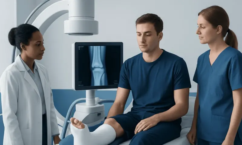
Shattered Talus Bone: Urgent Ankle Fracture Care

A shattered talus bone usually means a comminuted talus fracture, in which this important ankle bone has broken into multiple pieces. Prompt orthopedic assessment is essential because the talus supports ankle and foot movement and has a vulnerable blood supply that can affect healing.
Overview: What Does a Shattered Talus Bone Mean?
A shattered talus bone generally refers to a comminuted talus fracture, meaning the talus has broken into several fragments. The talus is a small but highly important bone at the top of the foot. It sits between the lower leg bones and the heel bone, transferring body weight through the ankle and helping the foot move on uneven ground.
Although the word “shattered” can sound frightening, treatment decisions are based on the exact fracture pattern rather than the description alone. Doctors consider which part of the talus is affected, whether pieces have moved out of place, whether nearby joints are involved and whether the skin or blood vessels have been injured. Early specialist care aims to restore alignment and reduce the risk of lasting ankle problems.
Talus fractures are less common than many other ankle fractures, but they deserve careful attention. Unlike most bones, much of the talus is covered in joint cartilage, and its blood supply can be disrupted by injury. This is why follow-up imaging and a gradual recovery plan are often important even after the initial fracture has been treated.
How the Talus Works and Where It Can Fracture
The talus has three main regions: the head at the front, the body in the middle and the neck between them. It forms the ankle joint with the tibia and fibula, the subtalar joint with the heel bone, and a joint with the navicular bone at the front of the foot. Together, these joints allow the foot to point up and down, adapt to surfaces and support walking.
A fracture can occur in the talar neck, body, head or one of the smaller projections at its sides or back. A shattered fracture often involves the talar body or neck, sometimes with dislocation of the ankle or subtalar joint. These injuries may occur alongside fractures of the heel, ankle or lower leg, particularly after a substantial impact.
The location matters because it influences stability, circulation and function. For example, a displaced fracture through the talar neck can affect blood flow to the body of the talus. A treating orthopedic specialist uses imaging and physical examination to explain the individual injury pattern and its likely implications.
Symptoms and Common Causes

A talus fracture usually causes sudden, severe ankle or hindfoot pain after an injury. Swelling, bruising and an inability to stand or walk are common. The ankle or foot may look misshapen if a fracture is displaced or a joint is dislocated. However, some fractures can be less obvious, especially if they involve a smaller part of the bone or develop from repeated stress.
Most serious talus fractures result from high-energy trauma, such as a road traffic collision, a fall from a height, a sports impact or a heavy object falling onto the foot. In these situations, the foot may be forced sharply upward, downward or twisted while bearing weight. A lower-energy injury can also cause a fracture in some people, particularly if bone strength is reduced.
Clinicians also consider symptoms such as numbness, tingling, coldness, increasing tightness or changes in skin color. These can suggest pressure on nerves or blood vessels and need prompt assessment. It is not safe to assume that an injury is only a sprain when walking is difficult, pain is severe or swelling follows a major impact.
- Intense pain around the ankle, heel or top of the foot
- Rapid swelling, bruising or tenderness
- Difficulty bearing weight or moving the ankle
- An unusual foot position or visible deformity
- Numbness, pale or blue toes, or a cold foot
How a Talus Fracture Is Diagnosed
Diagnosis begins with a history of how the injury happened and an examination of the foot and ankle. The clinician checks swelling, skin condition, pulses, sensation, toe movement and the position of the joints. This assessment is especially important after a high-impact injury because urgent treatment may be needed if there is a dislocation, an open wound or reduced circulation.
Plain X-rays are commonly the first imaging test. They can show many fractures and whether the ankle or subtalar joint is out of alignment. However, the talus has a complex shape, and small or comminuted fracture lines may not be fully visible on standard X-rays.
A CT scan is frequently used to map the fracture in greater detail. It helps the orthopedic team evaluate the number and position of bone fragments, the amount of joint surface involvement and whether surgery may be beneficial. MRI may be considered in selected cases, such as when an occult fracture or later changes in bone blood supply are suspected. Repeat X-rays or other scans may be used during recovery to monitor healing.
Treatment Options and Early Care
Initial care focuses on protecting the injured foot, controlling pain and swelling, and checking circulation and nerve function. The foot may be placed in a splint, kept elevated and protected from weight-bearing. If a joint is dislocated, clinicians may need to realign it urgently. An open fracture, in which bone communicates with a wound, requires emergency treatment because of infection risk.
Some nondisplaced, stable talus fractures can be managed without surgery using a cast or removable boot and a period of strict non-weight-bearing. Follow-up appointments and imaging are important because even a fracture that initially appears well aligned can occasionally shift. The length of immobilization and restrictions varies according to the fracture location and healing progress.
Displaced, unstable or comminuted fractures often require surgery to restore the shape of the bone and joint surfaces. A surgeon may use screws, plates or other fixation devices to hold fragments in alignment while healing occurs. If swelling is substantial, temporary stabilization or delayed definitive surgery may be safer than operating immediately. The goal is to create the best possible conditions for bone healing and joint function, not to promise a particular outcome.
For international patients, Acıbadem Health Point’s multidisciplinary orthopedic specialists and JCI-accredited hospitals diagnose and treat complex fractures, coordinating imaging, surgery and rehabilitation when appropriate.
Recovery, Rehabilitation and Possible Long-Term Effects
Healing after a shattered talus bone can require patience. Many people need to avoid putting weight on the injured foot for a number of weeks, sometimes longer, depending on fracture severity and surgical findings. Crutches, a walker, knee scooter or other mobility aid may help during this phase. The treating team gives individualized guidance on when it is safe to begin partial and then full weight-bearing.
Rehabilitation commonly starts with gentle movement when the fracture is stable enough. A physiotherapist may guide exercises to regain ankle range of motion, strengthen the calf and foot muscles, improve balance and gradually return to walking. Following the recommended plan matters: loading the joint too early may affect healing, while prolonged immobility without guidance can contribute to stiffness and weakness.
Potential complications include delayed union, nonunion, malunion, stiffness and post-traumatic arthritis. One specific concern is avascular necrosis, in which part of the talus loses blood supply and the bone weakens. This does not occur after every talus fracture, but the risk can be higher in severe displaced injuries. Regular review allows the team to identify healing concerns and discuss options early.
Return to work, driving, sport and higher-impact activities varies widely. It depends on pain, strength, mobility, imaging findings, the physical demands involved and whether complications develop. A clinician should clear these activities rather than relying on a fixed timeline from another person’s recovery.
Protecting Recovery and Supporting Bone Health
There is no guaranteed way to prevent a traumatic talus fracture, but practical safety measures can lower the chance of serious falls and sports injuries. These include using appropriate footwear, seat belts and protective equipment, following workplace safety procedures, and gradually building strength and balance when returning to activity after an injury.
During recovery, people should follow instructions about casts, boots, wound care and weight-bearing. Keeping appointments is particularly important after a talus fracture because clinicians may need to assess alignment and bone circulation over time. Smoking and nicotine use can impair bone healing, so discussing support to stop with a healthcare professional can be helpful.
A balanced diet with adequate protein, calcium and vitamin D supports general bone health. Supplements are not appropriate for everyone, so it is sensible to ask a clinician before starting them, especially for people with kidney disease, digestive conditions or other long-term health concerns. Medications and health conditions that affect bone strength should also be reviewed when relevant.
When to Seek Medical Care
Urgent medical assessment is needed after a major ankle or foot injury when there is severe pain, obvious deformity, inability to bear weight, an open wound or rapidly increasing swelling. Emergency care is also important if the toes are numb, pale, blue, cold, weak or difficult to move. These symptoms can indicate a problem with circulation, nerves or joint alignment.
People should arrange medical review if ankle or foot pain, swelling or difficulty walking persists after an injury, even if initial symptoms seem manageable. A suspected fracture should not be walked on or “tested” repeatedly. Until assessed, keeping the foot still, avoiding weight-bearing and using a wrapped cold pack briefly for comfort may be reasonable if it does not delay care.
Frequently asked questions
01Is a shattered talus bone an emergency?
It can be, particularly when the foot is deformed, circulation is affected, the fracture is open or a joint is dislocated. A suspected talus fracture should be assessed urgently because correct alignment and blood supply are important to healing.
02Can a shattered talus bone heal without surgery?
Some stable fractures that have not moved out of position can heal with immobilization and strict avoidance of weight-bearing. However, shattered or displaced fractures commonly need surgical assessment because several bone fragments or damaged joint surfaces may need to be realigned.
03How long does recovery from a talus fracture take?
Recovery varies considerably with the location and severity of the fracture, whether surgery is needed and how the bone heals. Protected weight-bearing may last weeks to months, and strength, mobility and confidence can continue improving for many months afterward.
04Will a talus fracture cause arthritis?
Not everyone develops arthritis after a talus fracture, but the risk is higher when the joint surface was damaged or the fracture was displaced. Long-term follow-up, rehabilitation and managing symptoms early can help clinicians address changes in the ankle or subtalar joint.
05What is avascular necrosis after a talus fracture?
Avascular necrosis means that part of a bone has lost enough blood supply to weaken or collapse over time. Because the talus has a relatively limited blood supply, this is a recognized risk after some severe fractures, and follow-up imaging can help monitor for it.
06When can someone walk again after a talus fracture?
Walking should restart only when the treating clinician confirms that the fracture is stable enough to bear weight. Beginning too early can place stress on healing bone, while a supervised gradual return helps restore movement and strength more safely.
This article is for general information only and is not a substitute for professional medical advice. Please consult a qualified doctor about your individual situation.
Joint and spine care in Turkey — expert assessment & treatment
JCI-accredited · board-certified surgeons · reply within 24h
Add us as a Preferred Source to see more of our trusted health content across Google Search, AI Overviews and Discover.