Testicular Ultrasound: Purpose, Procedure and Results

Key Takeaways
- A testicular ultrasound uses sound waves to create images of the testicles and scrotum without radiation.
- It is often ordered for pain, swelling, a lump, trauma, or concerns about infection, torsion, varicocele, or a mass.
- The test is usually straightforward, painless, and performed with a small handheld probe on the skin.
- Results help doctors decide whether reassurance, monitoring, treatment, or urgent care is needed.
- A normal or reassuring scan can still be followed by further evaluation if symptoms continue or change.
A testicular ultrasound is a quick, noninvasive imaging test that helps doctors examine the testicles and surrounding scrotal structures. It is commonly used to look into pain, swelling, lumps, injury, fertility concerns, or changes found during a physical exam.
Overview
A testicular ultrasound is one of the most useful imaging tests for checking the testicles, epididymis, and nearby scrotal tissues. It uses high-frequency sound waves to produce real-time pictures, which helps clinicians see what may be causing pain, swelling, a lump, or an abnormal feeling on examination. Because it does not use radiation, it is a common first-line test when the source of symptoms is not clear.
For many patients, the main concern is not the scan itself but what it may reveal. That is understandable, especially when symptoms appear suddenly or when the examination was done far from home or during an international trip. A testicular ultrasound is designed to answer practical questions: Is there inflammation? Is blood flow normal? Is there a cyst, fluid collection, hernia, or solid mass? The images help narrow those possibilities and guide the next step.
In men’s health care, this test is especially valuable because some scrotal conditions look similar from the outside but require very different treatment. A prompt, accurate ultrasound can help distinguish a problem that can be monitored from one that needs urgent care. For patients seeking evaluation abroad, it may also be a reassuring way to gain clarity quickly and avoid unnecessary delay.
What the Test Can Help Detect

Doctors may request a testicular ultrasound for several reasons. It can help evaluate a new lump, testicular tenderness, one-sided swelling, a heavy or dragging sensation, or pain that has not improved. It is also used after injury, when infection is suspected, or when a physical examination suggests something that should be looked at more closely.
The scan can provide useful information about common scrotal conditions, including:
- Hydrocele, which is a fluid buildup around the testicle
- Varicocele, which is enlargement of veins in the scrotum
- Epididymitis or orchitis, which are inflammatory or infectious conditions
- Testicular torsion, a sudden twisting that may reduce blood flow and needs urgent assessment
- Benign cysts and other noncancerous changes
- Solid masses that may need further evaluation
It is important to note that the scan does not replace a full medical assessment. A doctor uses the ultrasound together with the history, physical examination, and sometimes blood or urine tests. When the visit happens during a stay in another country, this combined approach can be especially helpful because it reduces guesswork and supports clear follow-up planning before travel home.
Common Symptoms and Reasons It Is Ordered

Many people are referred for a testicular ultrasound after noticing symptoms that are difficult to interpret on their own. Common Causes, Related Conditions, and When to See a Doctor" class="ahp-ilk">Testicular pain can range from a dull ache to sharp discomfort, and swelling may be gradual or sudden. Some men notice a firm area, a change in shape, or a feeling of heaviness in the scrotum.
The test may be ordered after a clinician finds something unusual during an examination, even if the patient has mild or no symptoms. This is often the case with a lump that feels distinct from the testicle itself, or with a suspected varicocele that becomes more noticeable when standing. In fertility care, ultrasound may also help evaluate the testicles and surrounding veins when there are signs that sperm production or testicular function could be affected.
Because symptoms can overlap, ultrasound is helpful in sorting through possibilities. For example, swelling can reflect infection, fluid, a hernia, or a vascular issue. A careful scan gives the doctor a better picture of what is happening and whether treatment should begin right away or can be planned more gradually.
Causes & Risk Factors
There is no single cause that leads to a testicular ultrasound; instead, the test is prompted by a set of symptoms, findings, or risk factors. Recent trauma, a history of torsion, urinary or genital infection, and fertility concerns may all lead a doctor to request imaging. A prior episode of testicular pain, even if it improved, may also justify an ultrasound if there is lingering uncertainty.
Some men are more likely to need this exam because of the way their symptoms appear. A painless lump, persistent one-sided enlargement, or a heavy feeling in the scrotum may prompt imaging even without acute pain. In adolescents and younger adults, doctors are often careful to check for torsion or varicocele; in older adults, they may be equally attentive to infection, fluid collections, or masses.
International patients sometimes come to evaluation after delays, especially if they have already tried to manage symptoms while traveling. In that setting, the ultrasound can be an efficient way to sort out what is urgent, what can be treated medically, and what may need specialist input such as urology follow-up.
Diagnosis and How the Procedure Is Performed
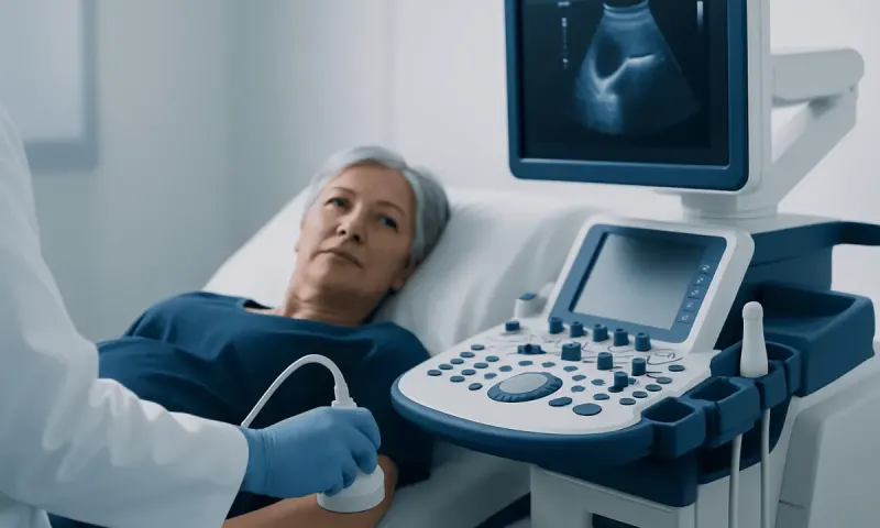
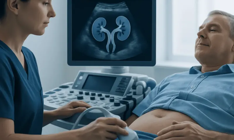
A testicular ultrasound is usually performed in a radiology or imaging unit. The patient typically lies on the back, and the scrotum may be supported with a towel to keep it still and comfortable. A clear gel is placed on the skin, and a small probe is moved gently over the scrotum to create images from different angles. If blood flow needs to be assessed, the sonographer may use Doppler ultrasound.
The procedure is generally quick and does not require injections or sedation. Most patients feel pressure from the probe and the coolness of the gel, but not pain from the scan itself. If the area is already tender, the staff usually works carefully to reduce discomfort. Patients are often able to return to normal activities immediately afterward.
Preparation is minimal in most cases. Clothing that is easy to remove and replace can make the visit simpler, and some centers may provide a gown. If the exam is being done because of an emergency concern, such as sudden severe pain, it is typically arranged promptly so the doctor can interpret the findings without delay.
Understanding the Results
Ultrasound results are interpreted by looking at the size, shape, texture, and blood flow of the testicles and surrounding structures. A normal scan may show no abnormal mass and normal blood flow, which can be reassuring. Even then, a doctor may still consider other causes of pain or discomfort if symptoms persist.
Some common findings are relatively straightforward. Fluid around the testicle may suggest a hydrocele. Enlarged veins can point to a varicocele. Increased blood flow and tissue change may support inflammation or infection. Reduced blood flow, especially with sudden pain, can raise concern for torsion and may require urgent action. A solid intratesticular mass is taken seriously and often leads to specialist evaluation and additional testing.
It is helpful to remember that an ultrasound report is only one part of the clinical picture. The meaning of a finding depends on age, symptoms, exam results, and timing. A small change seen after an injury, for example, may be watched differently from a similar finding in someone with a painless lump. A qualified doctor should explain the result in context and outline the next step clearly.
Treatment Options After the Scan
Treatment depends on what the ultrasound shows. Some conditions are treated with rest, anti-inflammatory measures, antibiotics, or planned follow-up. Others may require a urologic procedure or surgery. In urgent settings, the scan may quickly point to a condition that needs immediate intervention, such as testicular torsion.
If the result is reassuring, the doctor may recommend observation, symptom control, or additional testing if there are ongoing concerns. When a varicocele, hydrocele, or epididymal inflammation is found, treatment is guided by symptoms, severity, and whether fertility is a concern. If a mass is seen, further evaluation may include blood tests, repeat imaging, or referral to a specialist.
For patients traveling for care, it is especially important to leave with a clear written plan. That plan should include what the scan showed, what symptoms should prompt reassessment, and how follow-up will happen once the patient returns home. Good coordination helps prevent gaps in care and reduces uncertainty after discharge.
Prevention & Self-care, and When to See a Doctor
Not every cause of scrotal symptoms can be prevented, but sensible self-care can help reduce discomfort and support recovery. Wearing supportive underwear may ease aching or heaviness. Following treatment instructions, resting when advised, and avoiding heavy lifting or strenuous activity during recovery can also help. If an infection is being treated, completing the prescribed course and attending follow-up are important.
Men should seek medical attention promptly for sudden testicular pain, a rapidly enlarging swelling, a new hard lump, redness with fever, nausea with severe scrotal pain, or any concern about testicular torsion. A painless lump should also be checked, even if it does not seem urgent. Early assessment is especially important when symptoms are new, unexplained, or different from anything experienced before.
For patients who are visiting from another country, the timing of follow-up matters. If symptoms worsen after the scan or before travel home, they should not wait for a routine appointment. Acibadem Health Point’s multidisciplinary specialists and JCI-accredited hospitals diagnose and treat this condition for international patients, with coordinated care that can support both the initial assessment and the handoff to ongoing treatment.
Frequently asked questions
01What does a testicular ultrasound check for?
It checks the testicles and nearby scrotal structures for causes of pain, swelling, lumps, or other changes. Doctors use it to look for fluid, inflammation, blood-flow problems, cysts, varicocele, or a mass.
02Is a testicular ultrasound painful?
The scan is usually not painful. Some pressure may be felt from the probe, and the area can be sensitive if it is already inflamed or injured, but the test itself is typically well tolerated.
03Do patients need to fast or prepare for the test?
In most cases, no special preparation is needed. Patients are usually asked to wear or bring clothing that is easy to remove so the scrotal area can be examined comfortably.
04Can ultrasound tell if a lump is cancer?
Ultrasound can show whether a lump appears solid or fluid-filled and whether it is inside or outside the testicle. While it can raise or lower concern, only a doctor can interpret the result in context, and some findings require further evaluation.
05What happens if the scan is normal but pain continues?
A normal ultrasound is reassuring, but it does not explain every cause of pain. The doctor may consider muscle strain, referred pain, or another condition and may suggest follow-up if symptoms persist or change.
06How soon are the results available?
That depends on the setting and the urgency of the case. In many facilities, the images are reviewed the same day or soon after, especially if the doctor needs quick answers to guide treatment.
This article is for general information only and is not a substitute for professional medical advice. Please consult a qualified doctor about your individual situation.
IVF & fertility treatment in Turkey — success rates and costs
JCI-accredited · board-certified surgeons · reply within 24h
Add us as a Preferred Source to see more of our trusted health content across Google Search, AI Overviews and Discover.