Skin Cancer on the Face: Signs, Diagnosis and Care
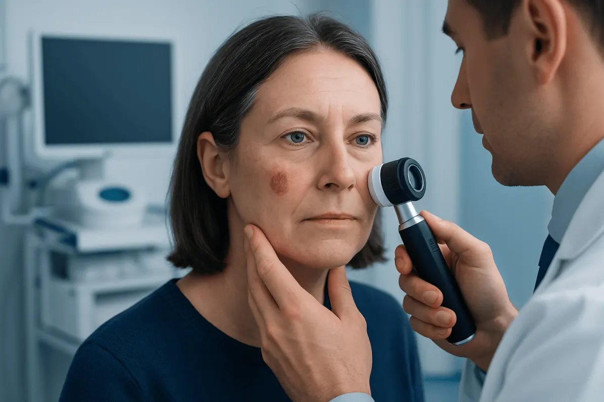
Skin cancer on the face can appear as a changing mole, a pearly bump, a scaly patch or a sore that does not heal. Early assessment by a dermatologist is important because most common facial skin cancers can be treated effectively while preserving as much healthy tissue and function as possible.
Skin Cancer on the Face: Overview
Skin cancer on the face is a growth of abnormal skin cells on areas that receive frequent sunlight, including the nose, ears, eyelids, lips, cheeks, forehead and scalp. It is often highly treatable when identified early. Because the face contains important structures for appearance, vision, speech and breathing, treatment planning aims to remove or control the cancer while protecting healthy tissue whenever possible.
The three main forms are basal cell carcinoma, squamous cell carcinoma and melanoma. Basal cell carcinoma is the most common and usually grows slowly. Squamous cell carcinoma can grow more quickly and, in some cases, spread beyond the skin. Melanoma is less common but can be more serious because it may spread if not detected and treated promptly.
Not every spot on the face is cancer. Acne, eczema, rosacea, infections and harmless age-related growths can look similar. However, a persistent or changing lesion deserves professional assessment rather than self-diagnosis.
What Does Skin Cancer Look Like When It Starts?
Early skin cancer on the face does not have one single appearance. Basal cell carcinoma may start as a shiny, pearly or flesh-coloured bump, sometimes with tiny visible blood vessels. It may also look like a pink patch, a scar-like area, or a sore that repeatedly crusts, bleeds or heals and returns.
Squamous cell carcinoma may appear as a rough, scaly or thickened red patch, a firm tender bump, or a crusted sore. A precancerous change called actinic keratosis can resemble a dry, sandpaper-like patch on sun-exposed skin. Melanoma may begin as a new or changing pigmented spot, although some melanomas are pink, red or skin-coloured rather than dark.
For moles and dark marks, clinicians often use the ABCDE reminder: Asymmetry, irregular Borders, varied Colour, increasing Diameter, and Evolving appearance. A lesion that looks noticeably different from a person’s other marks—sometimes called an “ugly duckling”—also warrants attention.
- A spot that grows, changes shape or changes colour
- A sore that does not heal within several weeks
- Repeated bleeding, crusting, itching, pain or tenderness
- A new persistent bump or scaly patch, especially on sun-exposed skin
Causes and Risk Factors for Facial Skin Cancer

Ultraviolet (UV) radiation from sunlight is a leading cause of skin cancer on the face. UV exposure can damage DNA in skin cells over time. The risk is associated with cumulative exposure as well as episodes of intense sun exposure and sunburn, particularly earlier in life. Tanning beds also expose skin to harmful UV radiation.
Risk is higher in people with fairer skin, light-coloured eyes or hair, freckles, a tendency to burn, or a history of significant sun exposure. However, skin cancer can affect people of every skin tone. In darker skin, changes may be less obvious or may arise in areas not usually exposed to the sun, so new or unusual lesions should still be assessed.
Other factors include increasing age, a personal or family history of skin cancer, many or atypical moles, a weakened immune system, previous radiation exposure and certain inherited conditions. Outdoor work, hobbies and living in high-UV environments can add to lifetime exposure.
Diagnosis and Assessment of a Facial Lesion
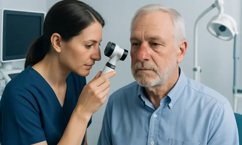
A dermatologist or other qualified clinician will ask about the lesion’s duration and changes, sun exposure, previous skin cancers and medical history. They will examine the area closely and may inspect the rest of the skin, including the scalp, ears, neck and areas that are less easily seen at home.
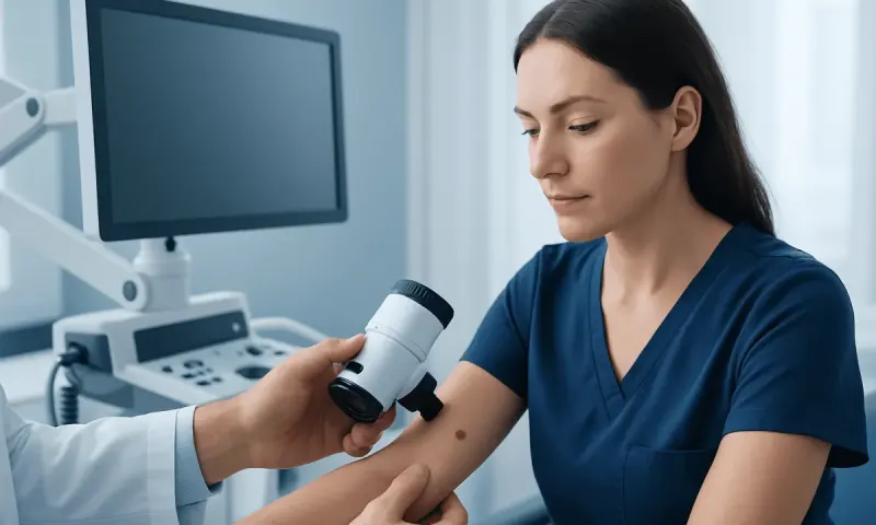
A dermatoscope, a handheld magnifying device with specialized lighting, may help the clinician assess pigment patterns and surface structures. If cancer is suspected, the diagnosis is confirmed with a biopsy. This involves removing a small sample or, when appropriate, the entire lesion for examination under a microscope by a pathologist.
The pathology result identifies the type of cancer and features that guide treatment. Imaging tests are not routinely needed for common early skin cancers, but may be considered when a lesion is large, recurrent, near deeper structures, involves lymph nodes, or has features suggesting a higher risk of spread.
How Treatable Is Skin Cancer on the Face?
Most basal cell carcinomas and many squamous cell carcinomas on the face are very treatable, especially when diagnosed before they grow deeply or spread. Melanoma outcomes also improve substantially when it is found at an early stage. The outlook depends on the exact cancer type, size, depth, location, whether it has spread, and whether it has been treated before.
Surgery is often the main treatment because it removes the cancer and allows the tissue to be examined. For cancers in cosmetically or functionally sensitive facial sites, a staged technique known as Mohs surgery may be recommended. It removes tissue in small layers and checks the edges during the procedure, helping preserve as much normal skin as possible while confirming that cancer cells have been cleared.
Other options may include standard surgical excision, curettage and electrosurgery for selected lesions, cryotherapy for some superficial precancerous lesions, topical medicines, photodynamic therapy or radiation therapy. Advanced, recurrent or metastatic cancers may require systemic treatments such as immunotherapy, targeted therapy or other oncology care. The best approach is individualized after pathology and specialist assessment.
After removal, reconstruction may be simple wound healing, stitches, a skin graft or a flap using nearby tissue. Planning considers the cancer margins as well as the natural contours and movement of the face.
Is Skin Cancer Serious if Caught Early?
Skin cancer can be serious, but early detection often makes treatment simpler and improves the chance of complete control. Many early basal cell and squamous cell cancers can be removed with local treatment. Early melanoma is also much more likely to be managed successfully before it reaches deeper layers of skin or other parts of the body.
It is still important not to delay assessment. Even slow-growing basal cell carcinoma can gradually damage nearby skin, cartilage, nerves or other tissues if left untreated, particularly around the nose, eyes and ears. Some squamous cell carcinomas and melanomas have a greater potential to spread, which is why a prompt diagnosis matters.
A clinician can explain the pathology report, stage where relevant, proposed treatment and expected follow-up. Questions about scarring, facial function and recovery are appropriate to discuss before treatment whenever time allows.
Can You Live a Normal Life with Skin Cancer?
Many people continue to live active, normal lives during and after treatment for skin cancer. Treatment may involve a short procedure and a period of wound care, while more complex facial surgery or advanced cancer treatment can require a longer recovery and closer follow-up. Support from a dermatology, surgery and oncology team can help patients understand what to expect.
Having one skin cancer increases the chance of developing another in the future, so ongoing skin awareness is important. Follow-up schedules vary according to the cancer type, stage and personal risk factors. A clinician may recommend regular full-skin examinations and checking the treated area for new changes.
Daily sun protection is a practical part of long-term care. This includes seeking shade when UV levels are high, wearing a broad-brimmed hat and protective clothing, using broad-spectrum sunscreen with SPF 30 or higher as directed, and avoiding tanning beds. Sunscreen is helpful but should complement, not replace, shade and protective clothing.
When to Seek Medical Care
A person should arrange a medical appointment for a new or changing mark on the face, especially if it bleeds, forms a recurring crust, becomes painful, or does not heal. Prompt review is particularly important for a changing mole, a rapidly enlarging lesion, a dark or irregular patch, or a persistent sore near the eye, lip, ear or nose.
People with a history of skin cancer, reduced immunity or many atypical moles should ask their clinician about an appropriate schedule for skin examinations. Taking a clear photograph of a spot over time may help document change, but it should not replace an in-person assessment.
At Acıbadem Health Point, multidisciplinary specialists in dermatology, pathology, surgical care and oncology assess and treat skin cancers for international patients in JCI-accredited hospitals. The appropriate care plan is based on the confirmed diagnosis, location and individual health needs.
Frequently asked questions
01Can skin cancer on the face be mistaken for a pimple?
Yes. Some early basal cell or squamous cell cancers can resemble a pimple, irritated bump or small sore. A spot that persists, repeatedly bleeds, crusts, grows or returns in the same place should be examined by a clinician.
02Does skin cancer on the face always look dark?
No. Many facial skin cancers are pink, red, pearly, scaly, skin-coloured or scar-like rather than dark. Although melanoma is often pigmented, it can occasionally have little or no visible pigment.
03How is skin cancer on the face removed?
Removal commonly involves a surgical procedure under local anaesthetic. Depending on the type and location, a specialist may use standard excision, Mohs surgery, topical treatment, radiation therapy or another suitable method.
04Will facial skin cancer treatment leave a scar?
Any procedure that removes skin can leave some degree of scarring. Specialists plan treatment to achieve cancer control while minimizing effects on appearance and function, and scars often continue to improve over time.
05Can skin cancer on the face come back after treatment?
Some skin cancers can recur, particularly if they were large, deeply invasive, previously treated or located in higher-risk areas. Regular follow-up and reporting any change at or near the treated site are important.
06How can a person check their face for skin cancer?
A person can use a mirror in good light to look at the forehead, nose, cheeks, lips, ears, eyelids and scalp line. It is helpful to ask another person or a hair professional to alert them to new scalp or ear lesions, but suspicious changes should be assessed by a medical professional.
This article is for general information only and is not a substitute for professional medical advice. Please consult a qualified doctor about your individual situation.
Hair transplant in Turkey — costs, graft counts & a free assessment
JCI-accredited · board-certified surgeons · reply within 24h
Add us as a Preferred Source to see more of our trusted health content across Google Search, AI Overviews and Discover.