Sacralization: A Congenital Spinal Variation
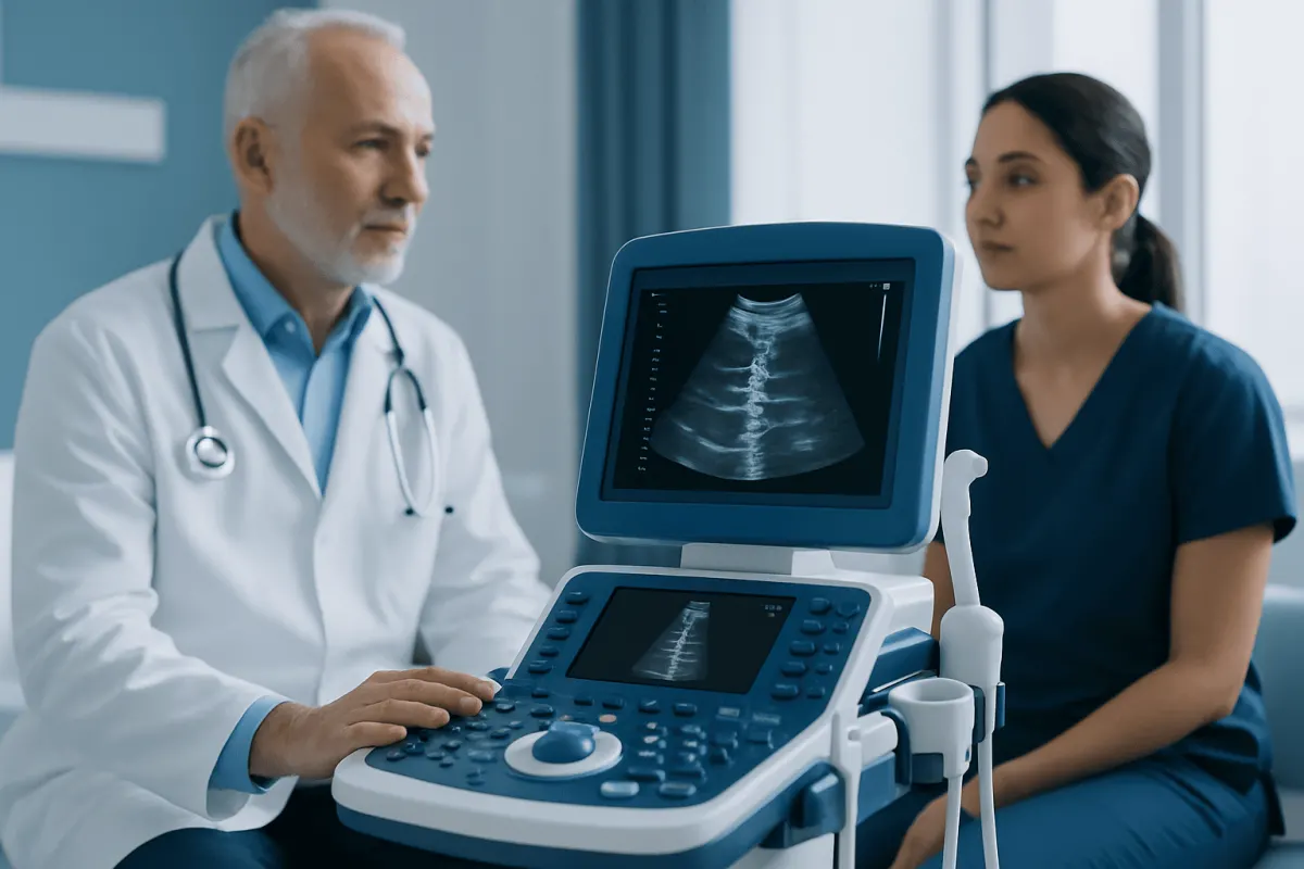
Sacralization is a congenital variation in which the fifth lumbar vertebra (L5) partly or completely connects with the sacrum. Many people never develop symptoms, but in some, altered spinal mechanics may contribute to low back, buttock, or leg pain.
Sacralization: what it means
Sacralization is a structural variation at the base of the spine. Normally, the lumbar spine has five movable vertebrae, numbered L1 to L5, above the sacrum, a triangular bone formed by fused vertebrae at the back of the pelvis. In sacralization, the lowest lumbar vertebra, usually L5, has an enlarged connection to the sacrum or pelvic bone. The connection may be on one side or both sides, and it may be partial or complete.
Clinicians use the broader term lumbosacral transitional vertebra (LSTV) because the anatomy can vary in either direction. In sacralization, L5 takes on some features of the sacrum. In the opposite variation, called lumbarization, the uppermost sacral segment appears more like an additional lumbar vertebra. These are developmental anatomical differences rather than diseases acquired later in life.
Sacralization is often found incidentally when an X-ray, CT scan, or MRI is performed for another reason. It does not necessarily require treatment. When a transitional vertebra is believed to be a clear source of ongoing low back pain, the situation may be called Bertolotti syndrome. This distinction matters: having sacralization on an image and having pain caused by sacralization are not always the same thing.
Evidence versus common myths
Myth: sacralization means the spine is damaged. Evidence does not support this. Sacralization is usually a congenital variation, meaning it develops before birth as the spine forms. It is not the result of poor posture, lifting, sports participation, a fall, or “wear and tear.” An injury may trigger pain in someone who has this anatomy, but it does not create the sacralization itself.
Myth: everyone with sacralization will have chronic back pain. Many people with a lumbosacral transitional vertebra have no pain and may never know it is present. Studies have found an association between some forms of transitional anatomy and low back pain, but an association does not prove that the variation is the cause in every individual. Back pain is common and can arise from muscles, joints, discs, nerves, or other structures.
Myth: surgery is the only solution. Most people do not need surgery. When symptoms occur, care usually begins with a careful diagnosis, activity guidance, rehabilitation, and non-surgical pain management. Surgery is considered only for selected people with persistent, function-limiting pain when clinical examination, imaging, and sometimes diagnostic injections consistently identify the transitional joint or a related structure as the pain generator.
Symptoms and possible effects on movement

When sacralization is symptomatic, the most common complaint is pain in the lower back, often close to one side of the pelvis. Pain may also be felt in the buttock, hip region, groin, or upper leg. It can become more noticeable with prolonged standing, walking, bending, twisting, or repetitive physical work. Some people describe stiffness or reduced comfort when changing position after sitting.
The transitional connection can change how forces move through the lower spine and pelvis. In certain patterns, this may place more stress on the disc or small facet joints above the transition, commonly at the L4-L5 level. A one-sided connection may also contribute to uneven loading around the sacroiliac region. These mechanical effects are possible explanations, but they must be considered alongside the person’s symptoms and examination findings.
Leg pain, tingling, numbness, or weakness are not inevitable features of sacralization. When present, they may suggest irritation or compression of a spinal nerve, often related to disc changes, narrowing around the nerve, or another coexisting spinal condition. A clinician can help distinguish pain referred from joints or muscles from pain that follows a nerve pathway.
Why sacralization occurs and who may be affected
Sacralization occurs during fetal skeletal development. The exact processes that lead to a transitional vertebra are not fully understood, but they involve normal variations in how the vertebrae at the lumbosacral junction segment and develop. It may be identified in children, teenagers, or adults, although imaging is more often performed when a person develops pain or another clinical concern.
It is not possible to prevent a congenital spinal variation after birth. Family patterns may occur in some cases, but sacralization is not generally approached as a simple inherited condition with predictable risks for relatives. A person who has sacralization does not need screening tests solely because a family member has it, unless a doctor recommends assessment for symptoms or another reason.
Symptoms, when they occur, may reflect more than the transitional vertebra alone. Muscle deconditioning, altered movement patterns, repetitive strain, disc degeneration, arthritis of the facet joints, and sacroiliac joint pain can coexist. For this reason, a complete assessment is more useful than assuming that an imaging finding is the single explanation for all low back discomfort.
How clinicians diagnose the source of pain
Diagnosis begins with a medical history and physical examination. A clinician will ask where the pain is felt, what movements worsen or relieve it, whether it travels into the leg, and whether there has been injury, fever, unexplained weight loss, or changes in bladder or bowel function. The examination may assess spinal movement, hip motion, posture, muscle strength, reflexes, sensation, and areas of tenderness.
Plain X-rays can often show a lumbosacral transitional vertebra and help classify the type of connection. However, accurately numbering spinal levels is particularly important in people with transitional anatomy, especially before injections or surgery. MRI may be used when there is concern about discs, nerves, inflammation, or other soft-tissue structures. CT can provide detailed images of bone when needed, although it is not routinely required for every patient.
If symptoms persist and the source remains uncertain, a specialist may recommend a targeted local anesthetic injection into the pseudo-joint, facet joint, sacroiliac joint, or another suspected pain source. Meaningful short-term relief after a precisely placed diagnostic injection can provide useful information. It is one part of the assessment, not a stand-alone diagnosis.
Treatment options and day-to-day self-care
Treatment is guided by symptoms, daily function, examination findings, and the likely pain source. For mild or recent symptoms, clinicians commonly recommend staying gently active rather than prolonged bed rest, adapting activities that clearly aggravate pain, and using heat or cold according to personal comfort. A doctor or pharmacist can advise whether pain-relief medicines are appropriate, taking into account other health conditions and medications.
Physical therapy is often central to care. An individualized program may focus on trunk and hip strength, flexibility, movement control, gradual return to activity, and strategies for lifting or working comfortably. The goal is not to “correct” the bone shape but to improve function and reduce stress on sensitive structures. Regular walking, swimming, cycling, or other low-impact activity can be useful when increased gradually and tolerated.
For pain that does not improve with rehabilitation and simple measures, image-guided injections may be considered. Depending on the suspected source, these may target the transitional articulation, nearby facet joints, sacroiliac joint, or irritated nerve root. Selected patients may be referred to a spine or pain specialist to discuss further options. Surgical removal of a painful pseudo-joint or spinal fusion is uncommon and requires careful selection because benefits and risks vary between individuals.
Acıbadem Health Point’s multidisciplinary specialists and JCI-accredited hospitals assess spinal symptoms and provide individualized diagnostic and treatment planning for international patients. A qualified clinician can help determine whether sacralization is relevant to a person’s pain and whether conservative care or specialist evaluation is appropriate.
When to seek medical care
A medical appointment is appropriate for low back pain that lasts several weeks, repeatedly returns, disrupts sleep or daily activities, or is accompanied by leg pain, numbness, tingling, or weakness. Assessment is also helpful when pain begins after an injury or when a person is unsure whether an imaging report showing sacralization explains their symptoms. Bringing prior images and reports, if available, may support an accurate review.
Urgent medical assessment is needed for new difficulty controlling the bladder or bowel, numbness around the genitals or inner thighs, rapidly progressive leg weakness, severe pain after significant trauma, or back pain accompanied by fever or feeling markedly unwell. These symptoms can indicate conditions that require prompt evaluation and should not be attributed to sacralization without medical review.
For most people, sacralization is manageable and does not prevent an active life. Understanding that it is an anatomical variation can reduce unnecessary worry, while a structured evaluation helps ensure that persistent pain receives the right attention.
Frequently asked questions
01Is sacralization serious?
Sacralization is usually not serious and is often found incidentally on spinal imaging. It is a congenital anatomical variation, not a progressive disease. Its importance depends on whether it is associated with symptoms and whether other causes of back pain have been assessed.
02Can sacralization cause sciatica?
Sacralization can sometimes contribute to changes in spinal mechanics that affect nearby discs, joints, or nerves. However, sciatica-like leg pain has many possible causes, including disc herniation and spinal narrowing. A clinical examination and, when appropriate, imaging are needed to identify the likely source.
03Can sacralization be corrected with exercise?
04Does sacralization get worse with age?
The anatomical variation itself does not progressively worsen. Like anyone else, a person with sacralization may develop age-related changes in discs or joints over time. Whether these changes cause symptoms varies widely from person to person.
05What test confirms sacralization?
A plain X-ray of the lower back or pelvis often identifies sacralization. MRI may be useful when symptoms suggest a disc, nerve, or soft-tissue problem, while CT may clarify detailed bone anatomy in selected situations. The imaging result should always be interpreted with the person’s symptoms and examination.
06When is surgery considered for sacralization?
Surgery is usually considered only after appropriate non-surgical treatment has not provided adequate relief and the pain source has been carefully confirmed. Specialists may use imaging and diagnostic injections to determine whether the transitional joint or another structure is responsible. The most suitable approach depends on the individual anatomy, symptoms, and overall spinal health.
This article is for general information only and is not a substitute for professional medical advice. Please consult a qualified doctor about your individual situation.
Joint and spine care in Turkey — expert assessment & treatment
JCI-accredited · board-certified surgeons · reply within 24h
Add us as a Preferred Source to see more of our trusted health content across Google Search, AI Overviews and Discover.