PSMA PET Scan: How It Helps Find Prostate Cancer
A PSMA PET scan is a specialised imaging examination used mainly to find, stage or assess possible return of prostate cancer. It combines a small amount of a prostate-specific tracer with PET and CT imaging to help clinicians identify areas that may need further evaluation or treatment planning.
PSMA PET Scan Overview: What It Shows
A PSMA PET scan is a nuclear medicine imaging test used primarily in people with prostate cancer. PSMA stands for prostate-specific membrane antigen, a protein often present at higher levels on prostate cancer cells. During the examination, a clinician injects a small amount of a radioactive tracer designed to bind to PSMA. A PET scanner detects the tracer, while CT imaging provides detailed anatomical information.
The scan may help show whether prostate cancer is confined to the prostate, has reached nearby lymph nodes or has spread to bones or other organs. It can also be useful when prostate-specific antigen (PSA) rises after surgery or radiotherapy and the location of recurrent disease is unclear. The findings help the multidisciplinary team decide whether local treatment, systemic treatment, monitoring or further testing is most appropriate.
Although highly sensitive in many settings, a PSMA PET scan does not diagnose every prostate abnormality with certainty. Some prostate cancers do not show much PSMA, and non-cancerous conditions can occasionally take up tracer. Results are therefore reviewed with PSA results, biopsy findings, examination and previous imaging.
How a PSMA PET Scan Works and Who May Need One
After the tracer enters the bloodstream, it circulates through the body and may attach to cells that express PSMA. Areas with greater tracer uptake can appear as bright spots on the PET images. The accompanying CT scan helps the radiologist or nuclear medicine physician identify the exact location of uptake and distinguish normal body structures from areas that may need attention.
A doctor may recommend a PSMA PET scan for a newly diagnosed prostate cancer that appears to carry a higher risk of spread, particularly when staging information could affect treatment decisions. It may also be considered for biochemical recurrence, meaning a rising PSA after previous treatment, or to assess the extent of known metastatic disease.
Not every person with prostate cancer needs this test. The decision depends on factors such as PSA level and pattern, Gleason grade group, cancer stage, previous treatment, symptoms and whether the scan result would change management. It is one component of a personalised prostate cancer assessment rather than a routine test for all prostate concerns.
PSMA PET Scan Preparation: What Should I Do to Prepare for a PSMA Test?

Patients should follow the instructions provided by the imaging centre because preparation can differ according to the specific PSMA tracer and whether CT contrast is planned. In many cases, patients can eat and drink normally, but some centres may request a short fast before the appointment. Water is often encouraged unless the care team gives different instructions, as hydration can support comfort and tracer clearance.
Before the visit, patients should tell the team about all medicines, supplements, allergies, kidney problems, diabetes, previous reactions to contrast material and any possibility of pregnancy or breastfeeding. Most regular medicines can be taken as usual, but the imaging team should confirm this. Bringing prior scans, pathology reports and a current medication list can help the reporting physician compare findings accurately.
Comfortable clothing without metal is practical. Metal items, such as jewellery, belts and some clothing fasteners, may need to be removed for scanning. Patients who are anxious in enclosed spaces, have pain that makes lying still difficult or need language support should mention this ahead of time so reasonable arrangements can be considered.
What happens if I eat carbs the day before a PET scan?
For most PSMA PET scans, eating carbohydrates the day before is not usually a problem. Carbohydrate restriction is more commonly associated with certain FDG PET scans, which use a different tracer and may require more specific dietary preparation. However, requirements vary, so the safest approach is to follow the exact instructions supplied for the scheduled scan and contact the imaging centre if there is uncertainty.
PSMA PET Scan Procedure: Step by Step
At the appointment, staff confirm the person’s identity, medical history and preparation. A small intravenous cannula is placed, and the PSMA tracer is injected. The injection itself is similar to a routine blood test or intravenous medication. Patients are usually asked to rest quietly during the uptake period, which often lasts about 45 to 90 minutes depending on the tracer and local protocol.
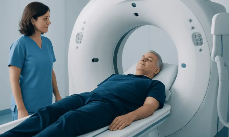
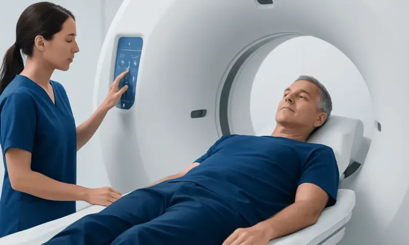
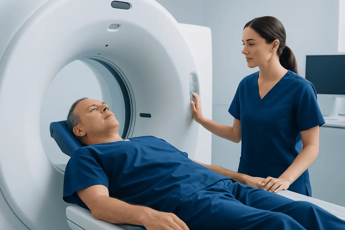
Before imaging, patients may be asked to empty their bladder. This can reduce tracer activity in the urinary system and improve image interpretation near the prostate bed and pelvis. During the scan, the person lies flat on a padded table that moves slowly through the scanner. Remaining still is important, but staff can communicate throughout the procedure.
The image-acquisition portion commonly takes around 20 to 40 minutes. The overall visit may take two to three hours, including registration, injection, uptake time and imaging. If a contrast-enhanced CT is part of the plan, the team will explain the additional steps and screen for relevant kidney disease or contrast allergies.
Results, Recovery and What the Findings Mean
Most people can leave soon after a PSMA PET scan and resume usual daily activities, unless they have received sedation or have been given separate instructions. Drinking fluids and urinating regularly for the rest of the day can help the small amount of remaining tracer leave the body. The radiation exposure is carefully controlled and is generally comparable to other medical nuclear imaging examinations.
The report describes where tracer uptake is seen and whether its pattern is suspicious for prostate cancer, while also noting limitations and any incidental findings. A positive area does not always mean cancer, and a negative scan does not exclude very small deposits of disease or cancer cells with low PSMA expression. The treating clinician explains what the findings mean in the context of the individual’s care.
How long does it take to get results from a PSMA PET scan?
Preliminary image review may begin on the day of the scan, but a final written report often takes several working days. Timing depends on the imaging centre, whether prior studies need comparison and the complexity of the findings. The referring urologist, oncologist or radiation oncologist should discuss the result and any next steps at a follow-up appointment.
For patients requiring coordinated evaluation, Cancer Treatment: Options for Every Stage" class="ahp-ilk">prostate cancer treatment planning may involve urology, medical oncology, radiation oncology, radiology and nuclear medicine specialists. Acıbadem Health Point’s multidisciplinary specialists and JCI-accredited hospitals evaluate and treat prostate cancer for international patients.
Benefits, Limitations and Possible Risks
The main benefit of a PSMA PET scan is improved localisation of PSMA-expressing prostate cancer. In the right clinical situation, it can identify disease that may not be visible on conventional imaging and may help avoid treatment that is unlikely to address the full extent of cancer. It can also support more precise planning of surgery, radiotherapy or systemic treatment.
Risks are generally low. The injection can cause brief discomfort, bruising or, rarely, infection at the injection site. Allergic reactions to the PSMA tracer are uncommon. If iodinated CT contrast is used, there is a small additional risk of allergic reaction or kidney-related complications in susceptible people; the team assesses this beforehand.
False-positive and false-negative results remain possible. Some normal tissues and benign conditions may show uptake, while very small lesions or tumours with little PSMA expression may not be detected. A scan should never be interpreted in isolation, and additional imaging, blood tests or biopsy may sometimes be recommended.
At what PSA level should a PSMA scan be done?
There is no single PSA level at which every person should have a PSMA PET scan. In suspected recurrence after treatment, detection rates generally rise as PSA rises, but scans can sometimes identify disease even at low PSA values. For newly diagnosed cancer, the decision is based more broadly on risk features such as tumour grade, stage and whether the result could alter treatment. The treating specialist can advise on the most appropriate timing.
When to Seek Medical Care
A PSMA PET scan is arranged by a clinician when there is a specific clinical question, such as staging a higher-risk prostate cancer or investigating a rising PSA after treatment. People should contact their treating team if they have missed preparation instructions, develop a new illness before the scan, have a history of severe contrast reaction or are unsure whether they can take usual medicines.
Anyone with urinary symptoms, pelvic pain, bone pain, unexplained weight loss or a newly abnormal PSA should seek medical assessment rather than assuming a scan is needed. These symptoms can have many possible causes, including benign prostate conditions, and a clinician can determine the appropriate tests.
After the scan, urgent medical care is appropriate for symptoms of a severe allergic reaction, such as trouble breathing, swelling of the face or throat, or widespread hives, though these reactions are rare. For less urgent questions about the report or next steps, patients should contact the clinician who requested the scan.
Frequently asked questions
01Is a PSMA PET scan painful?
The scan is usually not painful. Patients may feel a brief pinch when the intravenous line is placed and need to lie still during imaging. Staff can help with positioning if lying flat is uncomfortable.
02Do I need to fast before a PSMA PET scan?
Many PSMA PET protocols do not require fasting, but instructions can vary by tracer and centre. Patients should follow their appointment guidance rather than using general PET scan advice. If instructions are unclear, they should contact the imaging department before the visit.
03Can I take my regular medication before a PSMA PET scan?
Most people can take regular medicines as prescribed, but the imaging team should confirm this. Patients should provide a complete list of medicines and supplements, especially if they have diabetes, kidney disease or are scheduled to receive CT contrast.
04How long will I be at the imaging centre?
The complete visit commonly takes two to three hours. This includes check-in, tracer injection, a period for tracer uptake and the scan itself. The exact duration depends on the tracer and the imaging protocol used.
05Does a negative PSMA PET scan mean there is no prostate cancer?
No. A negative result means no PSMA-avid disease was identified at the resolution of that scan, but very small lesions or tumours with low PSMA expression may not be visible. The result should be considered together with PSA, pathology and other clinical information.
06Is it safe to be around family after a PSMA PET scan?
In most cases, normal contact with other people can continue after the scan. The tracer amount is small and decreases naturally over time. The imaging centre may provide specific temporary precautions, particularly around close contact with pregnant people or infants, depending on the tracer used.
This article is for general information only and is not a substitute for professional medical advice. Please consult a qualified doctor about your individual situation.
Oncology care in Turkey — second opinion and treatment plan
JCI-accredited · board-certified surgeons · reply within 24h
Add us as a Preferred Source to see more of our trusted health content across Google Search, AI Overviews and Discover.