Private Treatment for Skin Cancer: Care and Outlook

Private treatment for skin cancer usually begins with a dermatologist-led assessment of a suspicious lesion, followed by biopsy when needed and treatment planned around the cancer type, location and stage. Most skin cancers can be treated effectively when identified early, and follow-up remains important because new skin cancers can develop over time.
Private treatment for skin cancer: how care is planned
Private treatment for skin cancer generally provides timely access to clinical assessment, diagnostic testing and a treatment plan tailored to the individual. The appropriate approach depends on the suspected cancer type, its size and depth, where it is located, whether it has spread, the person’s medical history and their preferences. A visible spot alone cannot confirm cancer, so specialist examination is the first important step.
Skin cancer is not one condition. Basal cell carcinoma is the most common type and usually grows slowly. Squamous cell carcinoma can behave more aggressively in some situations, while melanoma is less common but has a greater potential to spread if not detected early. A private care pathway may involve dermatology, dermatopathology, plastic or reconstructive surgery, medical oncology and radiation oncology when appropriate.
Many lesions turn out to be harmless, but a new or changing mark deserves professional review. The aim is to establish an accurate diagnosis, remove or control confirmed cancer effectively, preserve function and appearance where possible, and arrange sensible long-term surveillance.
Assessment, dermoscopy and biopsy for diagnosis
A clinician will ask about the lesion’s history, including when it appeared, whether it has changed, and whether it itches, hurts, crusts or bleeds. They will also consider sun exposure, previous skin cancers, immune suppression, family history, skin type and use of medicines that may affect immunity. Examination may include the whole skin surface, not only the spot that prompted the appointment.
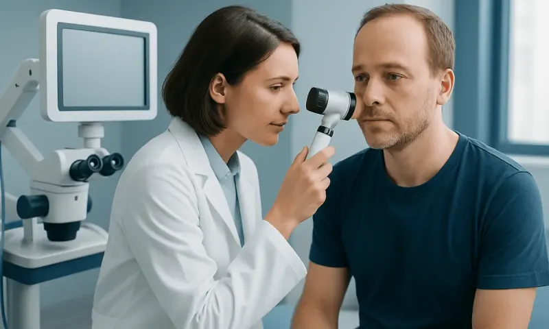
Dermoscopy is a painless examination using a handheld magnifying device and light. It helps clinicians assess pigment patterns, blood vessels and surface changes that may not be visible to the naked eye. Photography or digital mole monitoring may be used for selected people with many atypical moles, although it does not replace biopsy when cancer is suspected.
A biopsy removes all or part of the lesion for microscopic analysis by a pathologist. Depending on the lesion, this may be a shave, punch, incisional or excisional biopsy. The pathology report identifies the cancer type and, for melanoma, important features such as thickness and ulceration. This information guides discussions about skin cancer treatment and outlook.
Can you show me pictures of skin cancer that looks like a wart?

Photographs can be useful for general education, but they cannot diagnose a skin growth safely. Some squamous cell carcinomas, keratoacanthomas and occasionally other skin cancers can look wart-like: they may be rough, thickened, scaly, crusted, firm or raised. However, common warts, seborrheic keratoses and other benign lesions can look similar.
A wart-like lesion should be examined if it is new in adulthood, enlarging, painful, repeatedly bleeding, ulcerated, persistently crusted or not responding as expected to over-the-counter wart treatment. Particular care is appropriate for lesions on sun-exposed skin, the lip, ear, scalp, hands or lower legs, and for people with a weakened immune system.
Online images may create false reassurance or unnecessary worry because colour, texture and size vary widely. Rather than comparing a lesion with photographs, it is safer to arrange a skin examination. A clinician can use dermoscopy and recommend biopsy when the appearance is uncertain.
Treatment options and what may replace Mohs surgery
Treatment is individualised after diagnosis. Standard surgical excision removes the cancer with a margin of surrounding skin and is suitable for many basal cell and squamous cell carcinomas, as well as most early melanomas. The removed tissue is examined in a laboratory to check the diagnosis and margins. Reconstruction may be straightforward closure, a skin graft or a local flap, depending on the site and size of the wound.
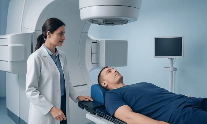
For selected superficial or low-risk non-melanoma skin cancers, alternatives may include curettage and cautery, cryotherapy, topical medicines, photodynamic therapy or radiation therapy. These options are not suitable for every lesion, and they are generally not used as a substitute for properly excising invasive melanoma. Advanced disease may require systemic medicines such as immunotherapy or targeted therapy under an oncology team.
Mohs surgery removes cancer in stages while examining the edges of each layer during the procedure. It can be especially useful for certain high-risk, recurrent or poorly defined cancers, and for sites where preserving healthy tissue is important, such as the nose, eyelids, ears, lips, hands or genitals.
What is the new procedure instead of Mohs surgery?
There is no single new procedure that replaces Mohs surgery for every person. Mohs remains a well-established option when complete margin control and tissue conservation are especially important. In other situations, standard excision with laboratory margin assessment is an appropriate and widely used alternative.
Some centres use specialised forms of margin-controlled excision, including staged excision with delayed reconstruction, or techniques that examine the peripheral and deep surgical margins in a more comprehensive way. Reflectance confocal microscopy and other imaging methods may help assess selected lesions before or during management, but availability and suitability vary. These approaches should not be viewed as interchangeable with Mohs surgery in all cases.
The best procedure depends on the pathology result, tumour subtype, location, size, recurrence risk and the clinician’s expertise. A dermatologist or skin cancer surgeon can explain whether a tissue-sparing technique, conventional excision, non-surgical treatment or referral for Mohs surgery offers the most appropriate balance of cancer control and cosmetic outcome.
What happens if you don't remove the basal cell?
If basal cell carcinoma is not treated, it usually continues to grow locally. It may become larger, develop a sore or ulcer, bleed, crust, become painful or damage nearby skin and deeper structures. Growth is often slow, but it is unpredictable enough that monitoring a confirmed basal cell carcinoma without a clinician-led plan is generally not advised.
Basal cell carcinoma only rarely spreads to distant parts of the body, but neglected tumours can invade cartilage, muscle, bone or structures around the eyes, nose or ears. Treatment may then become more complex and may require a larger operation, reconstruction, radiation therapy or systemic treatment. Earlier treatment is often simpler and can preserve more healthy tissue.
People who are not fit for surgery, or whose cancer is in a difficult location, may have other treatment options. The care team can discuss the benefits and limitations of surgery, radiation, topical treatment or other approaches according to the individual cancer. Continuing follow-up is important after treatment because future basal cell carcinomas can occur.
What does stage 1 melanoma look like?
Stage 1 melanoma does not have one reliable visual appearance. It is an early melanoma defined mainly by pathology findings, particularly its thickness and whether ulceration is present, rather than by how alarming it looks. It may appear as a new dark spot, a mole that is changing, or a lesion with uneven colour, asymmetry or an irregular border. Some melanomas are pink, red, skin-coloured or only lightly pigmented.
The ABCDE approach can support awareness: Asymmetry, Border irregularity, Colour variation, Diameter that is increasing or larger than many ordinary moles, and Evolution or change. The “ugly duckling” sign is also helpful: a mole that looks noticeably different from a person’s other marks should be checked. These signs are not diagnostic, and some melanomas do not follow them.
Stage 1 melanoma is usually managed with a wider local excision around the original biopsy site. Depending on features such as thickness, a specialist may discuss whether additional tests or sentinel lymph node biopsy are relevant. Early melanoma often has a favourable outlook after appropriate treatment, but continued skin and lymph-node follow-up is still recommended. More information about melanoma can help patients prepare for a specialist consultation.
When to seek medical care and protect skin health
Medical assessment is advisable for a new growth or a mole that changes in size, shape, colour or sensation. A lesion that bleeds without clear injury, forms a sore that does not heal, repeatedly crusts, becomes tender, or persists for several weeks should be reviewed. People with a previous skin cancer, many atypical moles, substantial ultraviolet exposure, a strong family history or reduced immunity may benefit from regular clinician-led skin checks.
Sun protection supports prevention but cannot replace skin examination. Practical measures include seeking shade during intense sunlight, wearing protective clothing and a broad-brimmed hat, using broad-spectrum sunscreen as directed, and avoiding deliberate tanning and sunbeds. Examine the skin periodically, including the scalp, back, soles, nails and areas not usually exposed to the sun.
After treatment, follow-up visits are tailored to the cancer type and individual risk. At Acıbadem Health Point, multidisciplinary specialists in JCI-accredited hospitals can assess and treat skin cancer for international patients, coordinating dermatology, pathology, surgery and oncology care when needed.
Frequently asked questions
01How quickly can skin cancer be diagnosed?
A specialist can often assess a suspicious lesion at the first appointment, but a biopsy is usually needed to confirm whether it is cancer. Pathology processing time varies by laboratory and the type of testing required. The treating team can explain the expected timeline and arrange further care if cancer is confirmed.
02Is a skin biopsy painful?
A skin biopsy is usually performed with local anaesthetic, so the area is numbed before tissue is removed. People may feel a brief sting from the anaesthetic and some pressure during the procedure. Mild soreness, bruising or a small scar can occur afterward, depending on the biopsy type and location.
03Can skin cancer be treated without surgery?
Some low-risk non-melanoma skin cancers may be treated with topical medicines, cryotherapy, photodynamic therapy, curettage or radiation therapy. The suitability of these treatments depends on the cancer type, depth, site and risk features. Surgery is often preferred when complete removal and detailed pathology assessment are needed.
04Does removing a mole cause cancer to spread?
No. Proper biopsy or surgical removal does not cause melanoma or other skin cancers to spread. Removing a suspicious lesion is an important way to establish a diagnosis and, in many cases, is part of definitive treatment.
05Can basal cell carcinoma return after treatment?
A treated basal cell carcinoma can occasionally recur, particularly if it had high-risk features or was difficult to remove completely. People who have had one basal cell carcinoma are also more likely to develop another skin cancer in the future. Scheduled follow-up and regular self-examination help identify new or recurrent lesions early.
06What should a person bring to a skin cancer consultation?
It can help to bring a list of medicines, previous biopsy or pathology reports, records of past skin cancer treatment and photographs showing how the lesion has changed. Information about family history, sun exposure and immune-related health conditions is also useful. The clinician may recommend a full-skin examination, so practical clothing can make the visit easier.
This article is for general information only and is not a substitute for professional medical advice. Please consult a qualified doctor about your individual situation.
Oncology care in Turkey — second opinion and treatment plan
JCI-accredited · board-certified surgeons · reply within 24h
Add us as a Preferred Source to see more of our trusted health content across Google Search, AI Overviews and Discover.