Private Chest X-Ray: What It Shows and How to Prepare
A private chest X-ray is an imaging examination that uses a small amount of radiation to create pictures of the lungs, heart, ribs and other structures in the chest. It may be requested to investigate respiratory symptoms, monitor a known condition, assess an injury or support pre-procedure evaluation.
What is a private chest X-ray?
A private chest X-ray is a diagnostic imaging examination arranged through a private healthcare provider rather than a public health service. It uses a controlled, low dose of X-ray radiation to produce images of the chest, including the lungs, heart outline, ribs, collarbones and diaphragm. The examination is commonly requested for symptoms such as a persistent cough, shortness of breath, chest discomfort, fever, or after a chest injury.
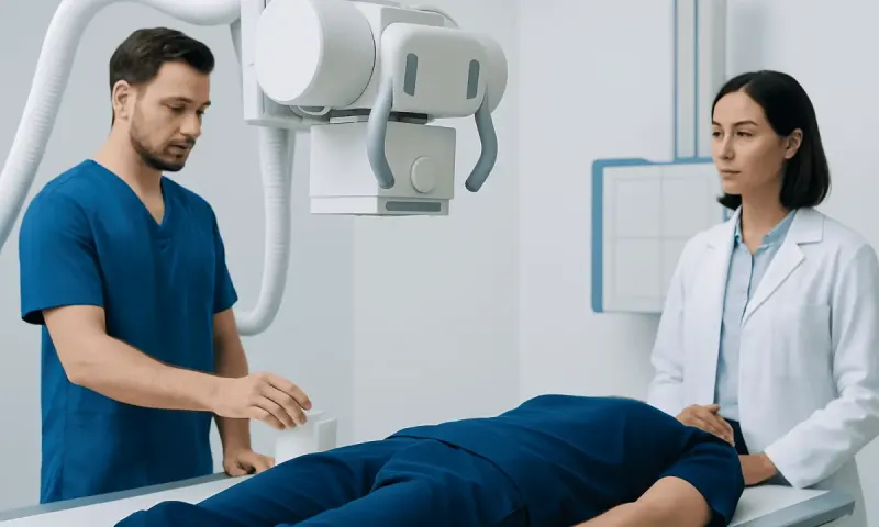
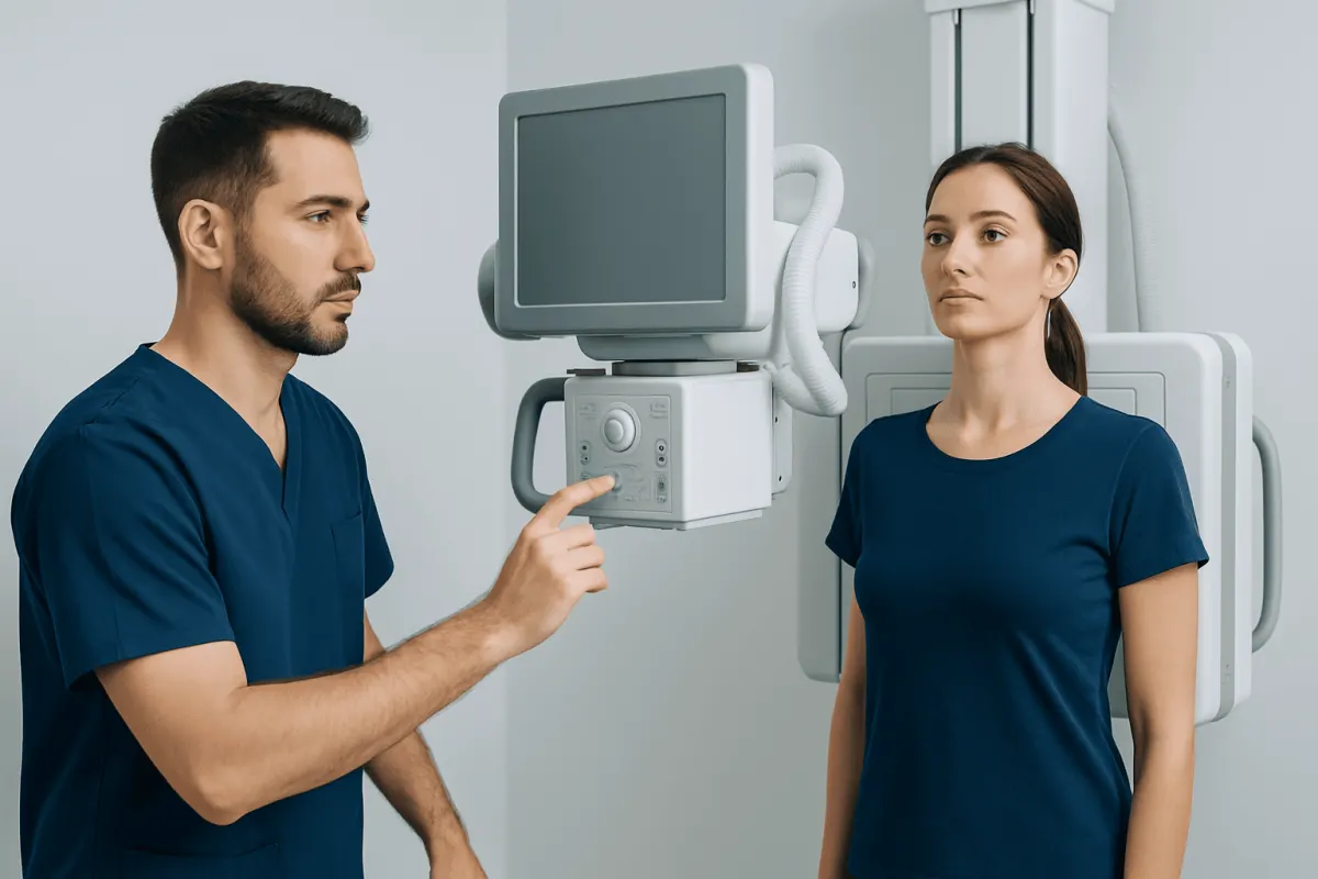
The test itself is usually simple. A radiographer positions the person standing against an imaging detector and takes one image from the front and, often, a second from the side. A radiologist reviews the images and sends a report to the referring clinician, who interprets the findings alongside symptoms, medical history and other test results.
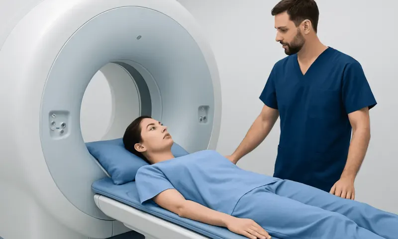
A chest X-ray can provide useful first-line information, but it is not a complete assessment of every structure in the chest. Depending on the clinical question, a clinician may recommend blood tests, an electrocardiogram, lung function testing, ultrasound, CT or MRI after reviewing the result.
Why might a clinician request a chest X-ray?
Chest X-rays are used in many clinical settings because they are widely available and can quickly identify certain changes in the lungs and chest. They may be requested when a person has a cough that does not settle, suspected infection, unexplained breathlessness, chest pain, coughing up blood, or an abnormal finding on examination.
The examination may also be used to assess an injury after a fall or impact, check the position of certain medical devices, or monitor selected lung and heart-related conditions. In some cases, it is part of a preoperative assessment when a clinician considers it medically appropriate.
An X-ray result is not usually considered in isolation. For example, a normal chest X-ray does not always exclude early infection, a small blood clot in the lungs, asthma, or coronary artery disease. The next step depends on the individual’s symptoms and level of risk.
What not to do before a chest X-ray?

There is usually no need to fast, stop normal meals or avoid drinking fluids before a chest X-ray. Unless a clinician gives different instructions, regular medicines can generally be taken as prescribed. People should not delay an urgently needed X-ray because they have eaten, exercised or taken medication.
Before the examination, it is helpful not to wear items that may interfere with the image. This includes necklaces, long chains, body piercings where removable, metal hair accessories, clothing with metal buttons or zips, and underwired bras. The imaging team may provide a gown or ask the person to change clothing for clear positioning.
It is important to tell the radiographer if pregnancy is possible or confirmed. Chest X-rays can still be performed when clinically necessary, but the care team will consider the reason for imaging and use appropriate precautions. A person should also mention recent imaging, previous chest surgery, implants or any difficulty standing still or taking a deep breath.
What diseases can show up on a chest X-ray?
A chest X-ray may show patterns that suggest lung infection, fluid around the lungs, lung collapse, chronic lung changes, some rib fractures, enlargement of the heart silhouette, or certain masses. It can support the assessment of conditions such as pneumonia, tuberculosis, chronic obstructive pulmonary disease and heart failure, although further testing may be needed to confirm a diagnosis.
Infection may appear as an area of increased whiteness in part of a lung, while fluid can produce a characteristic change at the lower edges of the chest. A pneumothorax, sometimes called a collapsed lung, may also be visible. Chest X-rays can occasionally identify findings that require urgent assessment, such as a large pleural effusion or significant lung collapse.
However, a chest X-ray has limitations. Small lung nodules, early cancers, blood clots in the lungs and subtle airway disease may not be seen clearly. If the X-ray is abnormal, or symptoms remain concerning despite a normal result, a clinician may recommend a CT scan or another appropriate investigation.
Can chest X-ray detect heart blockage?
No. A chest X-ray cannot directly detect blockages in the coronary arteries, which are the blood vessels that supply the heart muscle. It does not show whether cholesterol plaque has narrowed these arteries and cannot rule out angina or a heart attack.
The X-ray may show an enlarged heart silhouette, fluid in the lungs or other findings that can occur with some heart conditions. These changes can help guide assessment, but they do not identify the cause on their own. A clinician may use an electrocardiogram, blood tests, echocardiography, stress testing, coronary CT angiography or invasive coronary angiography when a heart blockage is suspected.
New, severe or persistent chest pressure, particularly with breathlessness, sweating, nausea, faintness, or pain spreading to the arm, jaw, back or shoulder, requires urgent medical assessment. It is safer not to wait for an outpatient X-ray appointment in this situation.
Does a chest X-ray include the abdomen?
A standard chest X-ray focuses on the chest. It usually includes the upper part of the abdomen, particularly the diaphragm and the area immediately below it, because these structures sit at the lower border of the chest. It is not designed to provide a full view or detailed assessment of abdominal organs such as the liver, gallbladder, kidneys, bowel or pelvis.
Occasionally, an abdominal issue may be partly visible on a chest X-ray. For example, free air beneath the diaphragm can sometimes be seen and may prompt urgent investigation. However, this is an incidental or limited observation rather than a complete abdominal examination.
If symptoms are mainly in the abdomen, a clinician may request an abdominal X-ray, ultrasound or CT scan depending on the suspected cause. The most suitable test is selected according to symptoms, examination findings and medical history.
Chest X-ray cost and insurance considerations
People often ask, “What does a chest X-ray cost?” There is no single answer. The price of a chest X-ray varies between countries, cities, hospitals and imaging centres. It can also differ according to whether one or two views are taken, whether a specialist consultation is included, how quickly a report is needed, and whether additional imaging is required.
The price of chest X-ray without insurance may be higher than the amount paid through an insurer, but insurance plans vary widely. The average cost of a chest X-ray with insurance can depend on deductibles, co-payments, referral rules, network status and prior authorisation requirements. Patients can ask the provider and insurer whether the quoted amount includes the radiographer’s examination, radiologist’s report and any clinician review.
For a private chest X-ray, it is reasonable to request a clear estimate before booking when the situation is not urgent. Cost should not delay emergency evaluation for severe chest pain, major breathing difficulty, coughing up blood or symptoms of a possible serious infection.
When to seek medical care
A clinician should assess a persistent cough, unexplained shortness of breath, fever with worsening chest symptoms, repeated chest infections, unexplained weight loss, or chest pain that does not clearly relate to a minor injury. A chest X-ray may be one part of that assessment, but the clinician will decide whether it is appropriate and how quickly it is needed.
Urgent medical care is needed for sudden or severe difficulty breathing, blue or grey lips, confusion, fainting, coughing up significant amounts of blood, or severe chest pressure. Emergency assessment is also important after a substantial chest injury, especially if breathing is painful or difficult.
Acıbadem Health Point’s multidisciplinary specialists and JCI-accredited hospitals can assess chest symptoms and arrange appropriate diagnostic imaging for international patients. After a chest X-ray, the referring clinician should explain the report, discuss whether any follow-up is needed and ensure that symptoms are addressed rather than relying on imaging alone.
Frequently asked questions
01How long does a private chest X-ray take?
The image-taking part usually takes only a few minutes. Allow extra time for registration, changing clothes if needed and receiving instructions from the radiographer. The time to receive the report varies by facility and clinical urgency.
02Is a chest X-ray painful?
A chest X-ray is not painful. The person may need to hold a deep breath briefly and stand in a specific position, which can be uncomfortable if there is chest or rib pain. The radiographer can help adapt positioning where possible.
03Do I need a referral for a private chest X-ray?
Requirements vary by country and provider. Many imaging services require a referral from a doctor or qualified clinician so the correct examination is selected and the result can be followed up appropriately. A provider can explain its local booking requirements.
04Can a chest X-ray show lung cancer?
A chest X-ray can sometimes show an abnormal area or mass that requires further investigation. It cannot reliably detect all lung cancers, especially small or early changes, and it cannot confirm cancer by itself. CT imaging and, when needed, tissue sampling are used to investigate suspicious findings.
05Is the radiation from a chest X-ray safe?
A chest X-ray uses a small amount of ionising radiation. Healthcare teams use the lowest practical exposure needed to obtain useful images, and the expected benefit is considered against the small radiation risk. People who may be pregnant should tell the imaging team before the test.
06What happens if a chest X-ray is abnormal?
An abnormal result does not always mean a serious condition. The clinician will consider the pattern on the X-ray together with symptoms, examination findings and medical history. They may recommend treatment, repeat imaging, blood tests, CT scanning or referral to a specialist depending on the finding.
This article is for general information only and is not a substitute for professional medical advice. Please consult a qualified doctor about your individual situation.
Explore treatments in Turkey — costs, top hospitals & a free quote
JCI-accredited · board-certified surgeons · reply within 24h
Add us as a Preferred Source to see more of our trusted health content across Google Search, AI Overviews and Discover.