Pressure Sores Stages: Images and Warning Signs
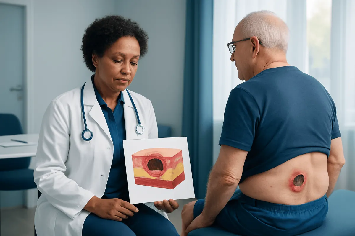
Pressure sores stages images are useful educational tools for recognizing patterns of skin damage caused by prolonged pressure, but photographs cannot reliably diagnose every wound. Skin tone, lighting, moisture, bruising, and deep tissue injury can make pressure sores look different from person to person.
What pressure sores stages images can and cannot show
Pressure sores stages images are commonly searched by people caring for someone who spends long periods in bed, uses a wheelchair, or has limited mobility. Images can show the typical appearance of different pressure injury categories, such as persistent discoloration, a shallow open wound, or deeper tissue loss. However, they are educational examples rather than a substitute for an in-person assessment.
A pressure sore may look different depending on skin tone, the body area involved, swelling, bruising, moisture, infection, and the person’s overall health. A photograph also cannot show important findings such as temperature changes, tenderness, tissue firmness, drainage odor, or the depth of damage beneath intact skin. Any new, persistent, or worsening skin change should be assessed by a qualified healthcare professional.
Pressure sores are also called pressure injuries, pressure ulcers, decubitus ulcers, or bed sores. They most often form over bony areas where the skin and deeper tissues are compressed between the body and a chair, bed, cast, medical device, or another firm surface.
How to read pressure injury stages safely
Healthcare professionals commonly use the National Pressure Injury Advisory Panel classification system. The stages describe the depth and type of tissue damage; they do not describe how severe a person feels, how long a wound has been present, or how quickly it will heal. A wound should not be “reverse staged” as it heals. For example, a healing stage 4 injury remains documented as a healing stage 4 injury.
In stage 1, the skin is intact but has localized discoloration that does not fade when pressure is briefly relieved. On lighter skin, this may appear red. On darker skin, it may appear darker than surrounding skin, purple, blue, or differently colored. The area may also feel warmer or cooler, firmer or softer, and may be tender, itchy, or painful.
Stage 2 involves partial-thickness loss of skin. It may look like a shallow pink or red open wound, a moist abrasion, or an intact or ruptured serum-filled blister. It does not show exposed fat, muscle, tendon, cartilage, or bone. Moisture-related skin damage, such as irritation from urine or stool, can resemble a stage 2 injury, so assessment of the cause matters.
Stage 3 means full-thickness skin loss. Fat tissue may be visible, and the wound may have granulation tissue or rolled edges. The depth varies by body location because some areas have more fatty tissue than others. Muscle, tendon, cartilage, and bone are not exposed in a stage 3 pressure injury.
Stage 4, unstageable, and deep tissue injuries
Stage 4 pressure injuries involve full-thickness skin and tissue loss with exposed or directly palpable fascia, muscle, tendon, cartilage, or bone. These wounds can have undermining or tunneling, meaning damage extends under intact-looking skin around the wound. They require prompt, specialized clinical assessment because complications may include serious infection or damage to deeper structures.
An unstageable pressure injury has full-thickness skin and tissue loss, but the true depth cannot be confirmed because slough or eschar covers the wound bed. Slough is moist yellow, tan, gray, green, or brown nonviable tissue. Eschar is dry, thick, dark brown or black tissue. A clinician may need to determine whether and when removal of nonviable tissue is appropriate; stable dry eschar on the heel is not routinely removed without clinical evaluation.
A deep tissue pressure injury may present as persistent deep red, maroon, or purple discoloration in intact or non-intact skin, sometimes with a blood-filled blister. It reflects damage in deeper soft tissues caused by pressure and shear. It can worsen quickly even when the surface initially looks only mildly changed, so it should be assessed without delay.
Not every wound near a pressure point is a pressure injury. Skin tears, diabetic foot ulcers, venous leg ulcers, arterial ulcers, surgical wounds, burns, and moisture-associated dermatitis have different causes and treatment needs. Correct identification helps guide safe care.
Why pressure sores develop and who is at risk
Pressure is the main cause of a pressure injury. When pressure is prolonged, blood flow can be reduced, depriving tissue of oxygen and nutrients. Shear is another important factor: it occurs when the skin stays in place while deeper tissues slide, such as when a person slides down in bed. Friction and moisture can make the skin more vulnerable, but do not usually cause deep pressure injury on their own.
People at greater risk include those who cannot reposition independently because of illness, injury, surgery, sedation, paralysis, frailty, or severe pain. Risk also increases with reduced sensation, poor circulation, swelling, incontinence, dehydration, inadequate nutrition, fever, diabetes, and use of medical devices that press on the skin.
Common locations include the tailbone, buttocks, hips, heels, ankles, elbows, shoulder blades, back of the head, knees, and ears. Devices such as oxygen tubing, masks, braces, splints, catheters, and compression garments can also create pressure points. A full skin check should include areas beneath and around devices when this can be done safely.
Assessment and treatment options
Assessment begins with a history of mobility, sensation, nutrition, continence, recent illness, and medical devices, followed by a careful skin and wound examination. The care team may measure the wound, document the tissue in the wound bed, check nearby skin, and assess for pain, drainage, odor, or signs of infection. Tests are not always needed, but clinicians may order them when they are concerned about infection, circulation, nutrition, or involvement of deeper tissue.
The first treatment priority is reducing pressure. This may include a regular repositioning plan, pressure-redistributing mattresses or cushions, heel off-loading, improved sitting support, and adjustment of devices. The right plan depends on the person’s mobility, comfort, medical condition, support surface, and ability to change position safely.
Wound care may include gentle cleansing, an appropriate dressing, moisture and incontinence management, pain relief, and treatment of contributing conditions. Some wounds require removal of nonviable tissue by trained clinicians, while others may need surgical evaluation. Antibiotics are not routinely used for every pressure sore; they are prescribed when there is evidence of a spreading or systemic infection, or another clinically confirmed infection.
Nutrition and hydration support healing, particularly when appetite is low or illness has caused weight loss. A doctor, wound-care clinician, dietitian, nurse, physical therapist, and occupational therapist may all contribute to an individualized care plan. Acıbadem Health Point’s multidisciplinary specialists and JCI-accredited hospitals can assess and treat pressure injuries for international patients.
Prevention and daily self-care
Prevention is most effective when it starts before skin breaks down. People at risk should have regular skin checks, especially over bony areas and under medical devices. Changes that persist after pressure is relieved should be taken seriously. For people with darker skin, comparing the area with nearby or opposite-side skin may help reveal subtle color, texture, or temperature differences.
A healthcare professional can advise on a safe repositioning schedule. It is important not to drag a person across sheets or a chair surface, as this can increase friction and shear. Transfers may require assistance, lifting devices, or slide sheets. Pillows, wedges, heel protectors, and pressure-redistributing cushions may help when selected and used correctly.
Keeping skin clean and dry is helpful, but overly frequent washing or harsh products can irritate it. Prompt cleansing after incontinence, protective barrier products when recommended, and breathable clothing or bedding can reduce moisture exposure. Skin should not be massaged over bony prominences, as massage may damage vulnerable tissue.
- Encourage regular fluids and balanced meals where medically appropriate.
- Support mobility and activity within the person’s abilities and care plan.
- Check that wheelchair cushions, mattresses, and medical devices fit correctly.
- Report pain, discoloration, blisters, or open areas early rather than waiting for the skin to worsen.
When to seek medical care
Medical advice should be sought promptly for any non-fading discoloration, blister, open wound, black or dark tissue, increasing pain, drainage, or wound that is becoming larger. Early assessment can identify whether the change is pressure-related and can help prevent deeper injury. People with diabetes, poor circulation, reduced sensation, or a weakened immune system should contact a clinician particularly early.
Urgent medical care is needed if there are signs of a serious infection or illness, such as fever, confusion, rapidly spreading redness, severe or escalating pain, pus-like drainage, a strong odor, or feeling suddenly unwell. These symptoms do not always mean infection, but they need timely evaluation.
Emergency care may be appropriate when a person is severely unwell, has difficulty breathing, faints, becomes confused, or has other acute symptoms alongside a wound. Caregivers should not attempt to cut away dead tissue, puncture blisters, or apply unadvised substances to a pressure sore. A clinician can recommend the safest next steps for the wound and the person’s overall health.
Frequently asked questions
01Can pressure sores stages images diagnose a wound?
No. Images can help a person recognize possible warning signs, but they cannot confirm the cause, depth, or severity of a wound. Lighting, skin tone, moisture, bruising, and deeper tissue damage can change the appearance, so a clinician should assess concerning skin changes.
02What does a stage 1 pressure sore look like?
A stage 1 pressure injury has intact skin with persistent localized discoloration that does not fade after pressure is relieved. It may be red on lighter skin and may appear darker, purple, blue, or otherwise different in color on darker skin. The area may also be painful, warm, cool, firm, or soft compared with nearby skin.
03Can a pressure sore be staged from 4 back to 3 as it heals?
No. Pressure injuries are not reverse staged during healing. A wound that was initially stage 4 should be documented as a healing stage 4 pressure injury, because it may still have lost deep tissue even when it becomes smaller or fills with new tissue.
04How quickly can a pressure sore develop?
A pressure injury can develop over a relatively short period when sustained pressure, shear, limited mobility, poor circulation, or moisture are present. The timing varies widely between individuals. Regular repositioning and skin checks are important for anyone at increased risk.
05Should a black pressure sore be removed at home?
No. Black tissue may be eschar, but whether it should be removed depends on the wound location, circulation, infection risk, and other clinical factors. A healthcare professional should examine it before any debridement is considered.
06What is the best way to prevent pressure sores in bed?
Key measures include regular repositioning, use of an appropriate pressure-redistributing mattress or support surface, keeping skin clean and protected from excess moisture, and checking pressure points daily. The safest repositioning plan should be tailored by a healthcare professional to the person’s condition and mobility.
This article is for general information only and is not a substitute for professional medical advice. Please consult a qualified doctor about your individual situation.
Persistent digestive symptoms? Get evaluated in Turkey
JCI-accredited · board-certified surgeons · reply within 24h
Add us as a Preferred Source to see more of our trusted health content across Google Search, AI Overviews and Discover.