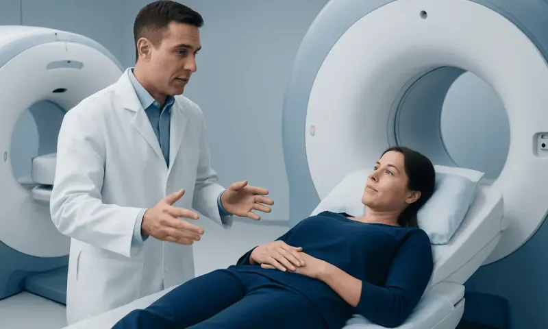
PET Scan vs CT Scan vs MRI: Which Image Is Needed?
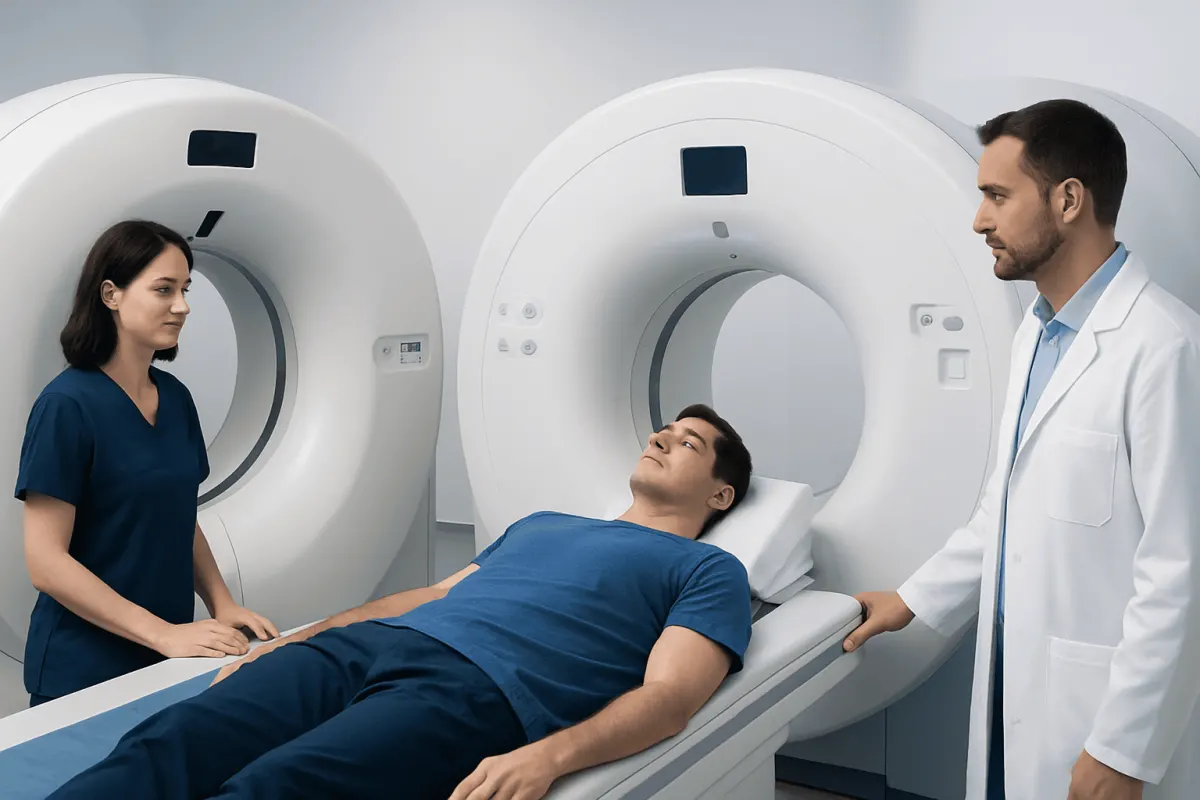
PET, CT and MRI scans answer different clinical questions rather than competing for the same role. PET highlights biological activity, CT rapidly shows internal anatomy and many urgent problems, while MRI provides highly detailed images of soft tissues such as the brain, spinal cord, joints and organs.
PET Scan vs CT Scan vs MRI: a side-by-side comparison
In PET scan vs CT scan vs MRI, the right test depends on what a clinician needs to learn. A PET scan is mainly used to show how tissues are functioning or using a tracer substance. A CT scan produces fast cross-sectional X-ray images of the body. An MRI scan uses strong magnets and radio waves to create particularly detailed pictures of soft tissues.
| Feature | PET scan | CT scan | MRI scan |
|---|---|---|---|
| Main strength | Cellular activity, metabolism and some molecular processes | Fast, detailed anatomy; lungs, bones, bleeding and many emergencies | Detailed soft tissue, brain, spinal cord, nerves, muscles and joints |
| How it works | Uses a small amount of radioactive tracer | Uses X-rays, sometimes with iodine contrast | Uses magnetic fields and radio waves, sometimes with gadolinium contrast |
| Radiation | Yes | Yes | No ionizing radiation |
| Typical duration | Often includes preparation and a scan lasting around 20 to 40 minutes | Usually only minutes | Often 20 to 60 minutes, depending on the body area and protocol |
| Common uses | Cancer staging or treatment monitoring, selected heart and brain conditions | Injury, chest symptoms, abdominal pain, kidney stones, lung disease and cancer assessment | Brain and spine symptoms, joint injuries, pelvic organs, liver lesions and some cancers |
These tests are often complementary. For example, PET/CT combines information about active tissue processes with a CT map of body structures. Results are interpreted alongside symptoms, examination findings, laboratory tests and, when needed, biopsy results.
How clinicians tell PET, CT and MRI apart
Clinicians begin with the clinical question rather than choosing the most advanced-looking scan. If there is concern about a fracture, internal bleeding, a blood clot in the lungs or an urgent abdominal problem, CT may be preferred because it is quick and widely useful in emergencies. CT is also very effective for assessing the lungs and many bone changes.
MRI is often selected when fine soft-tissue detail matters. It can help assess the brain after new neurological symptoms, the spinal cord and discs, ligaments and tendons, and many pelvic or abdominal structures. It may take longer than CT and requires the person to lie still in a narrow scanner, although some facilities offer wider-bore systems or supportive options for anxiety.
PET is usually considered when there is a need to understand whether tissue is unusually active. In oncology, it can help assess the extent of some cancers, look for recurrence, or evaluate response to treatment. It may also be used in selected cardiac and neurological situations. A radiologist or nuclear medicine physician determines whether PET alone, PET/CT, PET/MRI, CT or MRI will provide the most useful answer.
What can a PET scan show that an MRI cannot?

A PET scan can show patterns of metabolic activity that MRI does not directly measure. Many PET examinations use a glucose-like tracer, because some cancer cells and inflamed tissues use glucose differently from surrounding tissue. Areas with increased or reduced tracer uptake can indicate altered biological activity before a visible structural change is clear on anatomical imaging.
This does not mean PET is automatically better than MRI. Increased tracer uptake can occur with infection, inflammation, healing tissue and some non-cancerous conditions. MRI may better define the exact shape, size and relationship of an abnormality to nearby nerves, blood vessels or organs, and it can reveal subtle soft-tissue changes that may not be apparent on PET.
When appropriate, combined imaging can provide a fuller picture. PET/CT is commonly used to align metabolic findings with anatomical landmarks, while PET/MRI may be useful in selected settings. The scan choice should be based on the suspected condition and the information needed for care planning.
Can a PET scan pick up all cancers?
No. A PET scan cannot detect all cancers, and a normal PET result does not completely rule out cancer. Detection depends on the cancer type, size, location, biological behavior and the type of tracer used. Very small tumors, slow-growing tumors and tumors that do not take up the tracer strongly may be difficult to see.
PET findings can also be non-specific. Inflammation, infection and recent surgery or treatment may produce uptake that resembles cancer activity. For these reasons, PET results are assessed with CT, MRI, ultrasound, laboratory tests and clinical history. A suspicious area may still require a biopsy for a definite diagnosis.
For people undergoing cancer assessment, clinicians may use PET as one part of a wider plan that can include cancer treatment planning and follow-up. The most appropriate imaging pathway varies considerably between cancer types and individual circumstances.
What can a MRI show that a CT scan cannot?
MRI can provide greater contrast between many soft tissues than CT. This makes it especially valuable for examining the brain, spinal cord, nerves, bone marrow, muscles, ligaments, tendons and cartilage. MRI can also help characterize certain abnormalities in organs such as the liver, uterus, ovaries, prostate and heart.
For example, MRI may identify subtle changes related to a stroke, inflammation, multiple sclerosis, disc problems or ligament tears more clearly than CT in the right setting. It can also show fluid, swelling and tissue composition in ways that complement CT images. However, CT remains superior or more practical for some questions, including many bone injuries, lung conditions and acute bleeding evaluation.
MRI is not suitable for everyone without careful screening. Some implanted devices, metal fragments or older types of clips may be unsafe in an MRI environment. Many modern medical implants are MRI-conditional, meaning scanning may be possible under specific conditions. Patients should always tell the imaging team about implants, prior surgery, metal exposure and any possibility of pregnancy.
What is a drawback of having a PET scan?
A key drawback of a PET scan is that it involves exposure to a small amount of ionizing radiation from the tracer, often alongside CT radiation when PET/CT is performed. The medical team weighs this exposure against the expected benefit of obtaining information that may affect diagnosis or treatment decisions.
PET also requires preparation and may take longer than CT. People are commonly asked to follow instructions about eating, exercise and blood sugar management before the examination, because these factors can affect tracer distribution. People with diabetes should receive individualized preparation instructions from the imaging department or their clinician.
Another limitation is that PET can produce uncertain findings. Active inflammation or infection may appear similar to some cancers, and not every tumor is visible with a standard PET tracer. PET results therefore need expert interpretation and are not a substitute for clinical assessment or tissue diagnosis when biopsy is needed.
What to do before, during and after each scan
Before any imaging test, patients should share their symptoms, prior scans, allergies, medicines, kidney problems, diabetes, pregnancy status and breastfeeding status with their healthcare team. They should also report implanted devices, metal fragments, pacemakers, cochlear implants and previous reactions to contrast material. This information helps the team select a safe and useful test.
CT and MRI may use contrast material to improve visibility of blood vessels, inflammation or certain lesions. Kidney function may be checked in selected patients before contrast is given. Most people tolerate contrast well, but any previous reaction should be discussed in advance. The imaging team can explain the expected sensations and monitoring steps.
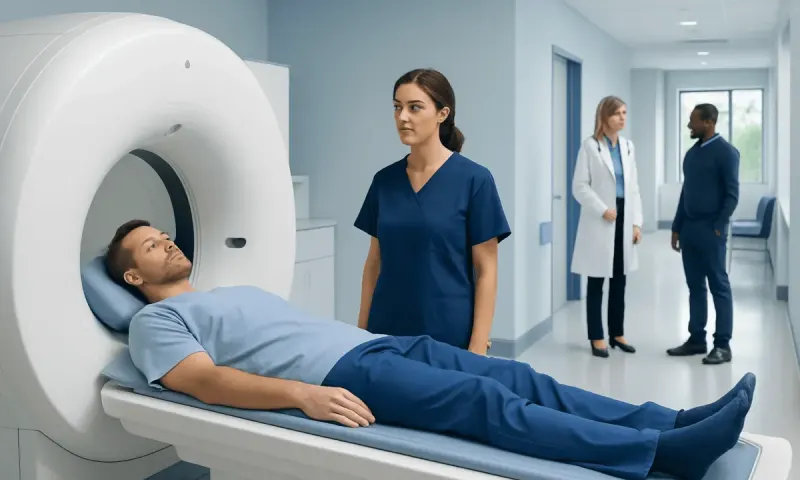
During a CT scan, the person usually lies still for a short time while the table moves through a ring-shaped scanner. MRI is noisier and longer, and ear protection is provided. PET typically involves an injection of tracer, a quiet waiting period for distribution, and then imaging. Following departmental instructions about food, fluids and activity is important for accurate PET results.
After the scan, many people can return to normal activities unless they have been given specific instructions. Drinking fluids may be advised after certain contrast studies, unless a clinician has recommended fluid restriction. Results are reviewed by a specialist and sent to the clinician who requested the scan, who can explain what they mean in the context of the person’s health.
When to seek medical care
Medical imaging should be arranged by a qualified clinician based on symptoms and clinical assessment. A person should seek urgent medical care for warning signs such as sudden weakness or speech difficulty, severe new headache, chest pain, serious trouble breathing, fainting, uncontrolled bleeding, major injury, or severe and worsening abdominal pain. Emergency clinicians can determine whether immediate imaging is necessary.
Non-urgent medical review is appropriate for persistent or unexplained symptoms, such as a new lump, unexplained weight loss, prolonged pain, a persistent cough, changes in bowel or bladder habits, or new neurological symptoms. These symptoms have many possible causes, and imaging is only one part of a safe diagnostic evaluation.
For international patients, Acıbadem Health Point’s multidisciplinary specialists and JCI-accredited hospitals can assess symptoms and coordinate appropriate diagnostic imaging and treatment planning. The best approach is individualized, with the least invasive test that can reasonably answer the clinical question.
Frequently asked questions
01Is PET, CT or MRI the most accurate scan?
No single scan is most accurate for every condition. Accuracy depends on the body area, suspected diagnosis and reason for testing. PET, CT and MRI may be used separately or together because each provides different information.
02Is MRI safer than CT or PET?
MRI does not use ionizing radiation, which is an important advantage for many patients. However, MRI has its own safety considerations, especially for certain implants or metal fragments. CT and PET use radiation, but are used when the expected medical benefit outweighs the potential risk.
03Why is PET often combined with CT?
PET identifies areas of altered tracer uptake, while CT shows detailed body anatomy. Combining the scans helps clinicians locate an active area more precisely and interpret whether it corresponds to a mass, lymph node, organ or normal structure.
04Can a CT scan replace an MRI?
Sometimes, but not always. CT may be the preferred rapid test for injuries, bleeding, lung disease and some abdominal conditions, while MRI is often better for detailed soft-tissue, brain, spinal cord and joint assessment. A clinician chooses according to the medical question.
05Do PET scans require fasting?
Many PET scans require temporary fasting because food intake and blood sugar can change how certain tracers are taken up by tissues. Requirements vary by tracer and health condition. The imaging center should provide personalized instructions, particularly for people with diabetes.
06How long does it take to get PET, CT or MRI results?
Timing varies by facility, scan complexity and whether comparison with earlier studies is needed. Images are generally reviewed by a radiologist or nuclear medicine physician before a report is sent to the requesting clinician. The clinician can explain the result and whether further testing is needed.
This article is for general information only and is not a substitute for professional medical advice. Please consult a qualified doctor about your individual situation.
Medical weight-loss options in Turkey — costs & eligibility
JCI-accredited · board-certified surgeons · reply within 24h
Add us as a Preferred Source to see more of our trusted health content across Google Search, AI Overviews and Discover.