Pelvic Ultrasound: Procedure, Uses and Results

Key Takeaways
- USG pelvis sonography uses sound waves, not radiation, to create images of the pelvic organs.
- It may be done through the abdomen, and sometimes with an internal probe for a clearer view of certain structures.
- The test can help evaluate pain, bleeding, cysts, fibroids, urinary concerns, and some fertility-related issues.
- Preparation depends on the type of scan and may include drinking water or having an empty bladder.
- Results should be interpreted by a qualified clinician in the context of symptoms and other tests.
USG pelvis sonography is a common, noninvasive imaging test that helps doctors look at organs and tissues in the pelvic area. It is often used to assess pelvic pain, abnormal bleeding, urinary symptoms, fertility concerns, and other conditions in women, men, and children when needed.
Overview
USG pelvis sonography, also called pelvic ultrasound, is a imaging test that helps clinicians see the organs and soft tissues inside the pelvis. It uses high-frequency sound waves, not ionizing radiation, which makes it a widely used first-line tool for many pelvic concerns.
The examination may focus on the uterus, ovaries, cervix, and surrounding structures in women; the bladder and nearby tissues in all patients; and, in some situations, the prostate or other pelvic organs in men. The exact target depends on the symptoms and the question the clinician is trying to answer.
For Myomectomy Recovery for International Patients: The Usual Travel Timeline" class="ahp-ilk">international patients, pelvic ultrasound is often one of the most practical tests to begin with because it is quick, noninvasive, and usually available in outpatient settings. It can help guide whether further imaging, laboratory tests, or specialist review is needed after travel or before a treatment plan is finalized.
Symptoms and Reasons It May Be Recommended

A pelvic ultrasound is often requested when symptoms point to the organs in the lower abdomen or pelvis. Common reasons include pelvic pain, pressure, bloating, heavy or irregular menstrual bleeding, pain during intercourse, urinary frequency, or trouble emptying the bladder.
In women, it is frequently used to evaluate conditions such as ovarian cysts, uterine fibroids, endometrial thickening, pelvic inflammatory changes, or early pregnancy concerns. It may also be part of a fertility workup when a doctor wants to assess the uterus, ovaries, or the timing of ovulation-related findings.
In men, pelvic sonography may be used to assess the bladder or, in specific cases, the prostate and nearby structures. In children and adolescents, it may be recommended for abdominal or pelvic pain, urinary symptoms, or congenital concerns, with the approach tailored to age and comfort.
Causes & Risk Factors

Pelvic ultrasound is not a diagnosis itself; it is a way to look for possible causes of symptoms. Findings may point toward structural conditions such as fibroids, cysts, polyps, enlarged prostate, bladder retention, or fluid collections, depending on the patient’s age and sex.
Some factors make pelvic conditions more likely. These can include a history of pelvic surgery, menstrual disorders, known endometriosis, repeated urinary tract problems, fertility difficulties, menopause-related bleeding, or a family history of certain gynecologic conditions. However, many people who undergo the scan have no major risk factors and simply need a careful evaluation of new symptoms.
Because symptoms can overlap, clinicians often use ultrasound alongside history, examination, and other tests. This is especially important for patients arriving from another country, where previous records may be incomplete or imaging methods may have varied.
Diagnosis: How the Scan Is Performed
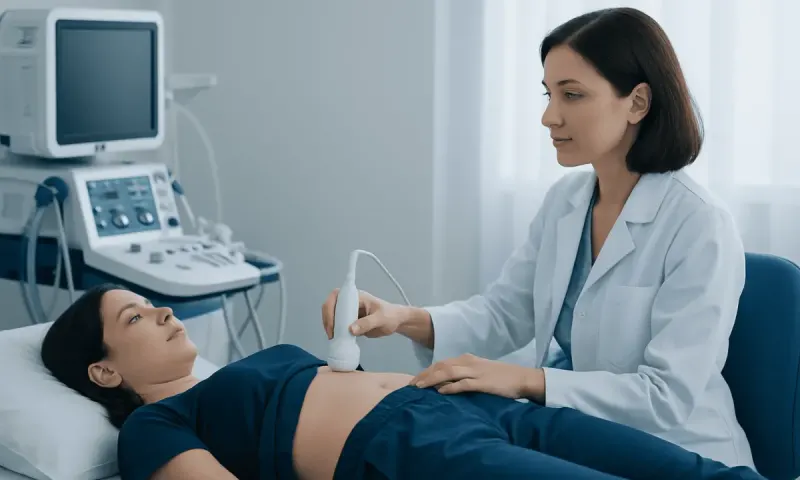
USG pelvis sonography is usually performed in one of two ways. A transabdominal scan is done from the outside of the body, with gel placed on the lower abdomen and a handheld probe moved over the skin. A transvaginal scan, used in selected patients, places a slim probe in the vagina to provide a closer view of the pelvic organs; this can offer clearer detail for some gynecologic questions.
Before the scan, preparation instructions depend on the method. A full bladder may be requested for a transabdominal study because it helps lift bowel gas and improves visibility. For some internal scans, the bladder may need to be emptied. The care team should explain what is needed in advance so the visit feels predictable and respectful.
During the exam, the patient usually lies on an examination table while the sonographer captures images and measurements. The test is typically painless, though pressure, mild discomfort, or a feeling of fullness can occur. Results are then reviewed by a radiologist or the treating physician, who interprets them together with symptoms and any prior tests.
Treatment Options
Ultrasound itself does not treat pelvic conditions, but it often helps identify the most appropriate next step. Depending on the findings, treatment may range from watchful waiting to medications, hormonal therapy, physical therapy, procedures, or surgery.
For example, simple ovarian cysts may only need follow-up imaging, while symptomatic fibroids, endometrial abnormalities, or suspicious masses may require further evaluation by a gynecologist or oncologic specialist. If urinary retention or prostate enlargement is suspected, a urologist may be involved. In fertility care, ultrasound findings may be combined with hormone testing and other reproductive assessments to shape treatment.
For patients traveling internationally, a clear ultrasound report can be especially helpful because it supports coordinated follow-up when returning home. If surgery or more advanced treatment is needed, the scan can help the clinical team plan safely and explain options in a stepwise way. Acibadem Health Point’s multidisciplinary specialists and JCI-accredited hospitals diagnose and treat this condition for international patients.
Prevention & Self-care
Most people cannot prevent the need for a pelvic ultrasound, but they can make the experience easier with a few practical steps. Following the preparation instructions exactly, bringing prior reports, and sharing current medications or pregnancy status can help the exam go smoothly.
After the scan, normal activities can usually be resumed right away. If the test was done internally, mild temporary spotting or sensitivity may occur in some patients; if anything feels unusual or persistent, the clinician should be informed. Keeping a personal copy of the report and images can be useful, especially for patients who will continue care in another country.
It also helps to note symptoms before the appointment: when pain started, whether bleeding patterns changed, and what makes symptoms better or worse. These details often improve the accuracy of interpretation more than a scan alone can do.
When to See a Doctor
Medical review is appropriate if pelvic symptoms are persistent, severe, or changing. Pain that interferes with daily activity, heavy or prolonged bleeding, bleeding after menopause, unexplained bloating, fever with pelvic discomfort, or urinary symptoms that do not settle should be assessed promptly.
Emergency care may be needed if there is sudden severe pain, fainting, heavy bleeding, or concern for pregnancy-related complications. Even when the ultrasound is normal, ongoing symptoms may still require follow-up because some conditions are better detected with repeat imaging or additional testing.
For international patients, it is reasonable to seek a second opinion when prior scans are unclear or reports use unfamiliar terminology. A qualified clinician can explain what the findings mean in plain language and decide whether more evaluation is needed.
What Results May Mean
Pelvic ultrasound reports usually describe the size, shape, and appearance of the organs seen during the scan. A report may mention normal findings, simple cysts, fibroids, thickened lining, free fluid, enlarged organs, or a structure that needs closer review.
Not every finding is urgent or dangerous. Some are common and benign, while others simply need monitoring over time. The important part is how the images fit with the patient’s age, symptoms, menstrual history, pregnancy status, and prior test results.
Because pelvic ultrasound is only one piece of the clinical picture, it should not be used in isolation to draw final conclusions. A careful discussion with the doctor helps turn the report into a clear plan, whether that means reassurance, follow-up imaging, or treatment.
Frequently asked questions
01Is USG pelvis sonography painful?
Most people find it comfortable or only mildly uncomfortable. A transabdominal scan usually feels like pressure from the probe, while an internal scan may cause brief pressure or sensitivity. The team can explain each step beforehand to reduce anxiety and improve comfort.
02Do patients need to drink water before the scan?
Sometimes, yes. A full bladder is often helpful for a transabdominal pelvic ultrasound because it improves visibility, but some internal scans may require an empty bladder. The clinic should give specific instructions based on the type of exam planned.
03Can pelvic ultrasound detect all problems?
No single test can detect every pelvic condition. Ultrasound is excellent for many structural issues, but some causes of pain or bleeding may need blood tests, urine tests, MRI, CT, or specialist evaluation. A doctor will decide what else is appropriate if symptoms continue.
04Is ultrasound safe during pregnancy?
Ultrasound is generally considered safe and is commonly used in pregnancy when medically indicated. It does not use radiation, which is one reason it is preferred for checking the uterus and early pregnancy-related concerns. The clinician will choose the safest approach for each situation.
05How long does the test take?
A pelvic ultrasound is usually a short outpatient procedure, though the exact time can vary with the reason for the exam and whether both transabdominal and internal views are needed. Extra time may also be used for preparation, review, and questions from the patient.
06What should a patient bring to the appointment?
It helps to bring prior imaging reports, referral notes, and a list of current symptoms and medications. Patients who are traveling from abroad may also want copies of earlier scans on a disc or USB if available. These details can help the clinician compare findings more accurately.
This article is for general information only and is not a substitute for professional medical advice. Please consult a qualified doctor about your individual situation.
Persistent digestive symptoms? Get evaluated in Turkey
JCI-accredited · board-certified surgeons · reply within 24h
Add us as a Preferred Source to see more of our trusted health content across Google Search, AI Overviews and Discover.