Omphalocele Is an Abdominal Wall Defect Found Before Birth

You went in for a routine pregnancy scan, and the sonographer paused a little longer than usual. Then came a word you had never heard before: omphalocele.
It means your baby’s intestines, liver, or other organs are developing outside the belly, held in a thin protective sac. It is a congenital abdominal wall defect — a structural birth difference. The good news is that it can often be spotted before birth, and it is managed with coordinated neonatal and surgical care.
Overview: what omphalocele means
In omphalocele, part of a baby’s intestine, liver, or other abdominal organs develop outside the abdomen, through an opening at the base of the umbilical cord. A thin, transparent sac covers those organs. Put simply: the abdominal wall does not close completely during fetal development.
This condition is not caused by anything a parent did or did not do. It forms early in pregnancy and is usually found on prenatal ultrasound or immediately after birth. The size can vary widely, from a small sac containing a short segment of intestine to a larger opening that includes several organs.
Omphalocele is often confused with other abdominal wall defects, so it helps to know the difference. Gastroschisis also involves organs outside the abdomen, but there the organs are usually not covered by a sac, and the opening sits beside the umbilical cord rather than at it. That difference can change evaluation, delivery planning, and treatment.
Omphalocele can occur on its own or alongside other congenital conditions. That is why medical teams look beyond the abdominal wall defect itself and also check the baby’s heart, chromosomes, and overall development.
How omphalocele appears before and after birth
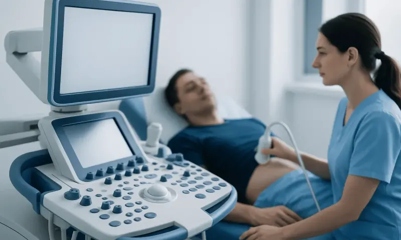
Before birth, omphalocele often appears on routine prenatal ultrasound as a membrane-covered sac extending from the baby’s abdomen at the umbilical area. Depending on the stage of pregnancy and the size of the defect, specialists may recommend repeat scans to monitor growth, the organs involved, and amniotic fluid levels.
After birth, the condition is usually visible right away. The abdomen may look smaller than expected because some of the organs have developed outside the abdominal cavity. The sac may contain only intestine, or it may also include the liver and other organs in larger omphaloceles.
Some newborns with omphalocele have no immediate breathing problems, while others may have difficulty because the abdominal cavity is small and the lungs may not be fully developed. Feeding may also need to wait until the baby is assessed and stabilized.
Doctors also look for signs of associated anomalies, because omphalocele can occur with heart defects, chromosomal differences, or syndromes that affect growth and development. This broader assessment helps guide both early treatment and longer-term follow-up.
Causes and risk factors

Omphalocele develops during early fetal growth, when the abdominal wall and internal organs are forming. During normal development, some of the intestine briefly moves outside the abdomen and then returns inside as the abdominal wall closes. In omphalocele, this process does not complete in the usual way.
In many families, no clear single cause is found. The condition can happen sporadically, meaning there is no known inherited pattern in that pregnancy. However, omphalocele can also be linked with chromosomal conditions, genetic syndromes, and other structural abnormalities, which is why prenatal genetic counseling and testing may be discussed.
Known risk factors are not specific enough to predict which pregnancy will be affected. What counts in practice is careful prenatal evaluation once the finding is suspected. This may include advanced ultrasound, fetal echocardiography, and discussion with maternal-fetal medicine, neonatology, genetics, and pediatric surgery teams.
Since related conditions can shape care decisions, doctors may also check the heart, digestive tract, urinary tract, and growth pattern. The aim is not just to confirm the omphalocele, but to understand your baby’s overall health needs.
How doctors diagnose omphalocele
Diagnosis commonly begins during pregnancy. Routine ultrasound can often identify omphalocele, especially in the second trimester. Once seen, more detailed imaging helps determine the size of the defect, which organs are in the sac, and whether other abnormalities are present.
Additional prenatal testing may include fetal echocardiography to assess the heart and, in some cases, genetic testing such as chorionic villus sampling or amniocentesis after counseling. These tests are not required in every case, but they can provide important information for planning birth and neonatal care.
After delivery, the diagnosis is usually confirmed by physical examination. Imaging such as X-rays or ultrasound may be used to better understand the anatomy or to look for associated issues. Blood tests and heart assessment may also be part of the early workup.
The diagnostic process often continues after birth because the treatment plan depends on more than the visible opening alone. Doctors consider the baby’s breathing, circulation, feeding readiness, the condition of the sac, and whether other organs or systems are affected.
Treatment options and surgical repair
Treatment starts with protecting the exposed organs and keeping the baby stable. Right after birth, the sac is carefully covered and monitored to reduce fluid loss, heat loss, and infection risk. The newborn is usually cared for in a neonatal intensive care setting while the team evaluates breathing, circulation, and any associated conditions.
Surgery is the main treatment, but the timing and method depend on the size of the omphalocele and the baby’s overall condition. Small omphaloceles may be repaired relatively soon by returning the organs to the abdomen and closing the opening. Larger defects may need staged repair, allowing the organs to move back gradually so the abdomen and breathing can adapt more safely. This may involve pediatric surgery and coordinated neonatal intensive care.
In some very large omphaloceles, immediate complete closure is not possible because the abdominal cavity is too small or the baby is medically fragile. In these situations, specialists may use a temporary protective approach and delay full repair until the baby grows stronger. Feeding support, breathing support, and careful monitoring are often part of this period.
Long-term follow-up may address feeding difficulties, reflux, growth, scar care, or weakness of the abdominal wall. If associated heart or genetic conditions are present, they are managed alongside the abdominal repair. In experienced centers, care is multidisciplinary and tailored to the child rather than based on a single standard timeline.
Daily care, recovery, and family support
Recovery after omphalocele treatment varies. Some babies recover steadily after a single operation, while others need a longer hospital stay because of breathing support, feeding transitions, or staged repair. Families are often guided step by step through tube feeding, wound care, and signs to watch for after discharge.
Nutrition is an important part of recovery. Some newborns begin feeding later than expected because the intestines need time to function normally after surgery. The care team may use intravenous nutrition at first and then gradually introduce milk feeds when it is safe to do so.
Parents may also need reassurance about appearance and healing. Scars, a differently shaped belly button area, or a small ventral hernia can occur after repair. Follow-up visits help monitor growth, abdominal wall strength, and developmental progress, and they give families a chance to ask practical questions.
Do not overlook your own well-being. Hearing that your baby has a congenital condition is stressful, even when treatment is going well. Clear communication, regular updates, and a coordinated plan usually help families feel more prepared and less overwhelmed in those first weeks.
When to seek medical care
Medical care should be sought promptly whenever omphalocele is suspected during pregnancy or seen after birth. A prenatal finding should be reviewed by an obstetric specialist and usually by a fetal medicine team so that delivery and newborn care can be planned in an appropriate hospital.
After birth, urgent medical attention is needed if the sac looks torn, leaking, darkened, or dry, or if the baby has trouble breathing, poor feeding, vomiting, a swollen abdomen, fever, or unusual sleepiness. None of these signs automatically means something serious, but each one needs to be looked at quickly.
Families should also contact the care team after surgery if there is redness around the wound, drainage, persistent vomiting, difficulty passing stool, poor weight gain, or signs of dehydration. Ongoing follow-up is important even when recovery seems smooth.
At Acıbadem Health Point, multidisciplinary specialists in neonatology, genetics, imaging, and surgery evaluate congenital abdominal wall conditions and provide treatment in JCI-accredited hospitals for international patients, including access to surgical care and advanced genetic testing when appropriate.
Frequently asked questions
01Is omphalocele the same as gastroschisis?
No. Both are abdominal wall defects, but omphalocele usually occurs at the base of the umbilical cord and the organs are covered by a sac. In gastroschisis, the opening is usually next to the umbilical cord and the organs are typically not covered.
02Can omphalocele be seen before a baby is born?
Yes. Many cases are found on prenatal ultrasound, often during routine screening. Once identified, doctors usually arrange more detailed imaging and may recommend evaluation for associated conditions.
03Does every baby with omphalocele need surgery?
Most babies will need surgical management, but the exact approach differs. Small defects may be repaired earlier, while large omphaloceles may need staged treatment and delayed closure depending on the baby's condition.
04What causes omphalocele?
Omphalocele happens during early fetal development when the abdominal wall does not close completely. Often there is no single clear cause, but it can sometimes occur with chromosomal conditions, syndromes, or other congenital differences.
05Can babies with omphalocele live healthy lives?
Many babies do well, especially when the omphalocele is isolated and treated in an experienced center. The outlook depends on the size of the defect, the baby's lung development, and whether there are associated medical conditions.
06Is vaginal delivery possible with omphalocele?
Delivery planning is individualized. Some pregnancies may proceed with vaginal birth, while others may need cesarean delivery depending on the size of the omphalocele, the organs involved, and other obstetric factors.
This article is for general information only and is not a substitute for professional medical advice. Please consult a qualified doctor about your individual situation.
Persistent digestive symptoms? Get evaluated in Turkey
JCI-accredited · board-certified surgeons · reply within 24h
Add us as a Preferred Source to see more of our trusted health content across Google Search, AI Overviews and Discover.