The Machines Used in an Eye Exam and What Each One Checks
You sit down, rest your chin on a little plastic support, and someone asks you to look at a tiny hot air balloon at the end of a tunnel. Then another machine. Then a puff of air. If you have ever wondered what all that equipment is actually measuring, you are not alone.
An eye exam machine is any device used during an eye check to measure vision or examine structures inside the eye. These machines help eye specialists detect common problems, guide diagnosis, and monitor conditions over time.
What an eye exam machine is
An eye exam machine is a medical device used to assess vision or examine the structures of the eye. In practice, the term does not refer to one single machine. It can describe several devices used during a routine eye check or a more detailed ophthalmology visit, including tools that measure focusing power, inspect the front of the eye, test eye pressure, or create images of the retina.
No device replaces the judgment of your eye specialist; it supports it. A complete eye assessment usually combines your medical history, your symptoms, vision testing, and findings from one or more devices. Put together, that picture can pick up everyday problems like nearsightedness as well as more serious disorders that threaten sight in the long run.
All this equipment can feel intimidating the first time. In reality, most of the tests are short, painless, and do not touch the inside of your eye. Knowing what each machine is for makes the visit feel more predictable, and it helps you ask better questions about your own eyes.
Common types of eye testing machines
Which machines you meet depends on why you came in. Some are for basic screening; others produce detailed images that let the doctor track a disease over time. Moving from one device to another in a single appointment is completely normal.
Common examples include:
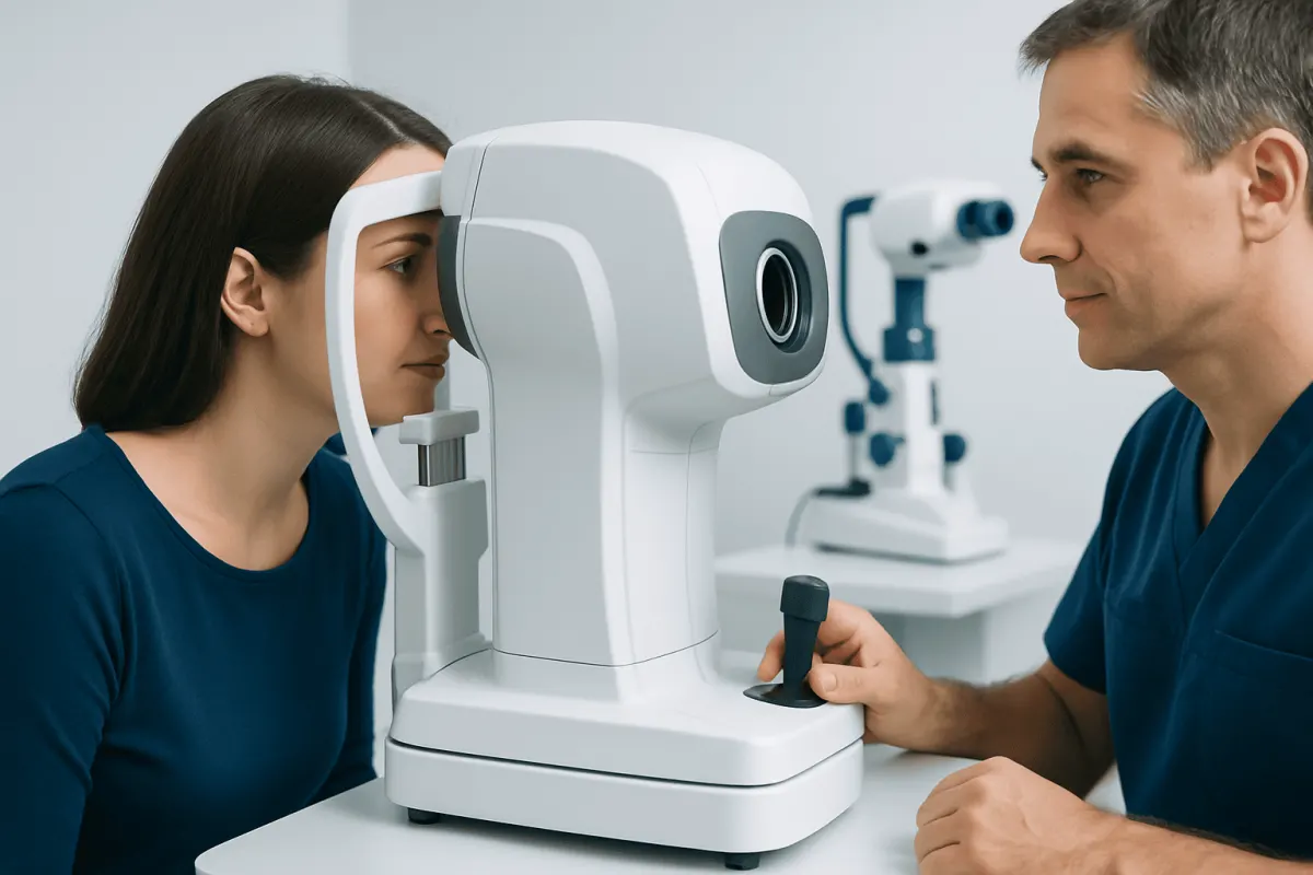
- Autorefractor: estimates the eye’s focusing power and helps assess whether glasses or contact lenses may be needed.
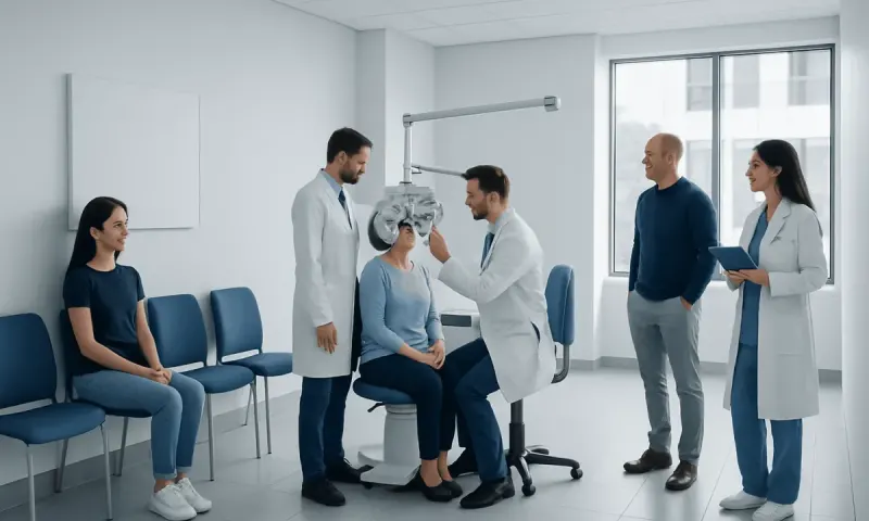
- Phoropter: the device with multiple lenses used during the familiar “which is clearer, one or two?” vision check.
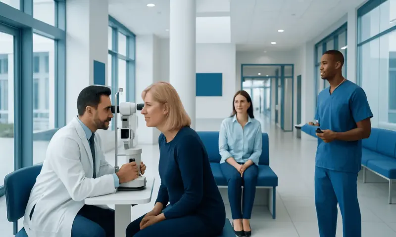
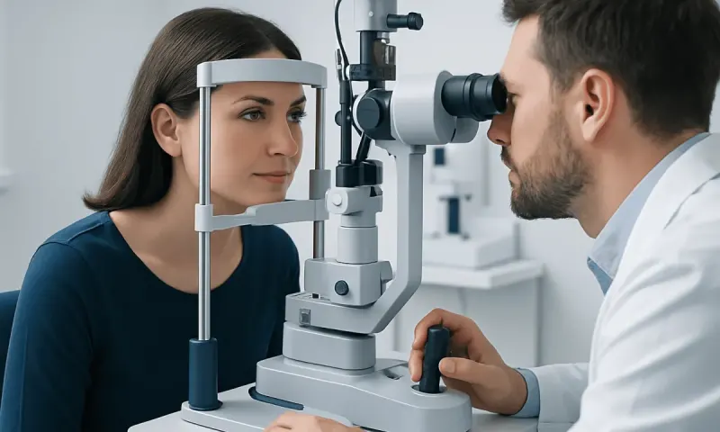
- Slit lamp: a microscope with a bright light that allows detailed examination of the cornea, iris, lens, and front of the eye.
- Tonometer: measures intraocular pressure, an important part of screening for glaucoma.
- Fundus camera or retinal camera: takes photographs of the retina, optic nerve, and blood vessels at the back of the eye.
- Optical coherence tomography (OCT): creates cross-sectional images of retinal layers and the optic nerve using light waves.
- Visual field analyzer: maps side vision and helps detect functional vision loss that may occur in glaucoma or neurological disease.
Some centers also use corneal topography devices to map the cornea’s surface, pachymeters to measure corneal thickness, and ultrasound-based tools when the inside of the eye cannot be seen clearly. The exact mix of machines depends on the patient’s age, symptoms, medical history, and whether the visit is for routine care, surgery planning, or follow-up of a known condition.
What these machines help diagnose

An eye exam machine can help detect many conditions before symptoms become severe. Devices that check refraction are used to identify nearsightedness, farsightedness, astigmatism, and age-related difficulty focusing up close. Imaging and pressure-testing machines can reveal early changes that may not be noticeable to the patient.
Eye testing machines are commonly used in the evaluation of cataracts, diabetic eye disease, retinal tears, macular disorders, glaucoma, and optic nerve problems. For example, retinal imaging and OCT may support assessment of retinal detachment or swelling in the retina, while tonometry and visual field testing help assess damage related to eye pressure.
In some cases, eye findings also provide clues about general health. Changes in retinal blood vessels may be seen in people with diabetes or high blood pressure. Because of this, a thorough eye examination can be part of broader preventive health care, not only a vision check.
What to expect during testing
From your side of the machine, almost nothing is required of you. With the autorefractor, you look at a target inside the device while it measures how light changes as it enters your eye. With the slit lamp, you rest your chin and forehead on the supports while the doctor looks at your eye under magnification.
For a tonometry test, the clinic may use a gentle puff of air or a small probe after numbing eye drops are placed. Retinal photography and OCT typically require the patient to focus on a target while images are captured in seconds. Some people may need dilating eye drops beforehand so the doctor can better see the retina and optic nerve.
Dilating drops can temporarily blur near vision and increase light sensitivity for several hours, so patients may wish to bring sunglasses and ask whether driving is advisable afterward. If surgery is being considered, additional imaging may be performed to support planning, such as measurements before cataract surgery or more detailed retinal scans before retinal detachment surgery.
Although testing is usually painless, a patient should tell the clinician if they feel discomfort, become very anxious, or have difficulty keeping the eyes still. Staff can often adjust positioning, explain the steps more clearly, or repeat the measurement if needed.
How doctors interpret the results
Numbers and images only mean something in context. One reading rarely tells the whole story. For example, an elevated eye pressure reading does not always mean glaucoma, and a normal pressure reading does not completely exclude it. The doctor considers pressure, optic nerve appearance, visual field testing, corneal thickness, symptoms, and medical history together.
Similarly, retinal photos and OCT scans are compared with examination findings and, when available, prior images. This helps the doctor see whether a condition is stable, improving, or progressing. In chronic disorders, repeated testing over time is often more informative than one isolated visit.
Children, older adults, and patients with communication or mobility challenges may need testing adapted to their needs. Machine readings can sometimes be affected by dry eye, blinking, eye movement, contact lenses, or poor positioning. If a result seems unclear, the specialist may repeat the test or use a different method to confirm the finding.
Benefits, limits, and safety
The main benefit of an eye exam machine is that it provides objective measurements and detailed images that may be difficult to judge by symptoms alone. These devices can improve early detection, help document subtle changes, and support treatment decisions. They are especially useful for monitoring diseases that progress gradually, such as glaucoma, diabetic retinopathy, or macular conditions.
Still, no machine diagnoses everything on its own. Screening devices may miss uncommon conditions, and abnormal findings do not always indicate serious disease. This is why machine-based testing works best as part of a complete examination by a qualified optometrist or ophthalmologist.
Most eye testing machines are very safe. The light used in retinal imaging and OCT is generally well tolerated, and pressure testing is routine in eye care. Some patients may have temporary discomfort from bright light or mild irritation after eye drops, but serious complications are uncommon. If a patient has had recent eye surgery, severe light sensitivity, or an allergy to specific eye drops, they should tell the clinic before testing begins.
When to seek medical care
Regular eye testing matters even when your vision seems fine, but some symptoms cannot wait. Get medical care promptly if you develop sudden vision loss, a curtain-like shadow over the vision, severe eye pain, rapid onset of flashes or many new floaters, marked redness with light sensitivity, or an eye injury. Any of these can point to a condition that needs urgent evaluation.
It is also worth being seen for gradual blurred vision, frequent headaches during visual tasks, worsening night vision, double vision, or trouble reading that interferes with daily life. If you have diabetes, high blood pressure, autoimmune disease, or a family history of serious eye disease, regular monitoring is worthwhile even when nothing feels wrong.
If a problem is identified, treatment depends on the cause. This may include glasses, monitoring, medication, laser procedures, or surgery such as glaucoma treatment. At Acıbadem Health Point, multidisciplinary specialists in JCI-accredited hospitals evaluate eye conditions and plan care for international patients based on the underlying diagnosis and the patient’s overall health.
Frequently asked questions
01Is an eye exam machine the same as a full eye exam?
No. An eye exam machine is one part of the testing process, while a full eye exam includes medical history, symptom review, visual assessment, and a doctor's examination. Machine results are most accurate and useful when interpreted together with the rest of the visit.
02Are eye testing machines painful?
Most are not painful. Patients may notice bright lights, a brief puff of air, or mild stinging from eye drops, but the tests are generally quick and well tolerated. If discomfort occurs, the patient should tell the clinician so the test can be adjusted.
03Why are several different machines used in one appointment?
Each machine measures something different. One may estimate glasses prescription, another may check eye pressure, and another may image the retina or optic nerve. Using more than one device helps build a more complete picture of eye health.
04Can an eye exam machine detect glaucoma?
It can help detect signs associated with glaucoma, but no single machine confirms the diagnosis by itself. Eye pressure testing, optic nerve imaging, and visual field testing are often combined with a clinical exam. This is important because glaucoma can be present even when pressure is not very high.
05Do I need dilating drops for every machine-based eye test?
Not always. Some tests can be done without dilation, while others are more accurate or more informative when the pupil is widened with drops. The need for dilation depends on the purpose of the visit and what the doctor needs to examine.
06How often should someone have eye testing?
The ideal schedule depends on age, symptoms, vision needs, and medical history. People with diabetes, a family history of eye disease, previous eye problems, or changing vision may need more frequent checks. A qualified eye professional can recommend a suitable interval.
This article is for general information only and is not a substitute for professional medical advice. Please consult a qualified doctor about your individual situation.
Explore treatments in Turkey — costs, top hospitals & a free quote
JCI-accredited · board-certified surgeons · reply within 24h
Add us as a Preferred Source to see more of our trusted health content across Google Search, AI Overviews and Discover.