Eosinophilic Granuloma: Bone Lesions and Care Options
Eosinophilic granuloma is a rare bone disorder and a localized form of Langerhans cell histiocytosis (LCH). It can cause bone pain, swelling, or a visible lump, but many single-site lesions have a favorable outlook when accurately diagnosed and appropriately monitored or treated.
Overview: What Is Eosinophilic Granuloma?
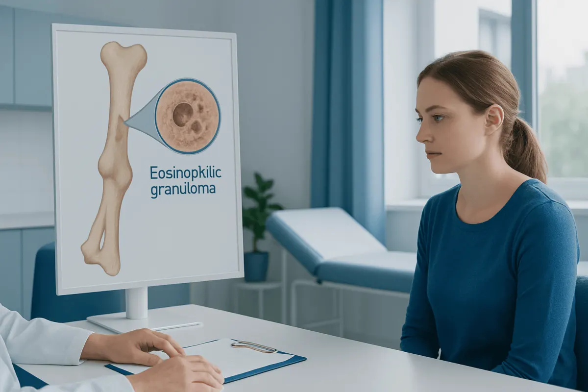
Eosinophilic granuloma is a localized form of Langerhans cell histiocytosis (LCH), a rare disorder in which certain immune cells, called Langerhans cells, build up in tissues and may cause inflammation and damage. In eosinophilic granuloma, this process most commonly creates one or a few lesions in bone. The term describes what may be seen under a microscope: Langerhans cells mixed with eosinophils, a type of white blood cell.
Although the word “granuloma” may sound concerning, eosinophilic granuloma is not the same as a typical infection-related granuloma, and it is not caused by an allergy. Its behavior varies. A single lesion in bone is often limited and may improve with relatively conservative care, while lesions affecting several bones or organs are considered part of a broader LCH picture and need more extensive evaluation.
The skull, jaw, ribs, pelvis, spine, and long bones of the arms or legs can be involved. The condition is seen most often in children, adolescents, and young adults, but adults can also be affected. Care is guided by where the lesion is located, whether other areas are involved, symptoms, and the risk of complications such as fracture or pressure on nearby nerves.
Symptoms and How the Condition May Present
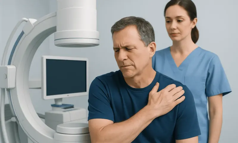
Symptoms depend mainly on the affected bone. Pain that persists or gradually worsens is common. There may be tenderness, localized swelling, or a lump over the lesion. Some people feel well otherwise, and a lesion is found incidentally when an X-ray or scan is performed for another reason.
Skull lesions may cause a tender bump on the scalp or headaches, although headaches have many more common causes. Jaw involvement can lead to gum swelling, loose teeth, jaw pain, or delayed healing after a dental procedure. A lesion in a rib may cause chest-wall pain, while a lesion in an arm or leg can become noticeable after minor injury or activity.
Spinal lesions require particular attention. They can cause back or neck pain and, in some cases, collapse or flattening of part of a vertebra. Numbness, weakness, difficulty walking, or changes in bladder or bowel function are uncommon but urgent symptoms because they can suggest pressure on the spinal cord or nerves.
- Persistent focal bone pain or tenderness
- A new lump or swelling over a bone
- Unexplained limp or reduced use of a limb in a child
- Jaw symptoms, including loose teeth or gum swelling
- Back pain with neurologic symptoms, which needs prompt assessment
Why Eosinophilic Granuloma Develops
The exact cause of eosinophilic granuloma is not fully understood. LCH was once viewed mainly as an inflammatory disorder, but research has shown that many LCH lesions contain acquired changes in genes that regulate cell growth, including changes in the MAPK signaling pathway. BRAF gene changes are found in some cases. These changes arise in affected cells during a person’s lifetime; they are generally not inherited from a parent and do not mean that family members are at increased risk.
It is not caused by poor hygiene, a particular food, exercise, or ordinary contact with another person. Parents should not blame themselves for a child’s diagnosis. Smoking is linked with some forms of LCH that affect the lungs in adults, but smoking does not explain most cases of eosinophilic granuloma involving bone.
A person’s clinical pattern is more useful than any single risk factor. Specialists look for whether disease is confined to one bone, occurs in multiple bones, or involves other organs such as the skin, pituitary gland, lungs, liver, spleen, or blood-forming system. This distinction helps determine the appropriate investigations, treatment plan, and follow-up schedule.
Diagnosis: Confirming the Lesion and Its Extent
Diagnosis begins with a medical history and physical examination. The clinician will ask about pain, swelling, injuries, fever, fatigue, thirst or frequent urination, skin changes, and other symptoms that may suggest broader LCH involvement. Blood tests may be used to assess general health and organ function, but they cannot diagnose eosinophilic granuloma by themselves.
Imaging is central to assessment. Plain X-rays can identify areas where bone has been damaged or thinned. CT scans provide detailed bone anatomy and may help plan a procedure, while MRI is particularly useful for soft tissues, the spine, brain-adjacent structures, and possible nerve involvement. Depending on the situation, doctors may also use whole-body imaging to look for additional lesions.
A biopsy is often needed to establish the diagnosis. During this procedure, a small sample of the lesion is examined by a pathologist. Langerhans cells have characteristic markers, commonly including CD1a and langerin (CD207), which help distinguish LCH from infection, benign bone cysts, lymphoma, other tumors, and metastatic disease. Molecular testing may also be considered, especially in multisystem, recurrent, or treatment-resistant disease.
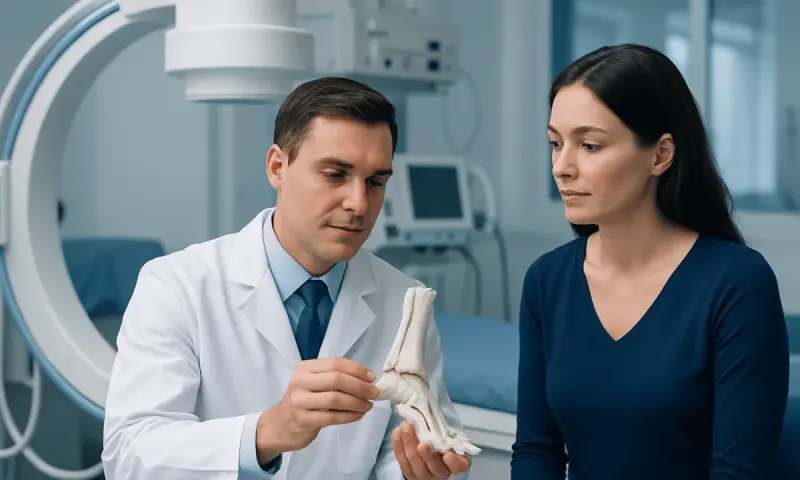
Because bone lesions have several possible causes, it is important not to assume that an abnormal scan represents eosinophilic granuloma. A coordinated review by radiology, pathology, orthopedic or pediatric specialists, and hematology-oncology teams can support an accurate diagnosis.
Modern Treatment Approaches
Treatment is individualized. For a single bone lesion that is not causing structural instability or neurologic risk, careful observation may be appropriate. Some lesions improve over time without extensive treatment, and scheduled clinical visits and imaging allow the team to confirm healing. Knee Pain Relief: Causes and Treatment" class="ahp-ilk">Pain relief, temporary activity changes, and protection of a vulnerable bone may be recommended during this period.
If treatment is needed, options may include biopsy with curettage, meaning removal of lesion tissue from the bone, sometimes with measures to support the bone’s structure. In selected cases, a local corticosteroid injection into the lesion may be considered. The choice depends on the lesion’s size, location, symptoms, and the likelihood of fracture or other local complications.
Lesions in difficult locations, including the spine or bones near the eye, ear, or central nervous system, should be managed by experienced specialists. Surgery may be used when there is instability, a fracture risk, compression of nearby structures, or a need for diagnosis. Radiation therapy is now used infrequently and only in carefully selected circumstances because other approaches are usually preferred, particularly for children.
When LCH affects multiple bones or organs, systemic therapy may be needed. This can include medicines that modify immune-cell activity or chemotherapy-based treatment plans. Targeted medicines may be considered for selected patients with an actionable molecular change, particularly when disease is persistent, recurrent, or more extensive. Treatment decisions should balance disease control with age, overall health, possible side effects, and long-term follow-up needs.
Outlook, Follow-Up, and Everyday Self-Care
The outlook for isolated eosinophilic granuloma of bone is often favorable. Many people recover well, especially when the lesion is localized and does not involve high-risk organs or critical structures. However, the course cannot be predicted from symptoms alone. Some people develop a new lesion later, and a smaller number have more widespread LCH, which is why follow-up remains important even after symptoms improve.
Follow-up may include physical examinations, symptom review, and repeat imaging at intervals chosen by the treating team. Children may need additional monitoring when lesions involve growth areas, the jaw, skull, or spine. People should report new bone pain, swelling, excessive thirst, frequent urination, persistent cough, skin rash, or unexplained changes in energy, as these symptoms may warrant reassessment.
At home, the most useful steps are practical rather than restrictive: follow activity guidance, attend imaging appointments, use prescribed pain relief safely, and avoid activities that could stress a weakened bone until a clinician confirms that it is safe. A balanced diet supports general bone health, but no special diet has been proven to treat eosinophilic granuloma. Smoking cessation is especially important for adults with lung involvement or smoking-related health risks.
Acıbadem Health Point’s multidisciplinary specialists and JCI-accredited hospitals can assess and treat LCH-related bone lesions for international patients, with care planned around the lesion location and the person’s overall clinical picture.
When to Seek Medical Care
A person should arrange medical assessment for persistent, localized bone pain, a growing lump over a bone, unexplained swelling, a limp, recurring jaw pain, or loose teeth without a clear dental explanation. These symptoms do not necessarily indicate eosinophilic granuloma, but they should be evaluated to identify the cause and prevent avoidable complications.
Prompt medical attention is appropriate for severe pain after minimal injury, inability to bear weight, new weakness or numbness, difficulty walking, or back or neck pain accompanied by changes in bladder or bowel control. These symptoms can indicate a fracture or possible nerve compression and should not be managed only at home.
People who have already been diagnosed should contact their care team if pain returns after improving, a new lump appears, or new symptoms develop in another part of the body. Timely review helps clinicians decide whether imaging, specialist assessment, or adjustment of the care plan is needed.
Frequently asked questions
01Is eosinophilic granuloma cancer?
Eosinophilic granuloma is a form of Langerhans cell histiocytosis, which has features of both inflammatory and abnormal cell-growth disorders. It is not usually discussed in the same way as common solid cancers, but it should be assessed and followed by clinicians experienced with LCH. The treatment approach depends on how extensive the disease is and which tissues are involved.
02Can eosinophilic granuloma heal on its own?
Some isolated bone lesions can improve over time with observation and supportive care. However, this decision should be made only after a clinician has confirmed the diagnosis and assessed the lesion’s location, stability, and potential risk to nearby structures. Follow-up imaging is often needed to document healing.
03How is eosinophilic granuloma diagnosed?
Doctors typically use imaging, such as X-ray, CT, or MRI, to assess the bone lesion. A biopsy is often performed to confirm Langerhans cell histiocytosis and exclude other causes of bone damage. Additional tests may be recommended to check for lesions elsewhere in the body.
04Can eosinophilic granuloma come back?
A treated lesion may recur, and some people can develop a new lesion at a different site later. The risk varies between individuals and is influenced by whether LCH is limited to one area or affects multiple sites. Regular follow-up helps detect changes early.
05Is eosinophilic granuloma hereditary?
In most cases, eosinophilic granuloma is not inherited. Some lesions have acquired gene changes in the affected cells, but these changes are generally not passed from parents to children. Family members usually do not need testing solely because one relative has LCH.
06What specialists treat eosinophilic granuloma?
Care may involve pediatricians or internists, orthopedic surgeons, radiologists, pathologists, hematologists-oncologists, and other specialists depending on the lesion location. Spine, jaw, skull, or multisystem disease may require additional expertise. A multidisciplinary team can coordinate diagnosis, treatment, and follow-up.
This article is for general information only and is not a substitute for professional medical advice. Please consult a qualified doctor about your individual situation.
Hair transplant in Turkey — costs, graft counts & a free assessment
JCI-accredited · board-certified surgeons · reply within 24h
Add us as a Preferred Source to see more of our trusted health content across Google Search, AI Overviews and Discover.