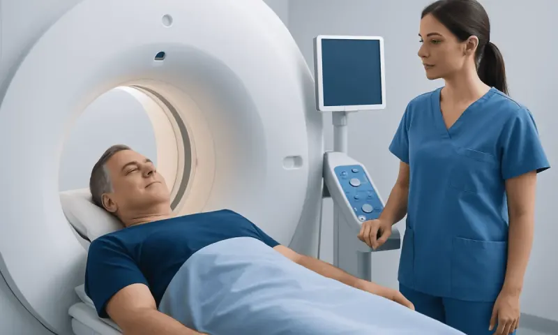
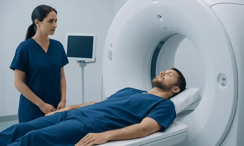
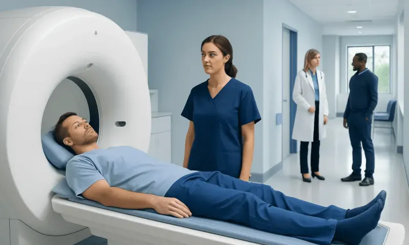
Does a CT Scan Show Soft Tissue Damage?
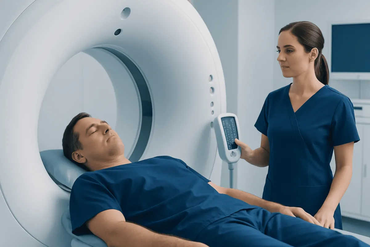
A CT scan can show certain types of soft tissue damage, including swelling, bleeding, fluid collections and some significant muscle injuries. However, MRI and ultrasound are often more useful for examining tendons, ligaments, muscles and other soft tissues in detail.
Does CT Scan Show Soft Tissue Damage?
Does CT scan show soft tissue damage? Yes, CT can show some soft tissue problems, particularly swelling, bruising, bleeding, fluid collections, infections, masses and major injuries after trauma. It is often used urgently because it produces detailed cross-sectional images quickly and can assess bones and nearby tissues at the same time.
However, CT is not always the most sensitive test for subtle soft tissue injury. Small muscle strains, partial tendon tears, ligament sprains, cartilage injuries and inflammation may not be clearly visible. Depending on the symptoms and body area, a clinician may recommend magnetic resonance imaging (MRI), ultrasound, examination over time, or a combination of these approaches.
Imaging is only one part of diagnosis. The location of pain, the mechanism of injury, physical examination findings, movement limitations and symptoms such as weakness or numbness all help determine whether further testing is needed.
What CT Can and Cannot Show
CT, or computed tomography, uses X-rays and computer processing to create detailed slice-like images of the body. It is particularly good at showing bones, lungs, the brain, internal organs and acute bleeding. In an emergency, it may help identify a fracture or dislocation that is contributing to pain and may also reveal associated soft tissue swelling or a hematoma, which is a collection of blood outside a blood vessel.
When people ask whether a CT scan can show soft tissue injury, the answer depends on the injury. CT may show a large muscle tear, marked swelling, gas in soft tissues, an abscess, a significant tendon abnormality in some locations, or bleeding around a damaged area. Contrast dye may sometimes be used to improve assessment of blood vessels, inflammation, infection or certain masses.
CT has lower soft-tissue contrast than MRI. This means that tissues with similar density can be harder to distinguish. A CT scan may therefore be normal in a person with a painful ligament sprain, a small tendon tear, early muscle injury or irritation of a joint-related structure.
- CT is commonly helpful for: fractures, complex trauma, acute bleeding, internal injuries and urgent evaluation.
- CT may be less definitive for: mild strains, sprains, partial tendon tears, subtle ligament damage and many cartilage injuries.
- Further imaging is selected individually: based on symptoms, examination and the suspected structure involved.
What Is the Best Test for Soft Tissue Damage?

What is the best test for soft tissue damage? MRI is often the best imaging test when a clinician needs detailed information about deeper muscles, tendons, ligaments, cartilage, nerves and other soft tissues. MRI does not use ionizing radiation and can show differences in water content within tissues, making edema and many types of injury easier to identify.
There is no single best test for every situation. Ultrasound can be very useful for superficial structures, including some shoulder tendons, muscles, ligaments and fluid collections. It can also show movement in real time and may guide injections or aspirations when these are clinically appropriate. Its usefulness depends partly on the structure being assessed and the experience of the operator.
CT may be the preferred first scan after high-energy injury, especially if a fracture, internal bleeding or injury to several body regions is possible. X-rays may be used first for many bone and joint concerns. A clinician chooses the test that is most likely to answer the specific clinical question while avoiding unnecessary radiation, delay or expense.
Does Inflammation Show on a CT Scan?
Does inflammation show on a CT scan? Sometimes. CT can show indirect or more obvious signs of inflammation, such as swelling, tissue thickening, fluid, enlarged lymph nodes, an abscess or changes in the fat surrounding an inflamed area. With contrast, inflamed tissue may enhance differently from surrounding tissue, which can help clinicians assess certain conditions.
Nevertheless, mild or early inflammation may not be visible on CT. CT findings are also not always specific: swelling can result from injury, infection, recent surgery or other causes. For this reason, scan results are interpreted alongside symptoms, physical examination, blood tests when appropriate and other imaging.
MRI can be more sensitive for inflammation in muscles, tendons, joints, bone marrow and soft tissues. Ultrasound may also identify fluid, thickened tendons, inflammation around joints and increased blood flow in certain superficial areas. The most suitable test depends on where the symptoms are located and what condition is being considered.
Does CT Scan Show Ligament or Muscle Damage?
People often ask, “Does CT scan show ligament damage?” CT can occasionally suggest a major ligament injury, particularly when there is an abnormal joint position, an avulsion fracture where a ligament pulls off a small piece of bone, substantial swelling or a clearly disrupted structure. Still, MRI is generally more accurate for directly evaluating many ligaments, especially around the knee, ankle, wrist and shoulder.
Similarly, a CT scan can show some muscle damage, such as a large tear, extensive bruising or a hematoma. But if the question is whether a CT will show a mild muscle strain or a subtle partial tear, MRI is usually more informative. Ultrasound may also assess selected muscle injuries and can be useful during follow-up.
Symptoms alone cannot reliably distinguish a strain from a tear, sprain, fracture or nerve-related problem. Pain after activity, tenderness, swelling, bruising, weakness and reduced range of motion can occur with multiple conditions. A careful assessment helps identify whether rest and rehabilitation are appropriate or whether imaging and specialist evaluation are needed.
How Do I Know If I Have Soft Tissue Damage?
How do I know if I have soft tissue damage? Soft tissue damage refers broadly to injury or disease affecting muscles, tendons, ligaments, fascia, skin, nerves, blood vessels or other non-bony structures. Common symptoms after an injury include pain, tenderness, swelling, bruising, stiffness, reduced movement, weakness or pain when using the affected area.
A muscle strain may cause pain when the muscle contracts or stretches. A ligament sprain may cause pain around a joint, swelling, bruising or a feeling of instability. Tendon problems can cause pain with specific movements, local tenderness and weakness. These patterns can provide clues, but they do not confirm the diagnosis without clinical assessment.
Initial care for a minor recent injury may include protecting the area from further injury, relative rest, gentle movement as tolerated, and cold or heat according to comfort and professional advice. Pain medicine should be used only as directed by a clinician or product instructions, especially for people with kidney disease, stomach ulcers, bleeding risks, pregnancy or medication interactions. A rehabilitation plan may be recommended once serious injury has been excluded.
What If CT Shows Nothing but Still in Pain?
What if CT shows nothing but still in pain? A normal CT result can be reassuring because it may exclude important problems such as many fractures, major bleeding or certain urgent internal injuries. It does not mean the pain is not real, and it does not exclude every soft tissue condition. Some injuries are too subtle for CT, develop clearer features over time, or require a different type of imaging.
Follow-up is important when pain persists, limits daily activities, causes weakness or does not improve as expected. A doctor may repeat the physical examination, review the timing and nature of symptoms, consider X-rays or MRI, arrange ultrasound, or refer to an orthopedic, sports medicine, rehabilitation or other appropriate specialist.
It can be helpful for the person to note when pain occurs, what movements make it worse, whether swelling changes, and whether there is numbness, tingling, weakness or instability. These details can guide the next clinical decision. Gradually returning to activity should be guided by symptoms and professional advice rather than by imaging alone.
When to Seek Medical Care
Prompt medical assessment is appropriate after a significant fall, collision, sports injury or other trauma, particularly when there is severe pain, visible deformity, inability to bear weight or use a limb, rapidly increasing swelling, or a deep wound. Emergency care is needed for symptoms such as uncontrolled bleeding, difficulty breathing, chest pain, sudden confusion, fainting, new weakness, loss of sensation, or a limb that becomes cold, pale or blue.
Medical advice should also be sought for pain that is worsening, persistent pain after a normal CT scan, fever with redness or swelling, unexplained bruising, a growing lump, or pain that interferes with sleep or daily activities. Early assessment can help clarify whether rehabilitation, monitoring, additional imaging or another treatment approach is appropriate.
Acıbadem Health Point’s multidisciplinary specialists and JCI-accredited hospitals assess musculoskeletal and other soft tissue concerns for international patients, using clinical examination and appropriate imaging when indicated. The goal is to identify the likely source of symptoms and develop an individual care plan with the patient.
Frequently asked questions
01Can a CT scan detect soft tissue damage?
A CT scan can detect some soft tissue damage, including substantial swelling, bleeding, fluid collections and some larger muscle injuries. It may not reliably identify subtle strains, sprains, partial tendon tears or ligament injuries. MRI or ultrasound may be recommended when those conditions are suspected.
02Will a CT scan show muscle damage?
CT may show major muscle tears, swelling or bleeding within a muscle. Mild strains and smaller tears can be difficult to see, particularly soon after injury. MRI is generally better for detailed assessment of muscle tissue.
03Do CT scans show soft tissue damage after a car accident?
CT is frequently used after a significant car accident because it can quickly look for fractures, internal bleeding and injuries involving the chest, abdomen, pelvis or head. It can show some associated soft tissue findings, but a normal CT does not exclude all muscle, tendon or ligament injuries. Follow-up may be needed if pain continues.
04Can a CT scan see tendons and ligaments?
CT can show some tendons and ligaments, especially when they are large or when there are related bone changes or severe swelling. However, it is usually not the preferred test for detailed tendon and ligament assessment. MRI is often more sensitive, while ultrasound may be useful for some superficial tendons.
05Why would a doctor order a CT instead of an MRI for an injury?
CT is faster, widely used in urgent settings and particularly effective for detecting fractures and acute bleeding. It may be safer or more practical for some people who cannot have an MRI. The choice also depends on the suspected injury, the body area and how urgently results are needed.
06Can pain continue even if a CT scan is normal?
Yes. Pain can continue because of a strain, sprain, tendon problem, nerve irritation, inflammation or another condition that CT does not show clearly. A clinician can reassess persistent symptoms and decide whether further examination, rehabilitation or different imaging is appropriate.
This article is for general information only and is not a substitute for professional medical advice. Please consult a qualified doctor about your individual situation.
Persistent digestive symptoms? Get evaluated in Turkey
JCI-accredited · board-certified surgeons · reply within 24h
Add us as a Preferred Source to see more of our trusted health content across Google Search, AI Overviews and Discover.