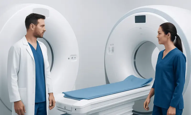
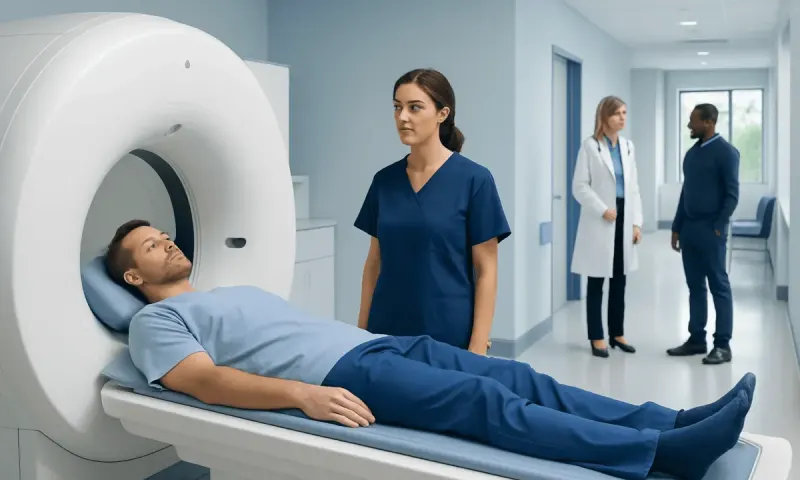
CT Scan Machine vs MRI Machine: Key Differences
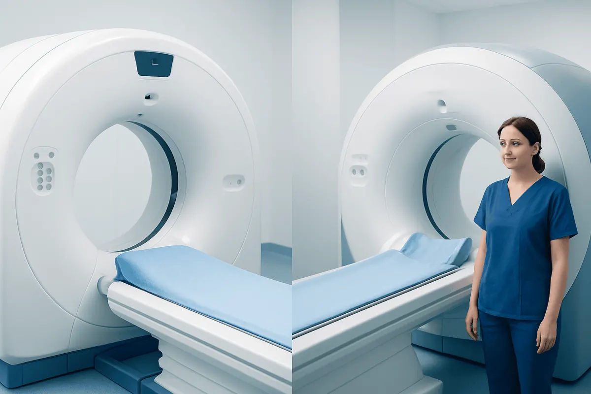
A CAT scan (CT) and an MRI are both imaging tests, but they create pictures in different ways and are chosen for different clinical needs. CT is often preferred for speed, bone injury and emergencies, while MRI provides especially detailed views of soft tissues, the brain, spinal cord and joints.
Cat Scan Machine vs MRI Machine: Side-by-Side Comparison
When comparing a cat scan machine vs mri machine, the main difference is the technology used. A CAT scan, more commonly called a CT scan, uses a series of X-ray images processed by a computer to create detailed cross-sectional pictures. MRI, or magnetic resonance imaging, uses a strong magnetic field and radio waves to produce images without using ionizing radiation.
Both tests can be highly valuable. The choice is not usually based on which machine is more advanced, but on what the clinician needs to see, how urgently information is needed, and whether the person can safely undergo the examination.
- CT: Usually fast; particularly useful for bones, lungs, bleeding, many abdominal problems and emergency assessment.
- MRI: Often takes longer; particularly useful for the brain, spinal cord, nerves, muscles, ligaments, cartilage and other soft tissues.
- Radiation: CT uses X-ray radiation. MRI does not use X-ray radiation.
- Comfort and access: CT is generally more open and quicker. MRI involves lying still in a longer, narrower scanner and can be noisy.
- Contrast: Both tests may use contrast material when additional detail is needed, although the contrast agents used are different.
How Clinicians Tell CT and MRI Apart
Clinicians begin with the medical question rather than the machine. For example, after a serious fall or road traffic injury, a CT scan may quickly identify fractures, bleeding or injury to organs. For persistent knee pain with suspected ligament damage, an MRI may show structures that are less visible on CT.
The body region also matters. CT is very effective for the chest, including the lungs, and for detailed assessment of many bone injuries. MRI provides excellent contrast between soft tissues and may be selected to assess the brain, spinal canal, joints, pelvic organs, liver lesions or muscle injuries. In some situations, both tests may be appropriate at different stages of care.
Safety and practical factors are considered as well. MRI may not be suitable for some people with certain implanted or metallic devices, although many modern implants are MRI-compatible. The imaging team carefully checks devices, previous surgery, metal fragments, pregnancy status, kidney function and prior reactions to contrast before proceeding.
How to Choose Between CT and MRI?
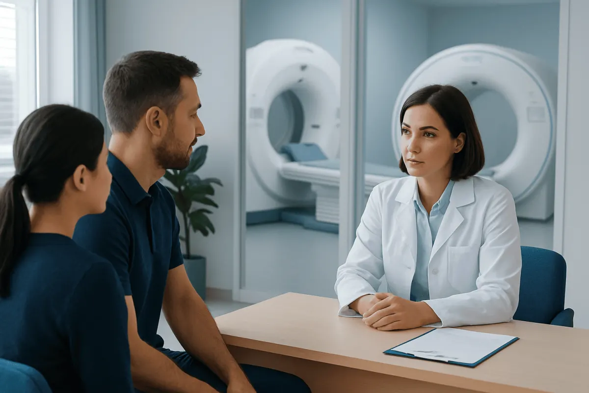
Patients generally should not need to choose independently between CT and MRI. A doctor selects the examination that is most likely to answer a specific clinical question while considering urgency, possible risks, previous imaging and whether the result is likely to change treatment.
CT may be chosen when rapid information is needed, such as after trauma or when a clinician needs to assess possible bleeding, a complicated infection, a blood clot in certain settings, or a lung condition. MRI may be selected when symptoms suggest a problem involving the brain, spinal cord, nerves, tendons, ligaments or cartilage. It may also help characterize some findings first seen on ultrasound or CT.
People should tell the referring clinician and radiology team about pregnancy or possible pregnancy, kidney disease, asthma or prior contrast reactions, claustrophobia, hearing devices, pacemakers, clips from prior surgery, medication pumps, joint replacements and any chance of metal in the eye or body. This information helps the team plan the safest and most useful examination.
What Does an MRI Show That a CT Scan Will Not?
MRI can often show soft-tissue differences with greater clarity than CT. It is especially helpful for examining the spinal cord, nerve roots, brain tissue, ligaments, tendons, cartilage, muscles, bone marrow and many pelvic structures. For instance, MRI may demonstrate a torn ligament, disc-related nerve compression, inflammation in soft tissues or subtle changes in brain tissue that CT may not show as clearly.
This does not mean MRI replaces CT. CT may be more informative for fine bone detail, calcium deposits, certain lung findings and rapid assessment of acute bleeding or traumatic injury. The usefulness of either scan depends on the timing of symptoms and the suspected condition.
For neurological symptoms, imaging is selected carefully. A CT scan may be performed first when a stroke or head injury is suspected because it is fast and can identify bleeding. MRI can provide additional information in selected cases, including some early ischemic changes and disorders affecting brain tissue or nerves. Evaluation of possible stroke should always be treated as urgent and guided by emergency clinicians.
Why Do Doctors Choose CT Over MRI?
Doctors often choose CT over MRI because CT is fast, widely useful in urgent care and particularly effective for several common clinical questions. It can quickly assess major injuries, fractures, internal bleeding, the chest and lungs, and many abdominal or pelvic conditions. CT may also be more practical when a person cannot lie still for a longer examination.
CT can be preferable for people who have an implant or device that is not safe in an MRI environment. It may also be easier for someone who experiences severe claustrophobia, although healthcare teams can sometimes offer support or alternatives for MRI when needed.
Because CT uses ionizing radiation, clinicians aim to use it only when the expected benefit outweighs the risk. Imaging protocols are tailored to the clinical situation, and unnecessary repeat scans are avoided. When appropriate, prior scans may be reviewed so that a new examination is not performed without a clear reason.
Which Shows More, an MRI or a CAT Scan?
Neither MRI nor a CAT scan always “shows more.” MRI generally shows more detail in many soft tissues, while CT may show more useful detail for bones, lungs, calcifications and certain urgent conditions. The most informative scan is the one that best matches the suspected cause of symptoms.
For example, CT may be the better first test for a complex facial fracture or suspected bleeding after an accident. MRI may be more revealing for a suspected rotator cuff tear, spinal nerve compression or certain brain disorders. A radiologist interprets the images in the context of the examination type, the reason for referral and the person’s medical history.
Sometimes an imaging report identifies an unexpected finding that needs follow-up. This does not automatically mean a serious condition is present. The treating clinician can explain whether the finding is likely related to symptoms, whether monitoring is appropriate, or whether further testing is needed.
Preparing for the Scan and Understanding Results
Preparation depends on the scan and whether contrast is planned. The imaging center may ask the person not to eat for a period beforehand, particularly for certain contrast-enhanced examinations. They may also request recent kidney-function blood tests when contrast is being considered. Instructions should be followed exactly, and regular medicines should only be changed if a clinician advises it.
For MRI, metal objects such as jewelry, watches, hairpins and some clothing items must be removed. The person is given hearing protection because the machine makes loud tapping sounds. During CT, it is important to remain still; if contrast is used, a brief warm sensation or metallic taste can occur and usually passes quickly.
A radiologist reviews the images and sends a report to the referring clinician. Results may be available quickly in urgent settings, while non-urgent examinations may take longer depending on the service. The referring clinician explains what the findings mean for the individual and whether treatment, follow-up imaging or referral is needed.
Acıbadem Health Point’s multidisciplinary specialists and JCI-accredited hospitals can assess symptoms, arrange appropriate imaging and coordinate care for international patients when CT or MRI evaluation is needed.
When to Seek Medical Care
Urgent medical assessment is important for sudden weakness or numbness on one side of the body, difficulty speaking, sudden severe headache, loss of consciousness, new seizures, severe chest pain, trouble breathing, major injury, or severe abdominal pain. These symptoms may require prompt evaluation, and emergency clinicians will decide whether CT, MRI or another test is needed.
A non-urgent medical appointment is appropriate for persistent or worsening headaches, ongoing back or neck pain with tingling or weakness, unexplained joint symptoms, recurrent abdominal pain, or symptoms that do not improve as expected. Imaging is not always necessary, but a clinician can determine whether it would help clarify the cause.
People should seek immediate help after a head injury if there is repeated vomiting, increasing drowsiness, confusion, worsening headache, seizures, weakness, unequal pupils or fluid leaking from the nose or ears. The safest next step is to contact local emergency services or attend an emergency department rather than waiting for an outpatient scan.
Frequently asked questions
01Is a CAT scan the same as a CT scan?
Yes. CAT stands for computed axial tomography, while CT stands for computed tomography. CT is the term used more commonly today, but both names refer to the same type of X-ray-based imaging examination.
02Is MRI safer than CT?
MRI does not use ionizing radiation, whereas CT uses X-rays. However, safety depends on the individual situation: MRI may not be suitable with some implants or metal objects, and both tests require special consideration if contrast is used. A clinician weighs the benefits and potential risks before recommending either scan.
03Can MRI and CT scans detect cancer?
Both CT and MRI can help detect, characterize and monitor some cancers, but imaging alone does not always confirm a diagnosis. The appropriate test depends on the body area and suspected cancer type. Further imaging, laboratory testing or a biopsy may sometimes be needed.
04Does a CT scan take less time than an MRI?
In most cases, yes. The image acquisition for CT is often very quick, which is one reason it is valuable in emergency care. MRI usually takes longer because it collects highly detailed images in several sequences.
05Can a person with claustrophobia have an MRI?
Many people with claustrophobia can still complete an MRI with planning and support. The care team may explain the process in advance, provide communication during the scan, consider relaxation strategies or discuss other options when medically appropriate. It is important to mention claustrophobia before the appointment.
06Why might contrast be used for a CT or MRI?
Contrast material can make blood vessels, inflammation, tumors and certain organs or tissues easier to distinguish on images. It is not needed for every scan. Before administering contrast, the care team checks relevant medical history, including kidney function and any prior contrast reaction.
This article is for general information only and is not a substitute for professional medical advice. Please consult a qualified doctor about your individual situation.
Joint and spine care in Turkey — expert assessment & treatment
JCI-accredited · board-certified surgeons · reply within 24h
Add us as a Preferred Source to see more of our trusted health content across Google Search, AI Overviews and Discover.