PET/CT Combines Two Scans to Show Structure and Activity
Your doctor mentions a “PET/CT” and you leave the appointment with the word written on a slip of paper and very little idea what it means. Here is the short version. It is one imaging examination that combines two scans, so it shows both the structure of your body and how active your cells are.
You may also see it written as CT PET CT. It is the same test. Doctors use it most often to investigate, stage or monitor cancer. But the pictures never stand alone: they have to be read alongside your symptoms, your medical history and your other Blood Test Results Explained: What CBC, Hemoglobin, and Platelets Mean" class="ahp-ilk">test results.
CT PET CT: what the examination means
CT PET CT usually refers to a PET/CT scan: one examination that combines positron emission tomography (PET) and computed tomography (CT). PET highlights areas of altered cellular activity, while CT creates detailed cross-sectional pictures of organs, bones and other body structures. Together, they can help clinicians identify where an abnormality is located and how active it appears.
For many PET/CT examinations, a small amount of a radiotracer is injected into a vein. The most frequently used tracer is fluorodeoxyglucose (FDG), a glucose-like substance. Cells that use more glucose may take up more FDG and appear as areas of increased uptake on the images. Cancer cells may do this, but so can normal tissues, healing areas, inflammation and infection.
So a PET/CT scan is a powerful tool, not an answer on its own. A radiologist or nuclear medicine physician interprets the images in the context of the reason for testing, previous scans, laboratory results and the person’s medical history. An unusual result does not automatically mean cancer, and a normal scan does not rule out every possible condition.
Why doctors request a PET/CT scan
PET/CT is most often used in oncology. It can help identify the extent of a known cancer, assess whether treatment is working, look for possible recurrence after treatment or help select the most informative area for a biopsy. It may also help distinguish active disease from scar tissue or other changes seen on earlier imaging.
How useful it is depends on which cancer is suspected or confirmed. For example, PET/CT may be useful in evaluating some lymphomas, lung cancers, head and neck cancers, colorectal cancers and melanomas. It is not equally useful for every cancer, because some tumors do not take up the usual tracer strongly or may be too small to detect.
Doctors may also use specialized PET/CT examinations in selected non-cancer situations. These include investigating certain infections or inflammatory disorders, assessing inflammation around prosthetic devices, evaluating some heart conditions, and supporting assessment of selected neurological disorders. The tracer and scanning protocol may differ according to the clinical question.
A PET/CT result often guides the next step rather than replacing it. Depending on the findings, the care team may recommend observation, additional imaging, a tissue biopsy, surgery, radiotherapy, systemic treatment or another form of evaluation.
How PET and CT work together

The CT portion uses X-rays to create detailed anatomical images. It can show the size, shape and location of tissues and may be performed with or without iodine-based contrast material. Contrast can make blood vessels and some organs easier to assess, but it is not needed for every PET/CT examination.
The PET portion detects signals from the radiotracer after it has circulated in the body. The scan does not directly “see” cancer cells. Instead, it maps tracer distribution, which reflects a biological process such as glucose use. The PET and CT images are digitally aligned, allowing the reporting doctor to identify whether increased uptake corresponds to a lymph node, lung nodule, bowel activity, muscle, bone or another structure.
This combined approach can improve accuracy compared with reviewing PET or CT alone in many clinical settings. However, image interpretation remains complex. Small lesions can be below the resolution of the scanner, high blood sugar can affect FDG imaging, and normal uptake in the brain, heart, kidneys, bladder or muscles can sometimes complicate interpretation.
That is why a report may call a finding likely benign, suspicious, indeterminate, or say it needs correlation with another test. This wording is routine in imaging. It reflects careful reading, not necessarily a serious diagnosis.
Preparing for a CT PET CT scan
Your imaging center will give you instructions of your own, and it is worth following them closely. For a standard FDG PET/CT, many people are asked to avoid eating for several hours beforehand while drinking plain water. Fasting helps reduce normal insulin-related changes in glucose use and can improve image quality.
Strenuous exercise is commonly avoided for about a day before the scan because active muscles may take up more tracer. Patients should usually keep warm and rest quietly after injection, as shivering and muscle tension can also affect the images. It is important to tell the team about all medicines, supplements and recent treatments rather than stopping prescribed medication without medical advice.
People with diabetes need personalized preparation because blood glucose management and fasting plans may require adjustment. The imaging team should also know about pregnancy, possible pregnancy, breastfeeding, kidney disease, prior reactions to contrast material, asthma, thyroid conditions and implanted medical devices. These factors do not always prevent scanning, but they may change the plan.
Wear comfortable clothing without metal, though you may be given a gown. Bringing previous scan images or reports can be helpful, particularly when the test is being performed to compare a finding over time.
What happens during the appointment
After check-in, staff may review the person’s history, check blood glucose when FDG is being used and place a small intravenous line. The radiotracer injection is usually quick. A waiting period, often around an hour but varying by tracer and protocol, allows the tracer to distribute through the body.
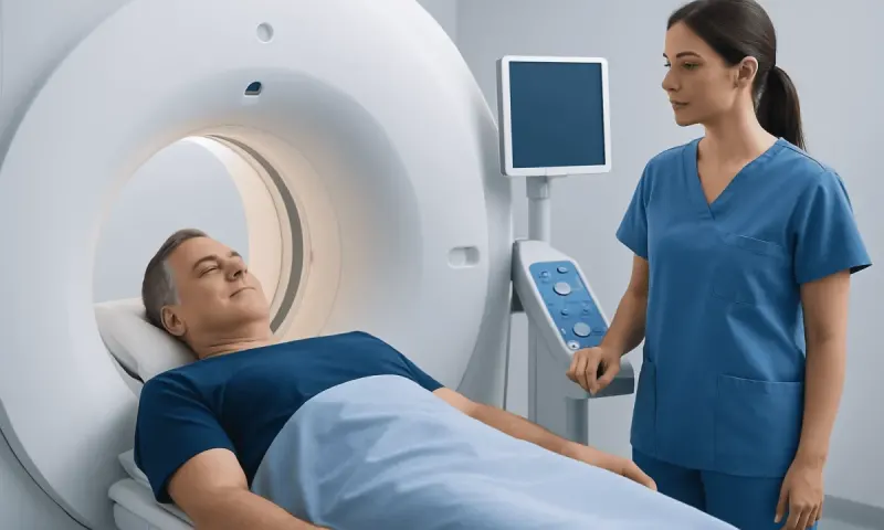
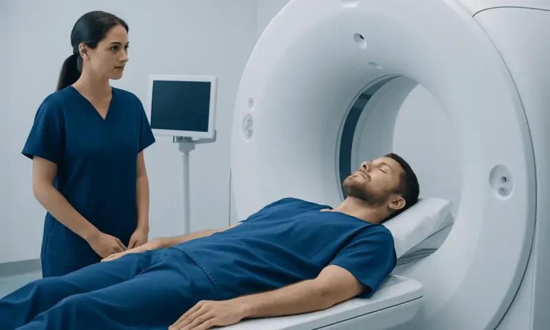
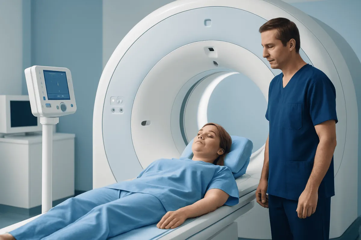
During the waiting period, patients are generally asked to relax quietly. The actual scanning time varies, but the person typically lies on a padded table that moves slowly through a large, open ring-shaped scanner. The examination is painless, although lying still can be uncomfortable for some people.
The CT scan itself is brief. If contrast is used, a temporary warm sensation or metallic taste can occur and usually passes quickly. Staff can see and speak with the patient throughout the scan. A person who has claustrophobia, significant pain, difficulty lying flat or concerns about the procedure should discuss this before the appointment, as the team may be able to help plan for comfort.
In many cases, normal activities can resume afterward. Drinking fluids may be advised to support elimination of the tracer, unless a clinician has recommended fluid restriction. Facilities may provide short-term instructions about limiting very close, prolonged contact with pregnant people or young children after certain radiotracers.
Safety, radiation and common misconceptions
PET/CT involves radiation from both the radiotracer and the CT component. The amount varies with the type of scan, body size and protocol. Medical teams use the lowest radiation level reasonably achievable while obtaining clinically useful images, and they request PET/CT when the expected information is likely to help guide care.
A common myth is that any area of high uptake proves cancer. In reality, infection, inflammation, recent surgery, vaccination, healing fractures and normal body functions can all produce increased uptake. Conversely, some cancers show little uptake on FDG PET, particularly when they are slow-growing, very small or biologically different from tumors that use glucose avidly.
People also assume a PET/CT can replace a biopsy. It cannot. Imaging can indicate where a problem may be and how widespread it appears, but tissue testing is often needed to confirm a cancer diagnosis and identify its precise type. The doctor will explain whether a biopsy is appropriate based on the full clinical picture.
Allergic reactions are uncommon with the PET tracer itself. If CT contrast is planned, there is a small risk of contrast reaction and a need for additional consideration in people with significantly reduced kidney function. Patients should report previous contrast reactions and relevant health conditions before the scan.
Understanding results and when to seek medical care
Results are commonly reviewed by the clinician who ordered the scan after a radiologist or nuclear medicine physician has issued a report. The timing varies by facility and clinical urgency. Do ask what the scan was meant to clarify, whether the findings need further tests, and how the result changes your care plan.
Medical care should be sought promptly for symptoms that may need urgent assessment, such as severe chest pain, sudden shortness of breath, coughing up blood, new weakness on one side of the body, confusion, seizures or severe allergic symptoms after contrast exposure. These symptoms should not be managed by waiting for a routine imaging appointment or result.
For non-urgent concerns, patients should contact their doctor if they have persistent unexplained weight loss, a new or enlarging lump, ongoing fever, unexplained bleeding, worsening pain, or symptoms that do not improve as expected. These symptoms have many possible causes, and early clinical assessment helps determine whether imaging or other tests are needed.
Acıbadem Health Point’s multidisciplinary specialists and JCI-accredited hospitals diagnose and treat complex conditions for international patients, with imaging findings reviewed as part of an individual clinical assessment. Patients should discuss PET/CT results and follow-up plans with a qualified doctor who knows their medical history.
Frequently asked questions
01Is CT PET CT the same as a PET/CT scan?
Yes. CT PET CT is commonly used to describe a PET/CT scan, which combines PET and CT imaging in one appointment. PET shows tracer activity in tissues, while CT provides anatomical detail.
02Can a PET/CT scan diagnose cancer by itself?
No. PET/CT can identify findings that may be suspicious for cancer and can show how extensive a known cancer may be. However, inflammation, infection and normal tissue activity can also cause PET uptake, so biopsy or other tests may be needed for confirmation.
03How long does a PET/CT scan take?
The total appointment often takes a few hours because there is usually a resting period after the tracer injection. The actual time on the scanner is generally shorter, but it varies according to the body area, tracer and imaging protocol.
04Do patients need to fast before a PET/CT scan?
Many FDG PET/CT scans require fasting for several hours, but instructions differ depending on the reason for the test and the tracer used. People with diabetes should receive individualized guidance from the imaging center or their doctor.
05Is PET/CT radiation dangerous?
PET/CT uses ionizing radiation, so it is not performed unnecessarily. The medical team balances the radiation exposure against the expected benefit of obtaining information that can guide diagnosis or treatment.
06What should patients tell the imaging team before PET/CT?
Patients should mention pregnancy or breastfeeding, diabetes, kidney disease, allergies or previous reactions to contrast, current medications and recent surgery, infection, vaccination or cancer treatment. This information can affect preparation and interpretation of the images.
This article is for general information only and is not a substitute for professional medical advice. Please consult a qualified doctor about your individual situation.
Oncology care in Turkey — second opinion and treatment plan
JCI-accredited · board-certified surgeons · reply within 24h
Add us as a Preferred Source to see more of our trusted health content across Google Search, AI Overviews and Discover.