Coats Disease Is Treated With Laser, Freezing or Surgery
Someone notices a white glow in a child’s pupil in a photograph, or an eye that drifts, or vision that has quietly slipped in one eye. That is often how Coats disease first comes to attention.
Treatment has a clear aim: close the abnormal leaking blood vessels in the retina, bring down the fluid buildup, and hold on to as much vision as possible. Which approach suits you or your child depends on the stage of the disease, how much of the retina has been damaged, and whether retinal detachment is present.
Coats Disease Treatment: How It Works
Coats disease treatment works by closing or destroying abnormal blood vessels in the retina, the light-sensitive tissue at the back of the eye. These fragile vessels can leak blood components and fluid, causing retinal swelling, fatty deposits called exudates and, in more advanced cases, retinal detachment. Treatment is planned to stop this leakage and protect the structure of the eye and any remaining useful vision.
The main treatments are laser photocoagulation and cryotherapy. Laser therapy applies focused light energy to seal abnormal vessels, while cryotherapy freezes vessels through the outer wall of the eye. Both methods may need to be repeated over several visits because Coats disease can involve multiple areas of the retina and may remain active over time.
When fluid accumulation is significant, an eye specialist may also recommend medicines injected into the eye to reduce swelling or decrease the activity of leaking vessels. More advanced disease may require retinal surgery to repair a detachment or manage complications. A retinal specialist will tailor the plan to your eye after a detailed examination and imaging.
Is Coats Disease Serious?
Coats disease can be serious because ongoing leakage can permanently damage the retina and reduce vision. Its severity varies greatly. Some people have a small area of abnormal vessels that can be treated before central vision is affected, while others are diagnosed after extensive leakage or retinal detachment has already developed.
Although Coats disease is not cancer and does not usually affect general physical health, it can threaten sight in the affected eye. In children, it can also lead to amblyopia, sometimes called “lazy eye,” if poor vision develops while the visual system is still maturing. So if you notice symptoms or something unusual about the eye’s appearance, see an ophthalmologist without delay.
Many cases affect one eye only. The other eye is usually healthy, but it should still be examined carefully. Ongoing monitoring helps clinicians identify treatment response, new leakage or complications at an early stage.
Candidacy and Evaluation Before Treatment

Anyone with confirmed or suspected Coats disease should be assessed by an ophthalmologist, ideally a retinal specialist. Treatment decisions are based on the disease stage rather than age alone. Laser treatment is generally most useful when the retina is still attached and the abnormal vessels can be reached safely. Cryotherapy may be preferred when lesions are farther forward in the eye or fluid prevents laser energy from reaching the target area.
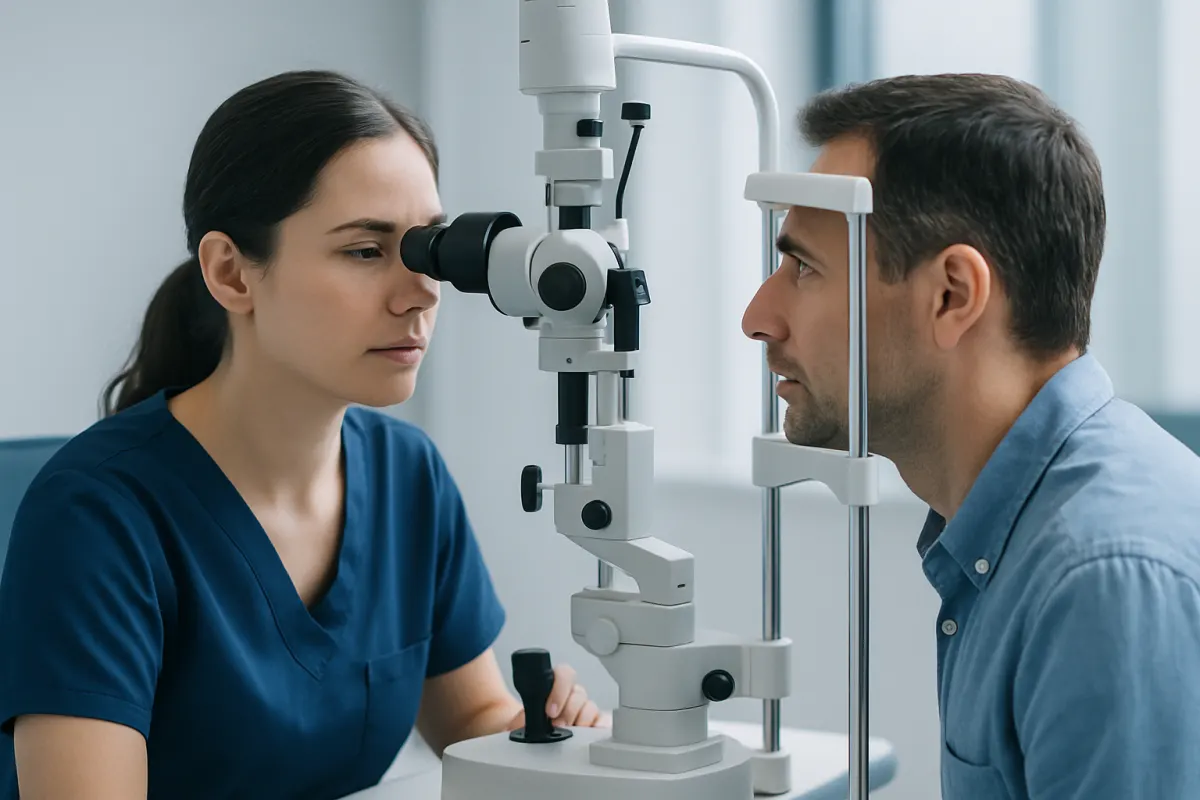
The evaluation usually includes visual acuity testing when possible, pupil dilation and a careful retinal examination. Retinal photography, optical coherence tomography and fluorescein angiography may be used to show fluid, exudates and leaking blood vessels in detail. Ultrasound can help assess the retina when the view into the eye is limited.
In children, doctors must also rule out other causes of a white pupil or retinal abnormality, particularly retinoblastoma. This distinction is essential because the management is very different. The care team may include pediatric ophthalmology, retina specialists, imaging professionals and, where needed, specialists in pediatric anesthesia.
- Earlier-stage disease: often treated with laser or cryotherapy.
- Fluid or marked exudation: may require an injection in addition to vessel-directed treatment.
- Retinal detachment: may require surgery as well as treatment of the abnormal vessels.
What Happens During Coats Disease Treatment?
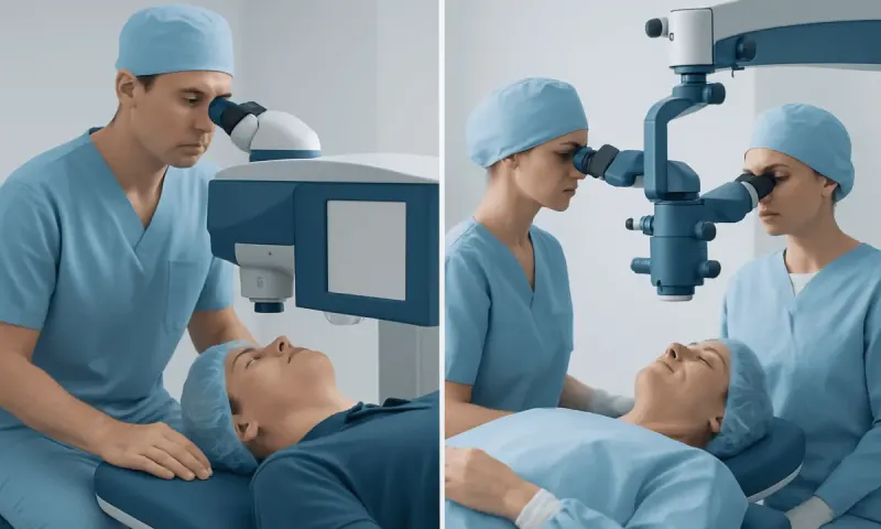
Laser photocoagulation is usually performed in an outpatient setting. After the pupil is dilated and the eye is numbed, the specialist applies laser spots to the abnormal retinal vessels. Children and some adults may need general anesthesia so the eye can remain still and the retina can be treated precisely. The laser creates controlled scarring that closes leaking vessels over time.
Cryotherapy is also performed by an eye specialist, often under anesthesia in younger children. A small freezing probe is placed on the outside of the eye over the area of abnormal retina. Freezing treatment causes the targeted vessels to close. Temporary inflammation and swelling can occur after cryotherapy, so a follow-up plan is especially important.
For selected patients, anti-VEGF medicines or corticosteroids may be injected into the eye to reduce leakage and fluid. These injections do not replace laser or cryotherapy; they are usually supportive treatments that may make definitive vessel treatment more effective or safer. In severe cases, vitreoretinal surgery may be used to address retinal detachment, drainage of fluid or other complications.
Each visit is shaped by how the eye is responding. Treatment is usually given in stages rather than trying to cover every area in one go. This approach can reduce inflammation and allow the retinal specialist to reassess the result between sessions.
Recovery Timeline, Benefits and Risks
Recovery depends on the treatment used, the age of the patient and the extent of retinal disease. After laser treatment, mild blurring, light sensitivity or irritation may be temporary. After cryotherapy or surgery, redness, soreness and swelling may last longer. The treating team will explain whether eye drops, a protective shield or activity limits are needed.
Follow-up visits are often scheduled within days or weeks after treatment, then at intervals based on disease activity. Imaging and retinal examination show whether leaking vessels have closed and whether fluid is improving. Some people need several treatment sessions over months. Longer-term monitoring remains important even after the retina appears stable.
The main potential benefit is preventing further leakage and preserving vision. If the macula, the central part of the retina responsible for detailed vision, has not been severely affected, the outlook for useful vision may be better. However, treatment cannot always reverse established retinal scarring, long-standing detachment or vision loss caused by amblyopia.
Risks vary by procedure and may include inflammation, increased eye pressure, bleeding, cataract, retinal scarring or worsening retinal detachment. Injections can rarely lead to infection or retinal complications. Retinal surgery has additional risks, including recurrent detachment. A retinal specialist can explain how these risks apply to an individual eye and why a particular option is recommended.
What Is the Treatment and Prognosis for Coats Disease?
The treatment and prognosis for Coats disease depend primarily on the stage at diagnosis. Early disease may respond well to laser photocoagulation or cryotherapy, sometimes with repeated sessions. When there is substantial retinal fluid, an injection may be used alongside these treatments. If retinal detachment is present, surgery may be considered to preserve the eye’s structure and, in selected cases, improve the chance of retaining vision.
Prognosis is generally better when the disease is found before central retinal involvement or complete retinal detachment. Even with successful control of leakage, vision may remain limited if the macula has been damaged or if amblyopia developed in childhood. That does not make treatment pointless. Controlling the disease protects the eye, lowers the risk of painful complications and helps hold on to the sight that remains.
Coats disease may become inactive after treatment, but recurrence or new areas of vascular leakage can occur. That is why regular retinal checks stay part of long-term care. Ask your care team how often you should be seen, which warning symptoms matter, and whether visual rehabilitation would help.
Can You Fix Coat Disease?
There is currently no single treatment that permanently “fixes” Coats disease in every person. However, the abnormal vessels and their leakage can often be controlled with laser therapy, cryotherapy, injections or surgery. The goal is to make the disease inactive, protect the retina and preserve as much vision and eye comfort as possible.
Whether vision improves depends on the condition of the retina before treatment. Fluid may decrease and vision may improve in some patients, especially when the central retina has not been severely damaged. In other cases, the main benefit is preventing further decline rather than restoring vision that has already been lost.
For children with reduced sight in one eye, ophthalmologists may also discuss amblyopia care, such as glasses or patching of the stronger eye when suitable. This type of treatment supports visual development but does not replace treatment of the retinal blood vessel problem. As the eye changes over time, the plan should be revisited.
Is Coats Disease a Rare Disease? When to Seek Medical Care
Yes. Coats disease is a rare retinal disorder. It is most commonly diagnosed in children and is more often seen in boys, although it can occur in girls and adults as well. Most cases are sporadic, meaning they are not usually inherited from a parent and are not typically passed on to future children.
Medical assessment should be arranged promptly if a child or adult develops reduced vision in one eye, a new squint, an abnormal red reflex in photographs, a white or yellow appearance in the pupil, eye pain or visible eye redness. A white pupil in a child should always receive urgent ophthalmic evaluation because several eye conditions can cause this sign and early diagnosis matters.
People already diagnosed with Coats disease should contact their eye team sooner than planned if vision suddenly worsens, flashes or floaters increase, pain develops, or the eye becomes notably red. Acıbadem Health Point’s multidisciplinary specialists and JCI-accredited hospitals evaluate and treat retinal conditions for international patients, with care coordinated according to the individual’s clinical needs.
Frequently asked questions
01What causes Coats disease?
Coats disease is caused by abnormal development of small blood vessels in the retina. These vessels become enlarged and leaky, allowing fluid and lipids to collect in or under the retina. The exact reason this happens is not fully understood, and most cases are not inherited.
02At what age is Coats disease diagnosed?
Coats disease is often diagnosed in childhood, commonly when vision changes, a squint or an unusual pupil appearance is noticed. It can also be diagnosed in teenagers and adults. Adult-onset cases may be milder, but they still need specialist retinal assessment.
03How many treatments are needed for Coats disease?
The number of treatments varies according to the location and extent of leaking vessels and the response of the retina. Some patients need several laser or cryotherapy sessions over time. Follow-up examinations guide whether further treatment is needed.
04Can Coats disease affect both eyes?
Coats disease usually affects one eye. Bilateral involvement is uncommon, but both eyes are examined carefully during assessment. If findings are present in both eyes, doctors may consider other retinal conditions as part of the evaluation.
05Does Coats disease always lead to blindness?
No. The outlook varies, and early treatment can often control leakage and help preserve vision. However, advanced disease can cause substantial, permanent sight loss in the affected eye, particularly if the central retina or retinal attachment has been severely affected.
06Is Coats disease cancer?
No. Coats disease is not cancer; it is a retinal blood vessel disorder. In young children, doctors may perform tests to distinguish it from retinoblastoma, an eye cancer that can produce similar signs such as a white pupil.
This article is for general information only and is not a substitute for professional medical advice. Please consult a qualified doctor about your individual situation.
Oncology care in Turkey — second opinion and treatment plan
JCI-accredited · board-certified surgeons · reply within 24h
Add us as a Preferred Source to see more of our trusted health content across Google Search, AI Overviews and Discover.