Auricular Reconstruction for Microtia: When Surgery Is Done and What to Expect

Auricular reconstruction for microtia is surgery to rebuild the outer ear when it is underdeveloped or absent. Timing, technique, and recovery depend on a child’s age, ear anatomy, hearing needs, and family preferences, so care is best planned with an experienced multidisciplinary team.
Overview: What auricular reconstruction for microtia means
Auricular reconstruction for microtia is an operation to rebuild the visible outer ear, called the auricle or pinna, when it has not fully formed. Microtia is a congenital condition, meaning it is present at birth. It can range from a small ear with recognizable parts to a more significantly underdeveloped ear, and in some children the ear canal is also narrow or absent.
The purpose of surgery is usually to create an ear that looks balanced with the opposite side and fits the proportions of the face. In some cases, treatment planning also relates to hearing care, but the surgery to rebuild the outer ear is different from procedures that address the ear canal or middle ear. A child may therefore need input from more than one specialist.
Families often first learn about microtia soon after birth. It is understandable to have questions about appearance, hearing, timing of treatment, and whether surgery is necessary. Auricular reconstruction is elective in many cases, which means the decision can be made thoughtfully after discussing options, expected outcomes, and the child’s needs.
When surgery is done
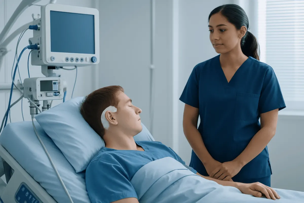
The best age for auricular reconstruction depends mainly on the technique being used and the child’s physical development. For reconstruction with the child’s own rib cartilage, surgeons often wait until later childhood, commonly around 8 to 10 years of age or sometimes later. This allows enough rib cartilage to be available for shaping the new ear and gives the opposite ear time to approach adult size for better matching.
For reconstruction using a synthetic framework, timing may be somewhat earlier in selected children because it does not depend on rib cartilage growth in the same way. Even so, surgeons still consider skin quality, overall health, emotional readiness, and whether the child can cooperate with postoperative care. The timing should be individualized rather than based on age alone.
If microtia affects hearing, hearing evaluation usually starts much earlier than reconstructive surgery. Babies and young children may need ongoing hearing support to help speech and language development. This is one reason treatment planning often involves pediatricians, audiologists, ENT specialists, and plastic surgeons working together.
Some families choose surgery in childhood, while others prefer to wait until the child is older and can participate more actively in the decision. Both approaches can be reasonable. The most important step is to discuss timing with a surgeon who has specific experience in auricular reconstruction and in caring for children with microtia.
Who may need it and what happens before surgery

Children with unilateral microtia, where one ear is affected, may seek reconstruction mainly for appearance and facial symmetry. Those with bilateral microtia, where both ears are affected, may have greater hearing-related needs and often require an even more coordinated care plan. Surgery may also be considered for older adolescents or adults who were not treated earlier.
Before surgery, the team will examine the shape of the ear area, skin condition, scalp hairline, jaw symmetry, and the unaffected ear if one is present. Hearing tests are important, especially if the ear canal is absent or narrow. In some patients, imaging studies may be used to understand the anatomy of the ear and nearby structures, particularly if additional hearing-related procedures are being considered.
The surgeon will also ask about previous operations, skin scarring, and any tendency toward problematic scar formation such as keloid scars. Families usually discuss the expected number of stages, where scars will be located, how long healing may take, and what daily care is needed afterward. Photographs and ear measurements help with planning.
Because microtia can occur along with other craniofacial differences, the evaluation may sometimes include additional specialists. Even when the condition is isolated, careful preparation helps families understand what surgery can and cannot achieve. The aim is a realistic plan that balances safety, appearance, and long-term satisfaction.
Main surgical options
There are two widely used approaches to auricular reconstruction for microtia. One uses the patient’s own rib cartilage, called autologous cartilage reconstruction. The surgeon removes a small amount of cartilage from the chest, sculpts it into the framework of an ear, and places it beneath the skin in the ear area. This method uses living tissue from the patient and can provide a durable, natural-feeling result.
The second approach uses a biocompatible synthetic framework, often covered with the patient’s own tissue and skin. In selected patients, this can allow reconstruction at a younger age and may avoid a chest incision. However, because it involves an implanted material, the surgeon must consider skin thickness, blood supply, the risk of exposure, and the child’s activity level.
Rib cartilage reconstruction is often done in multiple stages. The first stage creates the framework, and later stages may lift the ear away from the head and refine contour details. Implant-based reconstruction may also involve staged procedures depending on the technique and the patient’s anatomy. The exact plan varies from one center to another.
Some families also ask about prosthetic ears, which are removable external ears made to match the other side. These are not surgical reconstructions of the patient’s own tissue, but they can be a suitable option for some people. During consultation, the surgeon may compare surgical reconstruction with other reconstructive choices used in plastic surgery, such as breast reconstruction, to explain how individualized tissue planning affects results and recovery.
What to expect on the day of surgery and during recovery
Auricular reconstruction is usually performed under general anesthesia. Depending on the technique, the operation may take several hours, and some children stay in the hospital overnight or longer for monitoring. The surgical area will be protected with dressings, and if rib cartilage is used there will also be care instructions for the chest incision.
After surgery, swelling, bruising, tenderness, and a feeling of tightness are common and usually improve gradually. Doctors provide pain control, wound care instructions, and guidance about sleeping position, bathing, school, and sports. Protecting the reconstructed ear from pressure or accidental trauma is especially important in the early healing period.
Follow-up visits help the surgical team assess healing, remove or change dressings, and plan any next stage if needed. Families should understand that the final shape develops over time as swelling settles and tissues heal. It is normal for the reconstructed ear not to look fully finished immediately after the first operation.
Recovery can be emotionally meaningful for both the child and family. Younger children may need reassurance and help adapting to temporary changes in appearance during healing. As with other reconstructive procedures in cosmetic and reconstructive surgery, clear communication and regular follow-up make the process smoother and safer.
Benefits, limits, and possible risks
The main benefit of auricular reconstruction for microtia is improved ear shape and facial balance. For many children and adults, this can support confidence and reduce self-consciousness about appearance. When reconstruction is part of broader care, it may also fit into a more complete plan that addresses hearing, speech, and social development.
It is equally important to understand the limits of surgery. A reconstructed ear can look very natural, but it may not be identical to a typical ear or to the ear on the other side. Fine details, skin texture, projection, and scars vary from person to person, and more than one procedure is often needed to reach the desired result.
Possible risks include bleeding, infection, delayed wound healing, unfavorable scarring, asymmetry, and the need for revision surgery. Rib cartilage reconstruction also involves chest discomfort and a chest scar. Implant-based reconstruction carries additional concerns such as framework exposure or damage if the overlying tissue does not heal as expected.
These risks are not the same for every patient, and careful selection helps reduce them. Families should ask the surgical team about experience with pediatric ear reconstruction, expected stages, activity restrictions, and how complications would be managed if they occur. Good planning leads to safer care and more realistic expectations.
Self-care, long-term follow-up, and support for families
Before surgery, families can help by keeping appointments, completing hearing evaluations, and preparing the child in an age-appropriate way. Simple explanations, pictures approved by the surgeon, and a calm discussion about bandages and follow-up visits can make the experience less stressful. If the child uses hearing devices, those should be reviewed as part of the surgical plan.
After surgery, careful wound care and protection of the reconstructed ear matter more than any home remedy. Children should avoid rough play, contact sports, or pressure on the ear until the surgeon says it is safe. Sun protection and scar care may also be recommended once healing has progressed.
Long-term follow-up is useful because the child’s face continues to grow, and the team may want to monitor ear symmetry, scar quality, and the need for refinements. If hearing support is part of the child’s care, audiology and ENT follow-up remain important even after the outer ear has been reconstructed. In some cases, families also discuss related congenital differences such as syndactyly or other developmental conditions if they are part of a broader syndrome.
For international patients seeking evaluation, Acıbadem Health Point’s multidisciplinary specialists and JCI-accredited hospitals diagnose and treat microtia and perform reconstructive procedures, including coordinated assessment by plastic surgery, ENT, and audiology teams when needed.
When to contact a doctor
Families should contact the surgical team promptly if there is increasing redness, warmth, drainage, bleeding, fever, worsening pain, or swelling that seems to be getting worse instead of better. These symptoms do not always mean a serious problem, but they should be assessed. It is better to ask early than to wait.
Medical advice is also important if a dressing becomes loose, the child accidentally bumps the reconstructed ear, or there are concerns about the chest incision after rib cartilage surgery. If a child seems unusually sleepy, has trouble breathing, or cannot drink fluids after anesthesia, urgent medical review is needed. The care team will explain which symptoms require immediate attention.
Even without warning signs, regular scheduled follow-up should not be skipped. Ear reconstruction is a process rather than a single event, and each visit helps the team support healing and decide whether additional stages are appropriate. Families should feel comfortable bringing questions about appearance, activity, school, or emotional adjustment to these visits.
Frequently asked questions
01What is microtia?
Microtia is a condition present at birth in which the outer ear does not fully develop. It may affect one ear or both ears, and some children also have differences in the ear canal that can affect hearing.
02At what age is auricular reconstruction usually done?
The timing depends on the technique and the child’s growth. Reconstruction with rib cartilage is often done in later childhood, while some implant-based options may be considered earlier in selected patients.
03Does ear reconstruction improve hearing?
Auricular reconstruction mainly rebuilds the appearance of the outer ear. Hearing improvement depends on the anatomy of the ear canal and middle ear, so separate hearing treatments or procedures may be needed.
04How many operations are usually needed?
Many children need more than one stage, especially with rib cartilage reconstruction. The exact number varies with the surgical method, the ear’s anatomy, and how healing progresses.
05Will the reconstructed ear look completely natural?
The goal is a natural-looking ear that matches the face as closely as possible. Results can be very good, but no reconstruction can promise a perfect copy of a typical ear or the ear on the other side.
06Is auricular reconstruction painful?
Some discomfort is expected after surgery, and there may be additional soreness in the chest if rib cartilage is used. The care team provides pain management and recovery guidance to keep the child as comfortable as possible.
07What should families ask at the surgical consultation?
Helpful questions include which technique is recommended, how many stages are expected, what scars to expect, how long recovery may take, and how hearing care fits into the overall plan. Families can also ask how often the surgeon performs microtia reconstruction and what follow-up will involve.
This article is for general information only and is not a substitute for professional medical advice. Please consult a qualified doctor about your individual situation.
Explore treatments in Turkey — costs, top hospitals & a free quote
JCI-accredited · board-certified surgeons · reply within 24h
Add us as a Preferred Source to see more of our trusted health content across Google Search, AI Overviews and Discover.