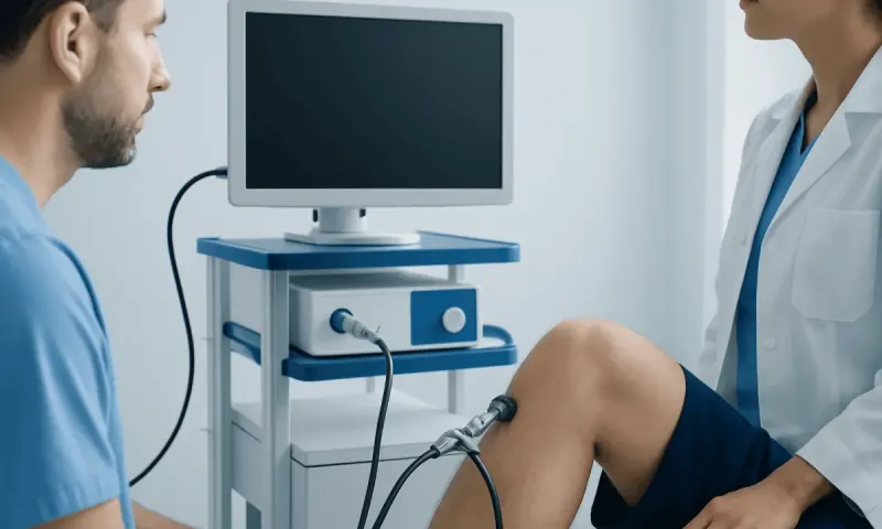
Ankylosing Spondylitis Diagnosis: Tests and Imaging

Ankylosing spondylitis diagnosis is based on a clinician’s assessment of symptoms, physical examination, blood tests and imaging of the sacroiliac joints and spine. No single test can confirm the condition in every person, so results are interpreted together over time.
Overview: How Ankylosing Spondylitis Is Diagnosed
Ankylosing spondylitis diagnosis usually involves a detailed discussion of symptoms, a physical examination, blood tests and imaging studies. The aim is to determine whether back or joint symptoms are caused by inflammation related to axial spondyloarthritis, rather than by muscle strain, disc problems, osteoarthritis or another health condition.
Ankylosing spondylitis is a form of inflammatory arthritis that mainly affects the sacroiliac joints, where the spine meets the pelvis, and the spine itself. It may also affect larger joints, tendons and areas outside the joints, including the eyes, skin and bowel. The term axial spondyloarthritis includes both ankylosing spondylitis, in which characteristic changes may be visible on X-ray, and non-radiographic axial spondyloarthritis, in which inflammation may be present without X-ray changes.
Diagnosis can take time because early symptoms may be subtle, may come and go, and can resemble common mechanical back pain. A rheumatologist is often the specialist best placed to bring together the medical history, examination and test results. A diagnosis does not depend on one result alone; it reflects the full clinical picture.
Symptoms and History That Guide Assessment
The consultation generally begins with questions about the pattern, duration and impact of symptoms. Inflammatory back pain often starts gradually before the age of 45, lasts for more than three months and feels worse after rest or during the second half of the night. Many people describe stiffness on waking that improves as they move, although these features alone do not confirm the condition.
The clinician may ask about pain in the buttocks that switches sides, neck or chest-wall discomfort, fatigue, and painful or swollen peripheral joints such as the hips, knees, ankles or shoulders. Pain at tendon attachment sites, especially the heel, can also be relevant. Symptoms such as a red, painful, light-sensitive eye, chronic diarrhea, abdominal symptoms or scaly skin rashes may provide useful clues because related inflammatory conditions can occur alongside axial spondyloarthritis.
Family history is important but is not required. The clinician may ask whether close relatives have ankylosing spondylitis, psoriasis, inflammatory bowel disease or recurrent eye inflammation. They will also consider daily activities, previous injuries, work demands, medications and other possible causes of pain.
Physical Examination and Clinical Findings

During the examination, the clinician assesses posture, walking pattern, spinal movement and the range of motion in the hips and other joints. They may observe how comfortably a person bends forward, turns the neck or expands the chest while breathing. These observations help identify areas of stiffness or restricted movement, but they are not sufficient on their own to establish a diagnosis.
The sacroiliac joints, spine, hips, heels and other symptomatic areas may be checked for tenderness, pain with movement, swelling or reduced flexibility. The clinician may also look for signs of psoriasis and ask about eye, bowel or urinary symptoms. Some examinations are used to monitor spinal mobility over time as well as to support the initial assessment.
Physical findings can be normal in early disease. This does not mean that symptoms are not real or that inflammation is excluded. Reassessment may be appropriate when symptoms persist, change or become more suggestive of inflammatory disease.
Blood Tests: What They Can and Cannot Show
Blood tests are used to look for evidence of inflammation and to help exclude other conditions. Erythrocyte sedimentation rate (ESR) and C-reactive protein (CRP) are common markers. Elevated values can support the presence of inflammation, but many people with ankylosing spondylitis have normal results, especially at certain stages of the condition.
Testing for HLA-B27, a genetic marker associated with axial spondyloarthritis, may be helpful when the symptoms and examination raise suspicion. However, many people who carry HLA-B27 never develop ankylosing spondylitis, and some people with the condition do not carry it. Therefore, an HLA-B27 result is interpreted alongside all other information rather than as a stand-alone diagnostic answer.
Other blood tests may be arranged to evaluate anemia, infection, autoimmune conditions or possible treatment considerations. A blood draw is brief and most people can return to normal activities immediately afterward. Results may be available at different times depending on the laboratory and the tests ordered.
Imaging: X-Rays, MRI and Other Scans
Imaging plays a central role in ankylosing spondylitis diagnosis, particularly imaging of the sacroiliac joints. Standard X-rays may show structural changes associated with sacroiliitis, including joint irregularity, narrowing or fusion. These changes can take years to become visible, so normal X-rays do not necessarily rule out early axial spondyloarthritis.
MRI can detect active inflammation and bone marrow changes before structural damage appears on X-ray. It is particularly useful when symptoms suggest inflammatory back pain but X-rays are inconclusive. MRI does not use ionizing radiation, but the scan requires lying still in a confined scanner for a period of time. People with implanted devices, metal fragments, severe claustrophobia or possible pregnancy should tell the imaging team in advance.
CT scans can show bone detail clearly but expose the body to more radiation than X-rays, so they are not routinely the first choice for early assessment. Ultrasound may help examine inflammation in peripheral joints or tendon attachment sites. Imaging results must be interpreted by experienced clinicians because not every abnormality seen on a scan is caused by inflammatory arthritis.
The Diagnostic Process, Recovery and Understanding Results
There is usually no physical recovery period after the diagnostic assessment. A clinical examination and blood tests are generally followed by normal daily activities. X-rays are quick, while MRI appointments take longer and may require screening questions beforehand. Sedation is not routinely needed for MRI, although individualized support may be discussed for people who are very anxious about the scan.
Results are reviewed in context. A clinician considers whether the symptoms have an inflammatory pattern, whether examination or imaging shows sacroiliac or spinal involvement, whether inflammatory markers are raised, and whether HLA-B27 or related conditions add support. It is possible to have inflammatory symptoms with normal blood tests, or to have HLA-B27 without having ankylosing spondylitis.
Some people receive a diagnosis of non-radiographic axial spondyloarthritis rather than ankylosing spondylitis when there are clinical features and MRI evidence of inflammation but no definite X-ray changes. Follow-up may be used to monitor symptoms and function, clarify uncertain findings and adjust management. The broader condition is explained in ankylosing spondylitis information for people seeking an overview of symptoms and care.
What Happens After Diagnosis
Once a diagnosis is made, the care plan is tailored to symptoms, inflammation, general health and personal goals. Regular movement, physiotherapy and targeted exercises are commonly important for maintaining posture, flexibility, strength and function. A physiotherapist can help select exercises that are safe and realistic for the individual.
Medication may be considered to reduce pain and inflammation and, for some people, to control ongoing active disease. Options can include anti-inflammatory medicines and, when appropriate, advanced therapies prescribed and monitored by a rheumatology team. Treatment decisions should account for other medical conditions, pregnancy plans, infection risk and potential side effects.
People may also benefit from coordinated care when eye, skin or bowel symptoms are present. Acıbadem Health Point’s multidisciplinary specialists and JCI-accredited hospitals diagnose and treat ankylosing spondylitis for international patients, with care plans developed around individual clinical needs.
When to Seek Medical Care
A medical assessment is advisable for back pain lasting more than three months, particularly if it began before age 45, causes prolonged morning stiffness, wakes a person during the night or improves more with movement than with rest. Prompt evaluation is also sensible for persistent buttock pain, unexplained joint swelling, recurring heel pain or a family history of spondyloarthritis-related conditions.
Urgent medical advice is needed for a painful red eye with blurred vision or sensitivity to light, as this may require prompt eye assessment. Immediate care is also appropriate for new weakness, numbness around the genital or anal area, loss of bladder or bowel control, fever with severe back pain, significant unexplained weight loss, or pain after major trauma. These symptoms may have causes other than ankylosing spondylitis and should not be self-managed.
Keeping a brief record of symptom timing, stiffness, sleep disruption, activity effects and other health changes can help make the consultation more productive. A qualified clinician can determine which tests are useful and whether referral to rheumatology is appropriate.
Frequently asked questions
01Is there one test that confirms ankylosing spondylitis?
No. Ankylosing spondylitis diagnosis is based on the combined findings from symptoms, physical examination, imaging and laboratory tests. HLA-B27 and inflammatory markers can provide useful information, but neither confirms nor excludes the condition by itself.
02Can an X-ray be normal if someone has ankylosing spondylitis?
Yes, especially early in the disease process. X-rays primarily show structural changes that can develop gradually. MRI may identify active inflammation in the sacroiliac joints before changes can be seen on an X-ray.
03What does a positive HLA-B27 test mean?
A positive HLA-B27 result means a person carries a genetic marker associated with a higher likelihood of some spondyloarthritis conditions. It does not mean that the person definitely has ankylosing spondylitis, because many HLA-B27-positive people never develop it. The result is most useful when interpreted with symptoms, examination and imaging.
04How long does ankylosing spondylitis diagnosis take?
The timing varies. Initial tests may be arranged promptly, but interpretation and specialist review can take longer, particularly if MRI or follow-up observation is needed. Diagnosis may be more straightforward when imaging and symptoms are clear, while early or atypical cases can require ongoing evaluation.
05Do normal CRP and ESR results rule out ankylosing spondylitis?
No. CRP and ESR can be normal in people with active axial spondyloarthritis or ankylosing spondylitis. Clinicians use these markers as part of the overall assessment rather than relying on them alone.
06Which doctor diagnoses ankylosing spondylitis?
A rheumatologist commonly confirms and manages ankylosing spondylitis because they specialize in inflammatory joint and connective tissue conditions. A primary care clinician, orthopedic specialist, physiotherapist, ophthalmologist or gastroenterologist may recognize suggestive symptoms and arrange referral. Collaborative care may be needed when symptoms affect the eyes, skin or digestive system.
This article is for general information only and is not a substitute for professional medical advice. Please consult a qualified doctor about your individual situation.
Persistent digestive symptoms? Get evaluated in Turkey
JCI-accredited · board-certified surgeons · reply within 24h
Add us as a Preferred Source to see more of our trusted health content across Google Search, AI Overviews and Discover.