Angiography vs Echocardiography: What Each Shows

Angiography and echocardiography answer different heart-health questions. Angiography is designed to show blood flow and narrowing in arteries, while echocardiography uses ultrasound to evaluate the heart muscle, chambers, valves and pumping function.
Angiography vs Echocardiography: a side-by-side comparison
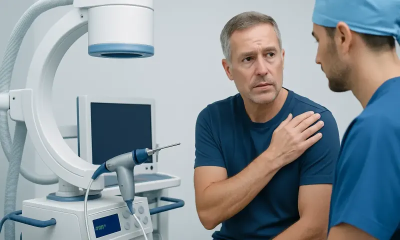
Angiography vs echocardiography is not usually a choice between two interchangeable tests. An echocardiogram is a noninvasive ultrasound examination that shows the heart’s structure and movement. Coronary angiography is an invasive imaging procedure that uses contrast dye and X-ray imaging to show the inside of the coronary arteries, which supply blood to the heart muscle.
The test a clinician recommends depends on what needs to be clarified: heart pumping ability, valve disease, an enlarged chamber, possible fluid around the heart, suspected artery narrowing, or another concern. Often, an echocardiogram is an early test, while angiography is considered when symptoms, risk assessment or other test findings suggest important coronary artery disease.
| Feature | Echocardiography | Coronary angiography |
|---|---|---|
| Main purpose | Assesses heart muscle, chambers, valves and pumping function | Shows narrowing or blockage in coronary arteries |
| How it works | Sound waves create moving images of the heart | Contrast dye and X-rays outline blood vessels |
| Invasiveness | Usually noninvasive; transesophageal echo uses a probe in the esophagus | Invasive; a catheter is passed through an artery |
| What it can identify | Reduced ejection fraction, valve problems, structural changes, fluid around the heart | Location and severity of coronary artery narrowing |
| Can treatment be performed at the same time? | No | Sometimes, such as angioplasty and stent placement when appropriate |
| Preparation and recovery | Usually minimal preparation and no recovery time for a standard scan | Requires preparation, monitoring and recovery after the procedure |
Both tests are widely used and are generally safe when ordered for an appropriate reason. They provide different types of information, so one test may complement rather than replace the other.
How clinicians tell the tests apart
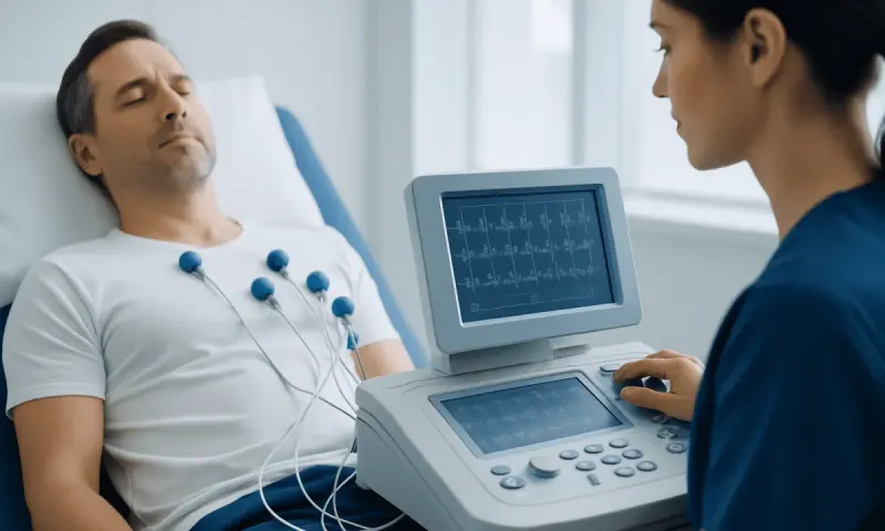
Clinicians begin with the person’s symptoms, medical history, examination findings and cardiovascular risk factors. Chest discomfort with exertion, shortness of breath, an abnormal electrocardiogram, a previous heart attack, diabetes, high blood pressure, smoking history or a strong family history may influence which investigations are useful.
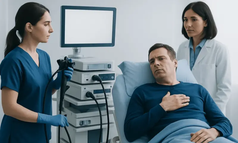
An echocardiogram is especially helpful when a clinician needs to understand how the heart is working. It can measure the strength of contraction, assess whether heart chambers are enlarged, examine valve opening and closing, and identify patterns of heart-muscle movement that may occur after an injury to the heart.
Angiography is used when there is a need to see the coronary arteries directly, particularly if the likelihood of clinically significant narrowing is high or an urgent intervention may be needed. Depending on the situation, a clinician may also recommend noninvasive coronary CT angiography, exercise testing, stress echocardiography, blood tests or other cardiac imaging before invasive angiography.
Which is better, echo or angiography?

Neither is inherently better because they are designed for different purposes. Echocardiography is usually the better test for evaluating a suspected valve condition, heart failure, an abnormal heart sound, a heart-muscle problem or the cause of some forms of breathlessness. It is often a convenient first assessment because standard transthoracic echocardiography does not require catheters, radiation or contrast dye.
Coronary angiography is the more appropriate test when the main question is whether coronary arteries have a narrowing that may be causing reduced blood supply to the heart. It is particularly valuable in some people with suspected acute coronary syndrome, persistent concerning symptoms, or high-risk findings on other tests.
A person should not try to select a test based only on symptoms found online. A cardiologist considers the urgency of symptoms, kidney function, allergy history, bleeding risk, prior test results and whether the result is likely to change treatment. In selected cases, coronary angiography can provide a diagnosis and allow treatment planning during the same episode of care.
Does an echocardiogram show if your arteries are clogged?
A standard echocardiogram does not directly show plaque or narrowing inside the coronary arteries. The coronary vessels are small and sit on the surface of the heart, so they are not routinely assessed in enough detail by standard ultrasound to confirm or rule out a blockage.
However, an echocardiogram may show indirect signs that reduced blood flow has affected the heart. For example, part of the heart muscle may move less strongly than expected, or the overall pumping function may be reduced. These findings can have several causes and do not, by themselves, prove that an artery is blocked.
A stress echocardiogram can provide additional information by comparing heart-muscle movement at rest and during exercise or medication-induced stress. If reduced blood supply is suspected, a clinician may use this result alongside other assessments to decide whether coronary CT angiography or invasive angiography is needed. Coronary artery disease may be discussed alongside related conditions such as coronary artery disease.
What is the most accurate test for a heart blockage?
For identifying and defining narrowing in the coronary arteries, invasive coronary angiography is commonly regarded as the reference test. It provides detailed, real-time images of the artery lumen and helps clinicians assess the location and apparent severity of narrowing. In some cases, additional catheter-based measurements or imaging can clarify whether a narrowing is limiting blood flow.
Accuracy must be considered in context. A narrowing seen on an angiogram does not always mean that it is causing symptoms or significantly reducing blood flow, and clinicians may use physiological measurements during the procedure when needed. Conversely, a person’s symptoms and overall risk profile remain important even when a single test is reassuring.
Coronary CT angiography is a useful noninvasive alternative for many people with stable symptoms and an appropriate likelihood of coronary disease. It can be highly informative, but image quality may be affected by factors such as heart rhythm, extensive coronary calcium or difficulty holding still. The best test is therefore the one that safely answers the relevant clinical question for the individual.
Which type of echocardiogram is the most accurate?
There is no single most accurate echocardiogram for every purpose. The standard transthoracic echocardiogram, performed with an ultrasound probe on the chest, is the usual starting point. It provides important information about heart size, pumping function, valve movement and fluid around the heart without sedation or invasive instruments.
Transesophageal echocardiography places an ultrasound probe in the esophagus, which lies close to the heart. This can produce more detailed views of certain structures, particularly some heart valves, the left atrium, the aorta and possible clots in selected situations. It is more invasive than a transthoracic scan and may require fasting and sedation.
Stress echocardiography is tailored to assess how the heart responds to increased workload and can help evaluate suspected reduced blood flow. Other advanced methods, including contrast echocardiography and three-dimensional echocardiography, may be used for specific questions. The most suitable technique is determined by the suspected condition and the quality of images obtained from the initial scan.
What to expect before, during and after each test
For a standard transthoracic echocardiogram, the person usually changes into a gown and lies on an examination couch. Gel is applied to the chest, and a sonographer moves a handheld probe across several positions. The scan is painless, typically takes less than an hour, and normal activities can generally resume immediately unless other tests or medical instructions apply.
Preparation for angiography may include fasting for a period, reviewing medications and checking kidney function and blood tests. A local anesthetic is used where the catheter enters an artery, commonly at the wrist or groin. The person may feel warmth when contrast dye is injected, but the procedure is closely monitored by a specialist team.
After angiography, staff monitor the access site, heart rhythm and general recovery. Instructions may include drinking fluids if appropriate, avoiding heavy lifting for a short period and watching for bleeding, swelling, increasing pain or color changes in the limb used for access. The treating team should provide individualized aftercare advice and explain when normal activities can resume.
When to seek medical care
New chest pressure, tightness, heaviness or pain should be assessed promptly, particularly if it occurs during exertion or is accompanied by shortness of breath, sweating, nausea, dizziness, fainting, or pain spreading to the arm, jaw, back or upper abdomen. Emergency services should be contacted immediately for severe, sudden or persistent symptoms, rather than driving oneself to a hospital.
A nonurgent medical appointment is appropriate for recurring chest discomfort, a decline in exercise tolerance, palpitations, unexplained breathlessness, ankle swelling or a newly detected heart murmur. These symptoms have many possible causes, and timely assessment can help identify the right next step.
Acıbadem Health Point’s multidisciplinary specialists and JCI-accredited hospitals evaluate and treat cardiovascular conditions for international patients, with testing selected according to individual clinical needs. A qualified clinician can explain the likely benefits, limitations and risks of echocardiography, angiography and alternative tests before a decision is made.
Frequently asked questions
01Is an echocardiogram painful?
A standard transthoracic echocardiogram is usually painless. The ultrasound probe may be pressed firmly against the chest to obtain clear images, but it does not enter the body. A transesophageal echocardiogram is more invasive and may cause temporary throat discomfort afterward.
02Is coronary angiography the same as a heart catheterization?
Coronary angiography is commonly performed as part of a cardiac catheterization procedure. A thin catheter is guided through an artery to the heart, and contrast dye is injected to make the coronary arteries visible on X-ray. Catheterization can also include other measurements or treatments when clinically needed.
03Can an echocardiogram detect a previous heart attack?
An echocardiogram may show an area of heart muscle that is not moving normally or is weaker, which can occur after a heart attack. These findings are not specific to a previous heart attack and need interpretation with symptoms, ECG findings and other tests. It cannot always identify the timing or exact cause of prior heart-muscle injury.
04Why would a doctor order an echocardiogram before angiography?
An echocardiogram can quickly assess heart pumping function, valves and structural conditions without using radiation, contrast dye or a catheter. Its findings may help clarify whether symptoms could be related to heart-muscle or valve disease. It may also help determine whether further coronary artery testing is appropriate.
05How long does it take to get angiography results?
The physician can often review angiography images during the procedure and discuss initial findings soon afterward. The timing of a complete explanation depends on the clinical situation and whether additional measurements, treatment or observation are required. The care team should explain the results and next steps before discharge whenever possible.
06Can a normal echocardiogram rule out heart disease?
No. A normal echocardiogram is reassuring about many structural and functional aspects of the heart, but it does not rule out every heart condition. In particular, it cannot reliably exclude coronary artery narrowing, early coronary artery disease or certain rhythm disorders. Further assessment may be needed if symptoms or risk factors remain concerning.
This article is for general information only and is not a substitute for professional medical advice. Please consult a qualified doctor about your individual situation.
Heart care in Turkey — expert evaluation and treatment
JCI-accredited · board-certified surgeons · reply within 24h
Add us as a Preferred Source to see more of our trusted health content across Google Search, AI Overviews and Discover.