Abdominal CT Scan for Pain, Injury, and Diagnosis

An abdominal CT scan is a fast imaging test that combines multiple X-ray images to show detailed cross-sectional views of the abdomen and nearby pelvic structures. It may help identify inflammation, injury, infection, stones, bleeding, tumors and other causes of symptoms, often with or without contrast material.
Overview: what is an abdominal CT scan?
An abdominal CT scan, also called computed tomography, uses a rotating X-ray scanner and computer processing to create detailed, slice-like images of the belly area. It can examine organs, muscles, fat, bones, blood vessels and lymph nodes in much greater detail than a standard X-ray. Depending on the reason for the test, the scan may include the pelvis as well.
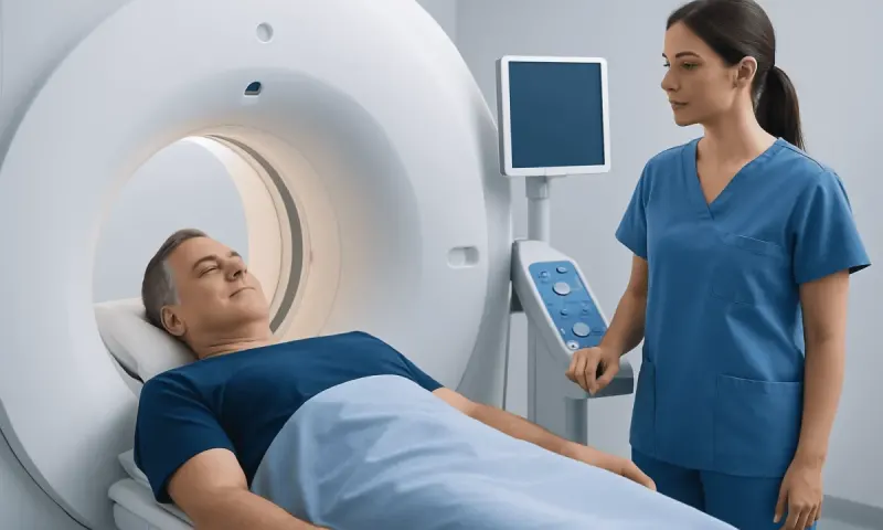
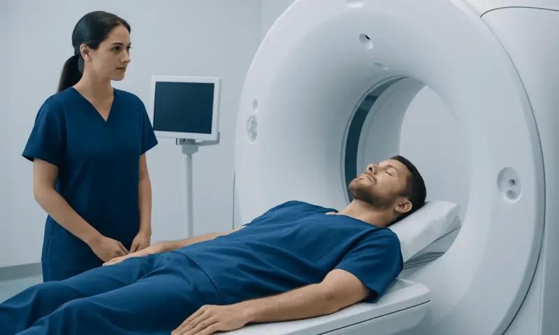
The test is commonly requested to investigate ongoing or sudden abdominal symptoms, assess an injury, look for infection or inflammation, or monitor a known condition. It is also used to help plan treatment or guide certain procedures. The scan is noninvasive: the person lies on a movable table that passes through a short, ring-shaped scanner.
Some abdominal CT examinations use contrast material, usually an iodine-based dye given through a vein and sometimes an oral liquid. Contrast can make blood vessels, organs and areas of inflammation easier to distinguish. The referring clinician and radiology team decide whether contrast is needed based on the clinical question and the person’s medical history.
What organs will a CT of the abdomen show?
A CT of the abdomen can show the liver, gallbladder, bile ducts, pancreas, spleen, stomach, small intestine, large intestine, kidneys, adrenal glands, bladder and upper reproductive structures. When the scan includes the pelvis, it may also show the lower urinary tract and pelvic organs in more detail.
It can also assess the abdominal aorta and other blood vessels, lymph nodes, abdominal wall, spine, ribs and surrounding soft tissues. The amount of detail for each structure depends on the scan protocol, whether contrast is used and factors such as bowel movement or body position.
CT imaging helps clinicians look for changes such as swelling, fluid collections, internal bleeding, stones, obstruction, injury, abscesses and masses. A scan result must always be interpreted together with symptoms, examination findings and, when relevant, blood or urine tests. An abnormal image does not by itself establish a diagnosis.
Why an abdominal CT scan may be recommended

A clinician may recommend an abdominal CT scan when symptoms need prompt clarification or when another test has not provided enough information. Common reasons include persistent or severe abdominal pain, unexplained fever, nausea and vomiting, changes in bowel habits, blood in the urine, jaundice, unexplained weight loss or concern after an abdominal injury.
CT can help evaluate conditions involving the digestive tract, urinary system, liver and biliary system. For example, it may support assessment of appendicitis, diverticulitis, kidney stones, pancreatitis, bowel obstruction or complications of inflammatory disease. The exact test chosen depends on the suspected condition; ultrasound, MRI, endoscopy or laboratory tests may be more suitable in some situations.
For people with a known diagnosis, CT may be used to assess the extent of disease, monitor a response to treatment or check for complications. Clinicians aim to use imaging only when the expected information is likely to influence care, particularly because CT involves a small amount of ionizing radiation.
What should you not do before an abdominal CT scan?
Patients should not assume that one preparation plan applies to every abdominal CT scan. Instructions vary according to the body area being studied and whether oral or intravenous contrast is planned. The imaging center may ask the patient not to eat for several hours beforehand, while clear fluids may still be allowed. Following the instructions provided by the care team is important.
Before the scan, patients should not forget to tell the team about a previous reaction to contrast material, asthma, kidney disease, thyroid disease, diabetes, pregnancy or possible pregnancy, and all medicines or supplements they take. Some medicines may need individual advice before contrast is administered; patients should not stop prescribed medicine unless their clinician tells them to do so.
Metal objects can affect image quality. Jewelry, belts, piercings and clothing with metal fasteners may need to be removed, and a hospital gown may be offered. Patients should also avoid arriving dehydrated if they have been permitted to drink fluids. If oral contrast is prescribed, it should be taken exactly as directed.
- Do not eat or drink contrary to the center’s specific instructions.
- Do not omit information about allergies, kidney problems or pregnancy.
- Do not stop regular medicines without professional advice.
- Do not wear removable metal items into the scanner room.
The procedure: what happens during the scan?
On arrival, the radiology team confirms the patient’s identity, reason for the test and relevant health information. If intravenous contrast is required, a small cannula is placed in a vein, usually in the arm or hand. The person lies on their back on a padded table, and positioning aids may be used to help them remain still.
During imaging, the table moves slowly through the open center of the CT scanner. The radiographer operates the scanner from an adjacent room but can see, hear and speak with the patient throughout. The team may ask the patient to hold their breath briefly, since movement can blur images.
When contrast enters the bloodstream, some people notice a warm flush, a metallic taste in the mouth or a brief feeling of needing to urinate. These sensations generally pass quickly. The scan itself does not hurt and commonly takes only minutes, although the full appointment may take longer because of preparation and observation.
What stomach problems show up on a CT scan?
A CT scan can identify some structural stomach problems, including thickening of the stomach wall, inflammation around the stomach, a perforation with free air, bleeding in certain circumstances, a large hernia, obstruction at the stomach outlet and a mass. It can also show effects on nearby organs, lymph nodes or tissues.
However, CT does not directly view the stomach lining as closely as an endoscopy. Small ulcers, mild gastritis and early lining changes may not be visible on CT. If symptoms suggest an ulcer, reflux-related damage, persistent indigestion or unexplained upper gastrointestinal bleeding, a clinician may recommend blood tests, stool testing or upper endoscopy in addition to, or instead of, CT.
CT findings are assessed in context. For example, abdominal pain and vomiting may arise from the stomach, pancreas, gallbladder, bowel or another organ, and imaging helps narrow the possibilities rather than replacing a clinical assessment. If stomach symptoms are persistent, recurrent or worsening, medical review is advisable.
What does an abdominal CT show that an ultrasound doesn't?
Both CT and ultrasound are useful, but they provide different information. Ultrasound uses sound waves and is especially valuable for examining the gallbladder, bile ducts, kidneys, pelvic organs and blood flow without radiation. It is often the first imaging test for many abdominal concerns.
An abdominal CT generally provides a wider field of view and more consistent detail of deep abdominal structures, bowel, pancreas, blood vessels, retroperitoneal tissues, bones and areas obscured by bowel gas. It can better show the extent of trauma, internal bleeding, abscesses, many tumors, complex infections, bowel obstruction and some complications of inflammatory conditions.
CT is not automatically better for every question. Ultrasound may be preferred in pregnancy, for children when appropriate, or when the clinical concern is well suited to ultrasound. MRI may provide added detail for certain liver, biliary, pancreatic or pelvic questions. The clinician selects the test that best balances diagnostic value, safety and the individual’s circumstances.
Recovery, results and when to seek medical care
Most people can return to their usual activities immediately after an abdominal CT scan. Those who received contrast are often advised to drink fluids afterward if this is medically appropriate, as hydration supports normal clearance through the kidneys. The radiology team will give individual instructions, especially for people with kidney disease, fluid restrictions or other health conditions.
A radiologist reviews the images and prepares a report for the clinician who ordered the examination. Timing varies by setting and urgency. The ordering clinician explains what the findings mean, whether additional tests are needed and what next steps, if any, are appropriate. A normal CT result can be reassuring, but it may not explain every symptom or rule out every condition.
Patients should seek urgent medical care for sudden severe or worsening abdominal pain, a rigid or markedly swollen abdomen, repeated vomiting, vomiting blood, black or bloody stools, fainting, chest pain, high fever with serious illness, or yellowing of the skin and eyes with severe symptoms. After contrast, urgent assessment is needed for trouble breathing, swelling of the face or throat, widespread hives or severe dizziness, as these may indicate a rare serious reaction.
For international patients, Acıbadem Health Point’s multidisciplinary specialists and JCI-accredited hospitals can coordinate diagnostic assessment and treatment planning when abdominal symptoms require further evaluation.
Frequently asked questions
01Is an abdominal CT scan painful?
The scan itself is painless. Lying still on the table and holding the breath briefly may be mildly uncomfortable for some people. If contrast is used, a short-lived warm sensation or metallic taste can occur, but this usually passes quickly.
02How long does an abdominal CT scan take?
The actual image acquisition often takes only a few minutes. The full appointment may take longer for registration, preparation, possible contrast administration and instructions afterward. The imaging center can provide a more specific estimate for the planned scan.
03Do you have to fast for an abdominal CT scan?
Fasting requirements depend on the type of CT scan and whether contrast material is needed. Some patients may be asked not to eat for several hours before the examination, while others may not need to fast. Patients should follow the instructions from their imaging provider rather than making changes on their own.
04Can an abdominal CT scan detect cancer?
An abdominal CT scan can identify masses, enlarged lymph nodes or other changes that may raise concern for cancer and can help assess their location and extent. It cannot always confirm the cause of an abnormality. Further imaging, blood tests, endoscopy or a biopsy may be needed to establish a diagnosis.
05Is contrast dye safe for an abdominal CT scan?
Contrast material is used routinely and is safe for most people, but reactions and kidney-related concerns can occur in some circumstances. The radiology team checks for prior contrast reactions, kidney problems and other relevant conditions beforehand. Patients should report any symptoms during or after the injection promptly.
06When will abdominal CT scan results be available?
A radiologist must review the images and issue a report, so results may be available the same day in urgent settings or after a longer interval for routine examinations. The clinician who ordered the scan is best placed to explain the findings in relation to the person’s symptoms and health history.
This article is for general information only and is not a substitute for professional medical advice. Please consult a qualified doctor about your individual situation.
Persistent digestive symptoms? Get evaluated in Turkey
JCI-accredited · board-certified surgeons · reply within 24h
Add us as a Preferred Source to see more of our trusted health content across Google Search, AI Overviews and Discover.