Abdomen Ultrasound vs CT Scan: Which Is Used?
For abdomen ultrasound vs CT scan, neither test is always better: ultrasound is often used first for organs such as the gallbladder, liver and kidneys, while CT gives broader, more detailed cross-sectional images. The right test depends on the symptoms, suspected condition, medical history and whether urgent information is needed.
Overview: Abdomen Ultrasound vs CT Scan
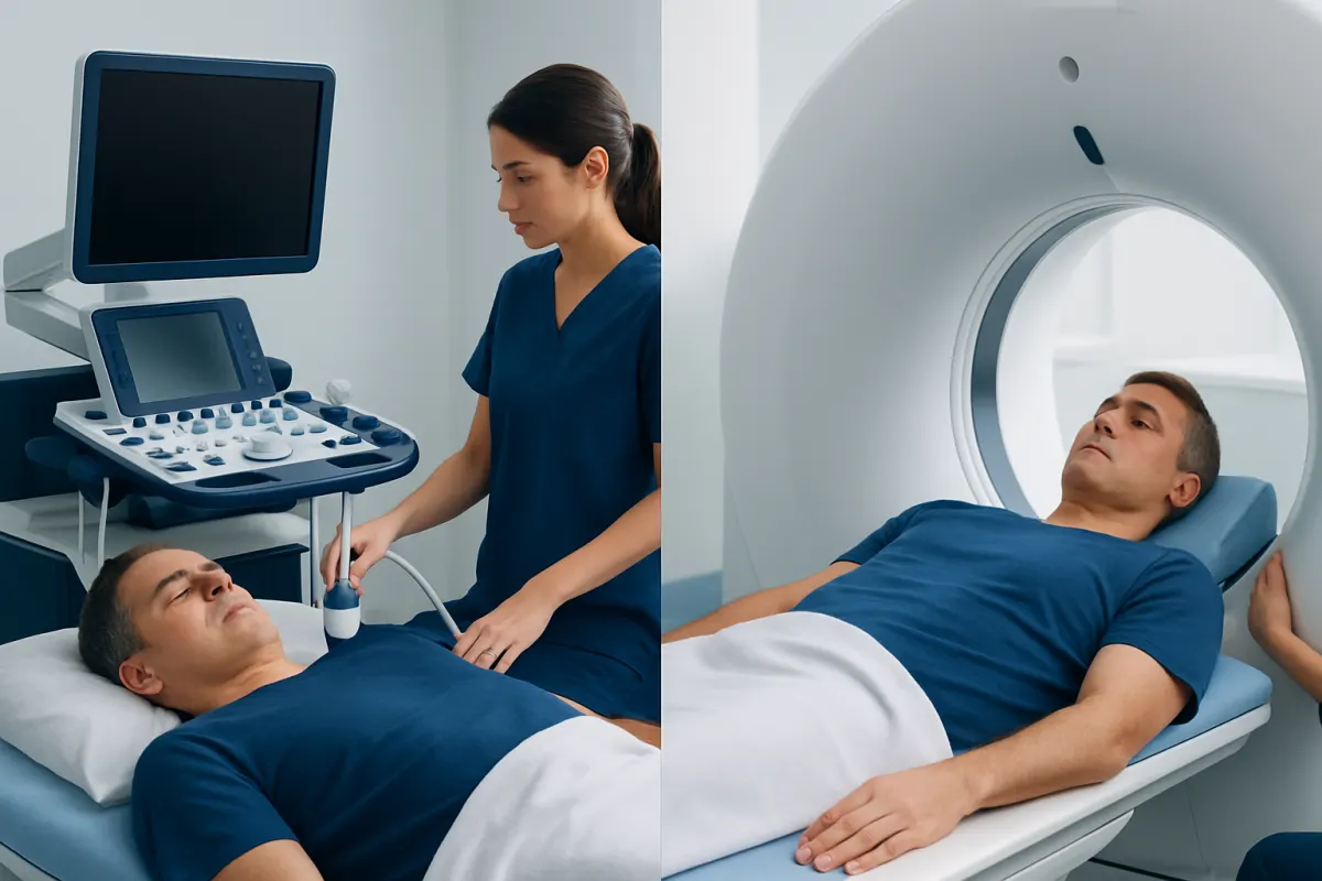
In an abdomen ultrasound vs CT scan comparison, ultrasound is usually the simpler, radiation-free examination, whereas computed tomography (CT) produces more detailed images of the abdomen and pelvis using X-rays. Both are valuable diagnostic tools, but they answer different clinical questions and may be used at different stages of assessment.
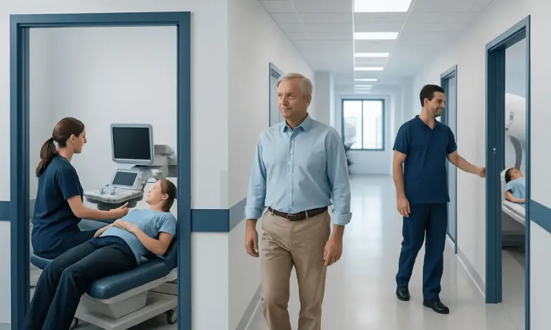
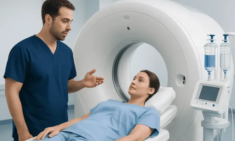
An abdominal ultrasound uses a handheld probe moved across the skin. Sound waves create real-time images of organs and blood flow. A CT scan involves lying still on a table that passes through a ring-shaped scanner, producing layered images that can be reviewed in multiple directions.
The best option depends on where the pain or other symptom is located, how suddenly it began, the person’s age, pregnancy status, kidney function, previous imaging and the condition the clinician is considering. In some situations, the two tests complement each other rather than compete.
Side-by-Side Comparison: Abdominal Ultrasound and CT
The following comparison can help patients understand why a clinician may recommend one scan before another. It does not replace an individual medical assessment.
- How it works: Ultrasound uses sound waves; CT uses X-rays and computer processing.
- Radiation: Ultrasound has no ionizing radiation. CT involves ionizing radiation.
- Detail and coverage: CT generally provides wider and more detailed views of the abdomen, pelvis, bowel, blood vessels and surrounding tissues.
- Strengths: Ultrasound is highly useful for gallbladder, bile duct, liver, kidneys, bladder, pelvic organs and fluid assessment. CT is often useful for appendicitis, bowel conditions, pancreatitis complications, trauma, abscesses, stones and internal bleeding.
- Speed: Both can be quick, but CT is commonly favored in emergencies because it can rapidly survey several organ systems.
- Preparation: Fasting may be requested before an abdominal ultrasound. CT may require oral or intravenous contrast, depending on the question being investigated.
Image quality with ultrasound can be affected by bowel gas, body shape and the depth of the organ being examined. CT is less limited by gas and can show deeper structures more consistently, although it may not be the first choice when avoiding radiation is particularly important.
Results are interpreted alongside the patient’s symptoms, physical examination and laboratory tests. A normal scan does not always rule out every possible cause of abdominal symptoms, and follow-up testing may occasionally be needed.
How Clinicians Choose Between the Tests

Clinicians begin by identifying the most likely source of symptoms. Pain in the right upper abdomen after meals, for example, may lead to ultrasound because it is very effective at detecting gallstones and assessing the gallbladder. Severe, diffuse or uncertain abdominal pain may lead to CT because it provides a broader overview of the abdomen and pelvis.
The timing and severity of symptoms also matter. Sudden severe pain, fever, repeated vomiting, significant tenderness, concern about infection or a possible injury may require rapid CT assessment. For non-urgent symptoms involving the liver, gallbladder, kidneys or pelvic structures, ultrasound may provide the needed information without radiation exposure.
Personal factors guide the decision as well. Pregnancy, childhood and a history of repeated radiation-based imaging may favor ultrasound when it can answer the clinical question. If contrast is being considered, the clinician will also review allergies, kidney health, diabetes medications and other relevant medical conditions.
For people with suspected gallbladder inflammation or stones, ultrasound is commonly the first imaging examination. Further evaluation may be needed if symptoms remain concerning despite unclear initial results, particularly when gallstones or bile duct blockage is suspected.
Which Is More Accurate for Diagnosing Abdominal Pain, Ultrasound or CT Scan?
CT is often more accurate overall when the cause of abdominal pain is uncertain because it can evaluate many organs, the bowel, blood vessels, lymph nodes and tissues outside the organs in one examination. It is frequently used when clinicians need to assess for appendicitis, diverticulitis, bowel obstruction, kidney stones, infection, injury or complications involving several parts of the abdomen.
However, accuracy depends on the suspected cause, not only on the technology. Ultrasound may be the more appropriate and highly accurate first test for gallstones, gallbladder inflammation, bile duct enlargement, fluid collections, certain kidney problems and many pelvic conditions. It can also assess movement and tenderness in real time while the probe is applied.
For abdominal pain, a clinician may start with ultrasound and then order CT if the result is inconclusive or does not explain ongoing symptoms. In other cases, CT is selected first because the pattern of symptoms suggests a condition that is better assessed with cross-sectional imaging.
Neither examination should be interpreted in isolation. Laboratory results, such as liver tests, inflammation markers or urine testing, can help make imaging findings more meaningful and guide next steps.
What Can a CT Scan Show That an Ultrasound Does Not?
A CT scan can provide a more complete view of deep abdominal and pelvic structures, particularly where bowel gas limits ultrasound images. It is often better at identifying inflammation around the appendix, bowel wall changes, bowel obstruction, perforation, abdominal abscesses, internal bleeding and injuries to organs after trauma.
CT can also give more detailed information about the pancreas, retroperitoneum, lymph nodes and tissues surrounding abdominal organs. It may reveal the size, location and relationship of a mass or fluid collection to nearby structures, which can be important when planning further testing or treatment.
When contrast is used appropriately, CT can assess blood vessels and organ blood supply. This may be relevant when clinicians are concerned about blood clots, aneurysms, reduced blood flow to the bowel or active bleeding. Contrast decisions are individualized because it may not be appropriate for everyone.
CT is also commonly used to investigate suspected appendicitis when symptoms and examination findings are concerning. The scan can identify an inflamed appendix and may also show alternative explanations for lower abdominal pain.
Why Don't Doctors Like to Do CT Scans?
Doctors do value CT scans and use them when the information is likely to change care. The main reason they avoid unnecessary CT is that it uses ionizing radiation. Although the benefit of an appropriately indicated CT can outweigh this risk, clinicians aim to keep radiation exposure as low as reasonably achievable, especially for children, pregnant patients and people likely to need repeated imaging.
Some CT examinations use iodine-based contrast material, which can rarely cause an allergic-type reaction. Contrast can also require extra caution in people with significantly reduced kidney function or certain other health conditions. The imaging team reviews relevant history and may adjust the plan when needed.
CT can sometimes detect unexpected findings that are harmless but need follow-up, creating worry or additional tests. This is another reason clinicians select CT thoughtfully rather than using it automatically for every episode of abdominal discomfort.
When CT is medically needed, it is a well-established test that can provide important, sometimes urgent information. Patients should tell the care team about pregnancy, possible pregnancy, prior contrast reactions, kidney disease and any medicines they take before the examination.
What Does an Abdominal Ultrasound Show That a CT Doesn't?
Ultrasound offers real-time imaging, which can show movement and blood flow in a way that is particularly useful for certain questions. Doppler ultrasound can assess the direction and speed of blood flow in vessels, and a sonographer may observe structures while the patient changes position or takes a breath.
For the gallbladder, ultrasound is particularly helpful because it can show small gallstones, sludge, gallbladder wall changes and tenderness directly under the probe. It may also show widening of bile ducts, which can suggest impaired bile flow. In many cases, this makes it the preferred starting scan for right upper abdominal pain.
Ultrasound can distinguish fluid-filled from solid areas in many organs and can guide procedures such as drainage or biopsy. It is portable, may be performed at the bedside, and does not require radiation. These features are valuable for pregnant people and for patients who need repeat imaging.
CT may still be used when ultrasound findings are uncertain, when a deeper or wider evaluation is required, or when clinicians are looking for complications beyond the area visible on ultrasound. The tests provide different kinds of information, and one is not universally superior to the other.
Preparing for Imaging and When to Seek Medical Care
Preparation instructions vary. For an abdominal ultrasound, patients may be asked not to eat for several hours because a full gallbladder and reduced bowel gas can improve images. For some pelvic ultrasound examinations, a full bladder may be requested. For CT, patients may need to avoid food for a period of time if contrast is planned, but they should follow the instructions provided by the imaging center.
Patients should bring information about prior scans and tell the team about allergies, kidney disease, pregnancy or possible pregnancy, and implanted medical devices. They should also ask whether contrast will be used and when results will be available. A qualified clinician can explain what the findings mean in the context of the individual’s symptoms.
Urgent medical assessment is appropriate for severe or worsening abdominal pain, pain with a rigid or very tender abdomen, fainting, confusion, persistent vomiting, vomiting blood, black or bloody stools, high fever, yellowing of the eyes or skin, chest pain, shortness of breath, or abdominal pain during pregnancy. New pain after a significant injury also needs prompt evaluation.
For international patients, Acıbadem Health Point’s multidisciplinary specialists and JCI-accredited hospitals can assess abdominal symptoms and arrange appropriate diagnostic imaging and treatment planning when needed.
Frequently asked questions
01Is an abdominal ultrasound safer than a CT scan?
Ultrasound does not use ionizing radiation, so it is often preferred when it can answer the clinical question, including during pregnancy. CT uses radiation, but it is considered appropriate when its diagnostic benefit is important. The care team weighs the expected benefit against potential risks for each patient.
02Can an ultrasound miss problems in the abdomen?
Yes. Bowel gas, body shape, deep structures and the location of a problem can limit ultrasound images. Ultrasound may be less informative for some bowel, pancreatic and deep pelvic conditions. If symptoms remain concerning, a clinician may recommend CT, MRI or other tests.
03Do all abdominal CT scans require contrast dye?
No. Whether contrast is used depends on the reason for the scan. Contrast may improve the assessment of blood vessels, inflammation, tumors and some organs, while non-contrast CT can be useful for concerns such as certain kidney stones. The radiologist and referring clinician select the protocol most suitable for the question.
04How long do abdominal ultrasound and CT scans take?
The actual scan is often completed within minutes, although appointment time may be longer because of registration, preparation and, for CT, possible contrast administration. Ultrasound duration can vary depending on how many organs need to be assessed and whether extra views are needed. The imaging center can provide more specific timing.
05Can I eat before an abdominal ultrasound or CT scan?
Patients may be asked to fast before an abdominal ultrasound, particularly when the gallbladder or upper abdomen is being evaluated. CT preparation varies according to whether contrast is planned and the type of scan requested. It is important to follow the specific instructions from the imaging department rather than making changes independently.
06Will a CT or ultrasound explain every type of abdominal pain?
Not always. Abdominal pain can arise from many conditions, including problems that may not be visible on a single imaging test. Medical history, examination, blood and urine tests, and sometimes repeat or different imaging help clinicians reach a diagnosis. Ongoing, severe or worsening pain should be reviewed by a healthcare professional.
This article is for general information only and is not a substitute for professional medical advice. Please consult a qualified doctor about your individual situation.
Persistent digestive symptoms? Get evaluated in Turkey
JCI-accredited · board-certified surgeons · reply within 24h
Add us as a Preferred Source to see more of our trusted health content across Google Search, AI Overviews and Discover.