Leukoplakia: Symptoms, Causes and Treatment

Key Takeaways
- Leukoplakia describes a white patch in the mouth that does not rub off easily.
- Tobacco use, alcohol, chronic irritation, and some oral habits can increase risk.
- A dentist or doctor may recommend close follow-up, a biopsy, or treatment depending on the appearance and location.
- Stopping tobacco, limiting alcohol, and addressing local irritation can help reduce ongoing risk.
- New, persistent, or changing oral white patches should be checked rather than watched indefinitely.
Medically reviewed by the Acıbadem clinical team — August 19, 2026
Leukoplakia is a term for white patches in the mouth that cannot be wiped away and should be evaluated by a dental or medical professional. Most cases are not cancer, but some require monitoring or treatment because certain changes can become precancerous over time.
Overview
Leukoplakia is a clinical term used for a white patch or plaque inside the mouth that cannot be easily wiped away and cannot be explained by another obvious cause at first glance. It is not a diagnosis of cancer, but it does mean the area deserves careful assessment because a small number of these patches can show precancerous change or, less commonly, contain early cancer cells.
The condition is usually noticed on the inner cheeks, gums, tongue, floor of the mouth, or along the lips. Some patches are smooth and flat, while others may feel thickened, rough, or slightly raised. Because the appearance alone cannot tell the full story, evaluation focuses on the person’s risk factors, the patch’s location, and whether it is changing over time.
For international patients, leukoplakia is often discovered during a routine dental visit, before travel plans, or while preparing for other treatment. That timing can be helpful, because a calm, step-by-step assessment can clarify whether the spot is likely due to irritation, a reversible habit, or something that needs closer monitoring.
Symptoms
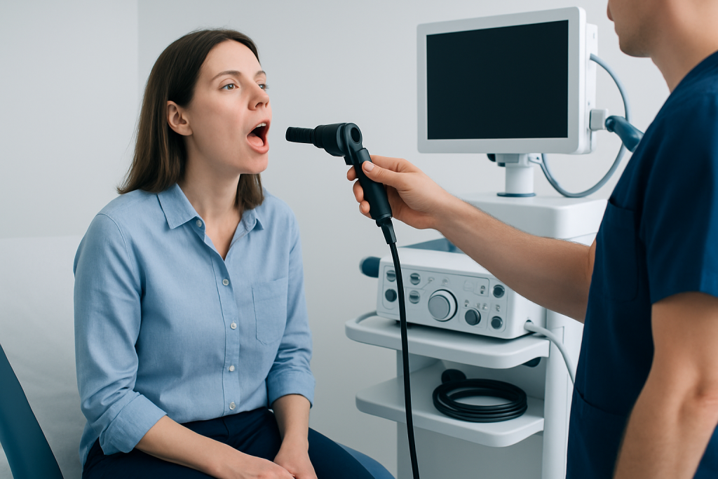
Leukoplakia may cause no symptoms at all, which is one reason it is often found by a clinician rather than by the person living with it. The patch may be white, grayish-white, thickened, or slightly wrinkled, and it usually remains in place when gently scraped. Some patches are well defined, while others have an irregular edge.
Symptoms, when present, are often subtle. A person may notice a rough feeling on the tongue, mild sensitivity to spicy foods, a persistent sore area, or a sensation that something in the mouth is “not quite right.” If the surface becomes red, ulcerated, bumpy, or painful, that does not automatically mean cancer, but it does make prompt evaluation more important.
- White or grayish patch in the mouth
- Patch that does not rub off
- Thickened, rough, or leathery surface
- Occasional mild burning or irritation
- Possible mixed red and white areas
Because many mouth conditions can look similar, it is best not to assume a white patch is harmless just because it is small or painless. Even a lesion that seems stable may still deserve a professional look if it has been present for more than two weeks.
Causes & Risk Factors

Leukoplakia is often linked to repeated irritation of the oral lining. Tobacco use, including smoking and smokeless forms, is one of the most recognized risk factors. Alcohol use can add to that risk, especially when combined with tobacco.
Other contributing factors may include rough teeth, ill-fitting dentures, chronic cheek biting, or repeated friction from dental appliances. Some cases are associated with fungal infection, certain medications, or long-term inflammation, although in many people no single cause is found. A related pattern called hairy leukoplakia can be seen in people with weakened immune function and has a different cause and clinical context.
Risk can also vary by the patch’s location and appearance. Lesions on the floor of the mouth, sides of the tongue, or areas with red or ulcerated changes are often watched more carefully because these locations may carry a higher likelihood of abnormal tissue changes.
- Tobacco exposure
- Heavy or regular alcohol use
- Chronic oral friction or irritation
- Poorly fitting dental appliances
- Long-standing inflammation or infection
- Weakened immune system in specific forms such as hairy leukoplakia
Diagnosis
Diagnosis begins with a careful examination of the mouth and a discussion of habits, symptoms, and medical history. A dentist, oral medicine specialist, ENT specialist, or doctor may ask how long the patch has been present, whether it has changed, and whether there are tobacco, alcohol, or dental irritation factors that could be contributing.
If the patch looks clearly related to irritation, the clinician may first remove the trigger and recheck the area after a short interval. If the patch persists, has concerning features, or appears in a higher-risk location, a biopsy may be recommended. In a biopsy, a small tissue sample is taken and examined under a microscope to look for dysplasia, which means abnormal cell change.
In some cases, photographs or regular reviews are used to track whether the lesion remains stable. This approach can be particularly useful for patients who travel for care, because it creates a clear follow-up plan that can be continued locally or with the treating team after returning home.
- Detailed oral examination
- Review of tobacco, alcohol, and dental irritation history
- Observation after removing possible triggers, when appropriate
- Biopsy for persistent or suspicious lesions
- Follow-up checks to monitor change over time
Treatment Options
Treatment depends on the cause, the appearance of the lesion, and whether abnormal cells are found. When an obvious irritation source is present, removing that trigger may allow the patch to improve. For example, smoothing a rough tooth edge, adjusting a denture, or stopping tobacco use may be part of the plan.
When biopsy shows dysplasia or when the lesion has suspicious features, a clinician may recommend removing the area. This can be done with surgical excision or, in selected cases, with other tissue-removal methods chosen by the care team. The goal is to eliminate abnormal tissue while preserving as much healthy oral function as possible.
Not every case needs immediate surgery. Some lesions are monitored with scheduled reviews, especially if the patch is low-risk and tissue sampling does not show concerning changes. The exact plan is individualized, because the safest approach depends on the person’s history, the lesion’s site, and the biopsy result.
- Remove or reduce local irritation
- Stop tobacco and avoid triggers that worsen the lesion
- Biopsy-guided treatment when dysplasia is present
- Surgical removal or other lesion-directed procedures in selected cases
- Ongoing monitoring when watchful follow-up is appropriate
Prevention & Self-care
There is no single guaranteed way to prevent leukoplakia, but there are practical steps that can lower risk and help the mouth heal. The most useful measure is to avoid tobacco in all forms and to reduce alcohol use, especially if oral white patches have already appeared. Good oral hygiene also matters, because chronic inflammation can make the lining of the mouth more vulnerable to irritation.
People who wear dentures or dental appliances should have them checked for fit if they cause rubbing or soreness. A gentle, consistent routine with brushing, flossing, and regular dental visits can catch small changes before they become larger issues. If a person already has a white patch, they should avoid trying to scrape or treat it at home, since this can irritate the area and delay proper assessment.
For patients planning treatment from another country, it helps to arrange clear follow-up before travel. Keeping photos, biopsy reports, and visit notes in one place makes it easier for the next clinician to compare findings and continue monitoring without interruption.
- Do not smoke or use tobacco products
- Limit alcohol intake
- Maintain regular dental care and clean oral hygiene
- Correct sources of friction, such as sharp teeth or ill-fitting dentures
- Keep records for follow-up if care continues across borders
When to See a Doctor
Any mouth white patch that lasts longer than two weeks should be evaluated, especially if it does not rub off or if the cause is not obvious. A prompt appointment is also wise if the patch changes in size, becomes rougher, develops red areas, or starts to ulcerate or bleed. These changes do not mean a serious diagnosis is certain, but they do mean the lesion should be examined without delay.
People with tobacco exposure, heavy alcohol use, a history of oral lesions, or a weakened immune system should be particularly consistent about follow-up. If swallowing becomes difficult, pain increases, or a lump appears in the neck, medical review should be arranged sooner.
For patients seeking care internationally, a coordinated evaluation can be especially reassuring because it helps determine whether the lesion needs biopsy, removal, or observation. Acibadem Health Point’s multidisciplinary specialists and JCI-accredited hospitals diagnose and treat leukoplakia for international patients as part of a structured, evidence-based care pathway.
- Patch lasts more than two weeks
- Patch changes color, texture, or size
- Red, ulcerated, or bleeding areas develop
- Pain, swallowing difficulty, or a neck lump appears
- There is a history of tobacco or significant alcohol use
What Living With Follow-Up Often Looks Like
After a diagnosis, the next step is often not one dramatic decision but a series of measured ones. Some people need a biopsy result explained in plain language. Others need a plan for monitoring a patch that appears low-risk but still deserves attention over time. Either way, the aim is clarity rather than urgency for its own sake.
Because oral tissues can change gradually, follow-up matters even when the first evaluation is reassuring. A clinician may schedule periodic reviews and compare the lesion with earlier photographs or measurements. That approach can fit both local care and travel-based care, as long as the patient knows which changes should prompt an earlier visit.
With proper assessment, many patients move forward with a simple and understandable plan: remove the trigger if possible, treat any abnormal tissue if needed, and stay alert to change. That combination is often the most practical path to protecting long-term oral health.
Frequently asked questions
Is leukoplakia the same as oral cancer?
No. Leukoplakia is a description of a white oral patch, not a cancer diagnosis. However, some leukoplakia lesions can contain precancerous changes, so a professional evaluation is important.
Can leukoplakia go away on its own?
Sometimes a patch improves if the source of irritation is removed, such as tobacco use or a rough denture. If it persists, a clinician may recommend biopsy or closer follow-up.
Does every leukoplakia patch need a biopsy?
Not every patch needs immediate biopsy, but many persistent or suspicious lesions do. The decision depends on the location, appearance, duration, and risk factors.
What is the most important thing a person can do at home?
Stopping tobacco use is one of the most helpful steps. It is also important to keep regular dental appointments and avoid trying to scrape or self-treat the patch.
How is leukoplakia monitored after diagnosis?
Follow-up usually involves repeat mouth examinations and sometimes photos or measurements to watch for change. If the lesion grows, changes texture, or becomes more symptomatic, further testing may be needed.
When should someone seek urgent review?
Urgent review is sensible if the patch becomes painful, ulcerated, bleeds, or is associated with difficulty swallowing or a neck lump. These signs warrant prompt professional assessment.
References
- American Dental Association
- National Cancer Institute
- World Health Organization
- Mayo Clinic
- American Academy of Oral and Maxillofacial Pathology
This article is for general information only and is not a substitute for professional medical advice. Please consult a qualified doctor about your individual situation.
Explore treatments in Turkey — costs, top hospitals & a free quote
JCI-accredited · board-certified surgeons · reply within 24h
Add us as a Preferred Source to see more of our trusted health content across Google Search, AI Overviews and Discover.