What Is the Difference Between an X-Ray and CT?
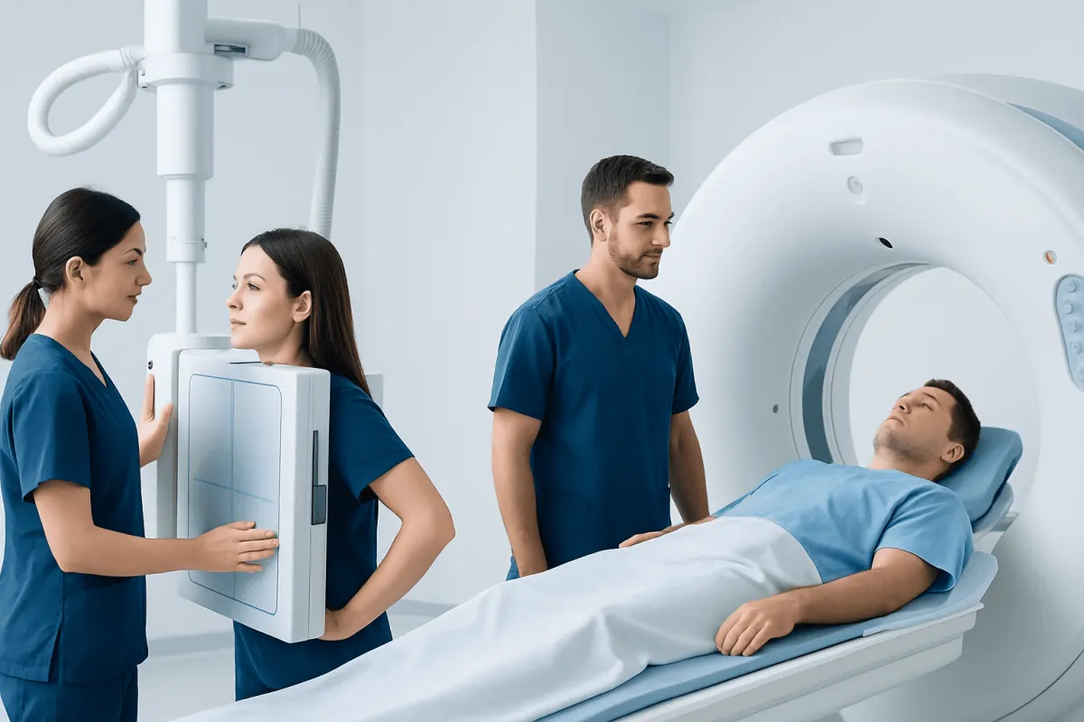
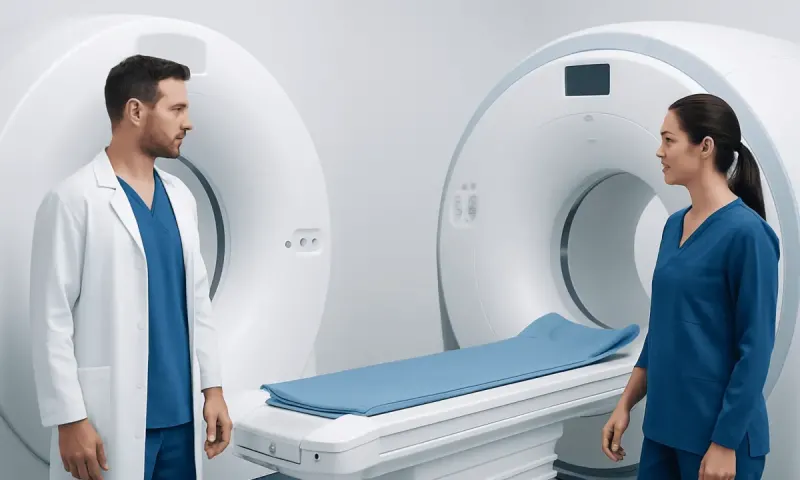
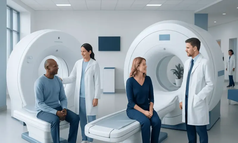
An X-ray and a CT scan both use ionizing radiation, but they produce different types of images. An X-ray is a fast, two-dimensional view that is often used first, while computed tomography (CT) combines many X-ray images to create detailed cross-sectional views of the body.
X-ray vs CT scan: a side-by-side comparison
What’s the difference between an X-ray and a CT scan? A conventional X-ray sends a small amount of radiation through the body to make one flat, two-dimensional image. A CT scan, also called a CAT scan or computed tomography scan, takes many X-ray measurements from different angles and uses a computer to build detailed cross-sectional images, sometimes called slices.
In simple terms, an X-ray is often useful for a quick overview, while CT provides a more complete internal map. Both tests are commonly performed by radiology teams and interpreted alongside symptoms, examination findings, and other Blood Test Results Explained: What CBC, Hemoglobin, and Platelets Mean" class="ahp-ilk">test results.
- Image type: X-ray produces a single 2D image; CT produces multiple detailed slices and may create 3D reconstructions.
- Best-known uses: X-ray is often used for suspected fractures and initial chest imaging; CT is often used for complex injuries, internal organs, bleeding, and detailed lung or brain assessment.
- Time: An X-ray usually takes only a few minutes. The scan itself in CT is often brief, although preparation and positioning can take longer.
- Radiation: Both use radiation, but CT usually delivers more radiation than a single X-ray because it gathers many images.
- Contrast material: Some CT scans use contrast dye to make blood vessels or organs easier to evaluate. Most standard X-rays do not require it.
What is the basic difference between conventional X-ray and CT scan?
The basic difference between conventional X-ray and CT scan is image detail and perspective. An X-ray is similar to looking at a shadow: structures overlap in a single image. Dense materials such as bone block more X-rays and usually appear lighter, while air-filled areas appear darker. This makes standard X-rays particularly useful for many bone problems and for an initial look at the lungs.
CT reduces the problem of overlapping structures by imaging the body in thin sections. A clinician can review these sections one at a time or reconstruct them in different planes. This allows clearer evaluation of tissues that may be difficult to separate on an X-ray, such as the brain, abdominal organs, blood vessels, lungs, and complex joints.
Whether a person asks what’s the difference between an X-ray and a CAT scan or what’s the difference between an X-ray and a CT scan, the answer is the same: “CAT” is an older term for CT, and both describe computed tomography imaging.
What can a CT scan show that an X-ray cannot?

A CT scan can reveal detail that may be hidden or unclear on an X-ray. For example, it can show small or complex fractures, internal bleeding, swelling in the brain, injuries to abdominal organs, kidney stones, certain blood vessel problems, and the size and location of many masses or infections. CT can also distinguish fat, fluid, air, bone, and many soft tissues more reliably than a plain X-ray.
For chest symptoms, the difference between a chest X-ray and a CT scan can be especially important. A chest X-ray gives a rapid overview of the lungs, heart silhouette, ribs, and pleural space. A chest CT can provide much more detailed views of lung tissue, airways, lymph nodes, blood vessels, and small abnormalities that may not be visible on an X-ray.
More detail does not automatically mean CT is needed. A clinician considers whether the result is likely to change care. For example, an uncomplicated injury may be assessed well with an X-ray, while a complicated fracture, significant trauma, or unclear result may warrant computed tomography (CT) imaging.
Which is best, an X-ray or CT scan?
Neither test is universally best. The right examination is the one that answers the medical question with an appropriate balance of accuracy, speed, radiation exposure, and practical considerations. A doctor may start with an X-ray when a quick first assessment is likely to be enough, particularly for many suspected fractures, joint concerns, or common chest complaints.
CT may be preferred when symptoms are severe, the situation is urgent, an X-ray result is uncertain, or the clinician needs a detailed view of structures beneath or behind one another. It is frequently useful after significant injury, for possible stroke or head trauma, for severe abdominal pain, and for suspected complications affecting internal organs.
A normal X-ray does not always rule out a condition. If symptoms persist or clinical concern remains high, further imaging may be appropriate. The radiology referral should be individualized, especially for children, pregnant people, and people who have had multiple imaging studies.
Why would a doctor order a CT scan instead of an MRI?
CT and MRI are different imaging methods. CT uses X-rays, whereas magnetic resonance imaging (MRI) uses powerful magnets and radiofrequency waves rather than ionizing radiation. When considering what’s the difference between X-ray, CT scan, and MRI, X-ray is the simplest and quickest 2D radiograph; CT is a detailed X-ray-based cross-sectional examination; and MRI often provides especially detailed images of soft tissues without radiation.
A doctor may order CT instead of MRI because CT is often faster, more available in urgent settings, and highly effective for many questions involving bone, lungs, acute bleeding, some abdominal problems, and traumatic injury. CT can also be suitable for people who cannot undergo MRI because of certain implanted devices, metal fragments, severe claustrophobia, or difficulty remaining still for a longer scan.
MRI may be better for other concerns, including many disorders of the brain, spinal cord, nerves, muscles, ligaments, and some pelvic organs. The choice is based on the suspected condition, the body part involved, the time available, individual safety factors, and the information needed for care.
What is the difference between X-ray, CT scan, and ultrasound?
Ultrasound differs from both X-ray and CT because it uses sound waves, not ionizing radiation. A handheld probe sends sound waves into the body and receives returning echoes to create real-time images. It is commonly used to assess pregnancy, abdominal organs, pelvic organs, blood flow, and some soft-tissue concerns.
Ultrasound has important strengths: it does not use radiation, can often be performed at the bedside, and can show movement and blood flow. However, image quality may be limited by air, bone, body shape, and the depth or location of the structure being examined. It is generally not the first test for looking through the skull, evaluating the lungs in the same way as CT, or comprehensively assessing major trauma.
Therefore, the difference between X-ray, CT scan, and ultrasound is not simply image quality. Each has a distinct role. The clinician selects the test that can best answer the question safely and efficiently.
What should you not do before a CT scan?
Preparation depends on the body area being scanned and whether contrast material will be used. Patients should not assume that the same instructions apply to every CT examination. They should follow the radiology department’s directions, which may include avoiding food or drink for a specified period before certain contrast-enhanced scans.
Before the appointment, patients should tell the healthcare team about pregnancy or a possible pregnancy, previous reactions to contrast material, kidney disease, diabetes, thyroid conditions, asthma, and all medicines or supplements they use. They should not stop prescribed medication unless a clinician specifically instructs them to do so. It is also important not to hide a prior contrast reaction, even if it was mild.
Patients may need to remove metal objects, jewelry, or clothing with metal components in the scan area. Drinking fluids after a contrast-enhanced CT may be advised for some people, but individual instructions can vary. The imaging team can explain what to expect and what to do after the scan.
When to seek medical care
Medical imaging should be guided by a qualified clinician rather than requested solely on the basis of a symptom. People should seek urgent medical care for symptoms such as sudden weakness or difficulty speaking, severe headache after an injury, chest pain, serious breathing difficulty, coughing blood, severe or worsening abdominal pain, loss of consciousness, or significant trauma.
Prompt medical assessment is also appropriate for persistent pain after an injury, fever with worsening respiratory symptoms, unexplained weight loss, or a symptom that does not improve as expected. The clinician may decide that examination, laboratory tests, X-ray, CT, ultrasound, MRI, or observation is most appropriate.
Acıbadem Health Point’s multidisciplinary specialists and JCI-accredited hospitals support international patients requiring diagnostic evaluation and treatment planning. Imaging results should always be discussed with the treating doctor, who can explain their meaning in the context of the person’s overall health.
Frequently asked questions
01Is a CT scan the same as an X-ray?
No. A CT scan uses X-rays, but it is more advanced than a conventional X-ray. It collects images from multiple angles and processes them into detailed cross-sectional views of the body.
02What is the difference between a chest X-ray and a CT scan?
A chest X-ray is a quick, two-dimensional image that provides a general view of the lungs, heart, ribs, and surrounding structures. A chest CT shows the chest in thin sections and can identify more subtle or complex findings in lung tissue, airways, blood vessels, and lymph nodes.
03Does a CT scan use more radiation than an X-ray?
In general, yes. A CT scan gathers many X-ray images, so its radiation exposure is usually higher than that from a single standard X-ray. Clinicians weigh this consideration against the value of the additional diagnostic information.
04Can an X-ray miss a fracture?
Yes, some fractures may not be visible on an initial X-ray, especially if they are small, nondisplaced, located in a complex area, or very recent. If symptoms and examination findings suggest a fracture despite a normal X-ray, a doctor may recommend repeat imaging, CT, MRI, or another test.
05Is CT better than MRI?
CT is not always better than MRI; the tests answer different questions. CT is often faster and especially useful for bone injuries, lung assessment, acute bleeding, and emergency situations, while MRI is often preferred for detailed assessment of many soft tissues, nerves, and the spinal cord.
06Will a CT scan hurt?
A CT scan is painless. The person usually lies still on a table while it moves through the scanner; some scans involve an injection of contrast material, which may cause a brief warm sensation or a metallic taste. Staff can explain the process and monitor the patient throughout the examination.
This article is for general information only and is not a substitute for professional medical advice. Please consult a qualified doctor about your individual situation.
Joint and spine care in Turkey — expert assessment & treatment
JCI-accredited · board-certified surgeons · reply within 24h
Add us as a Preferred Source to see more of our trusted health content across Google Search, AI Overviews and Discover.