What Does Vulvar Cancer Look Like on the Skin?
Vulvar cancer can look like a new lump, a sore that does not heal, a thickened or discolored skin patch, or an area that persistently itches, burns, or bleeds. Many vulvar changes are not cancer, but any lasting change should be examined by a qualified clinician.
What does vulvar cancer look like?
Vulvar cancer may look like a new lump, raised growth, thickened patch of skin, open sore, or an area that has changed color. It can be skin-colored, white, red, pink, dark brown, or black, and it may resemble a wart, mole, rash, or ulcer. Some changes are painless, while others itch, burn, feel tender, or bleed.
The vulva includes the outer female genital structures, such as the labia, clitoris, and opening of the vagina. Most visible or uncomfortable vulvar changes are caused by noncancerous conditions, including irritation, infections, cysts, inflammatory skin disorders, or benign growths. However, a change that persists, enlarges, recurs, or does not heal should be assessed rather than self-diagnosed.
Vulvar cancer is uncommon and is most often diagnosed in older adults, although it can occur at other ages. The most common form is squamous cell carcinoma, which begins in the cells lining the vulvar skin. Less common types include melanoma, basal cell carcinoma, adenocarcinoma, and sarcoma, and their appearance may differ.
Signs and symptoms that may need assessment
Symptoms can develop gradually and may be subtle at first. Persistent itch is one of the more common concerns, particularly when it affects one specific area and does not improve with appropriate care. Not every person has symptoms, and a lesion may first be noticed during routine gynecologic examination.
Possible signs and symptoms include:
- a new lump, bump, thickened area, or wart-like growth
- a sore, crack, ulcer, or raw patch that does not heal
- ongoing itching, burning, pain, or tenderness
- skin that becomes white, red, darker than surrounding skin, scaly, or velvety
- bleeding, spotting, or discharge not related to menstruation
- pain with urination or sexual activity
- a new or changing mole-like spot on the vulva
- a lump in the groin, which may reflect enlarged lymph nodes
Symptoms such as itching and discoloration can also occur with conditions such as eczema, psoriasis, yeast infection, lichen sclerosus, or contact dermatitis. The key issue is persistence or change over time. A clinician can examine the area and determine whether testing is needed.
Why vulvar changes happen: causes and risk factors
Vulvar cancer develops when cells acquire changes that allow them to grow abnormally. In many cases, there is no single identifiable cause. Two broad pathways are recognized: some cancers are associated with persistent infection by certain high-risk types of human papillomavirus (HPV), while others are associated with long-standing inflammatory skin conditions, especially lichen sclerosus.
Factors that may increase risk include older age, a history of high-grade vulvar precancerous changes, smoking, a weakened immune system, persistent high-risk HPV infection, and chronic vulvar skin disease. HPV-related cancers are more likely to occur at younger ages than cancers associated with lichen sclerosus, although individual patterns vary.
Having a risk factor does not mean a person will develop cancer, and people without recognized risk factors can still be diagnosed. Regular attention to vulvar health, prompt assessment of symptoms, and recommended HPV vaccination can all play a role in prevention and early detection.
How vulvar cancer is diagnosed
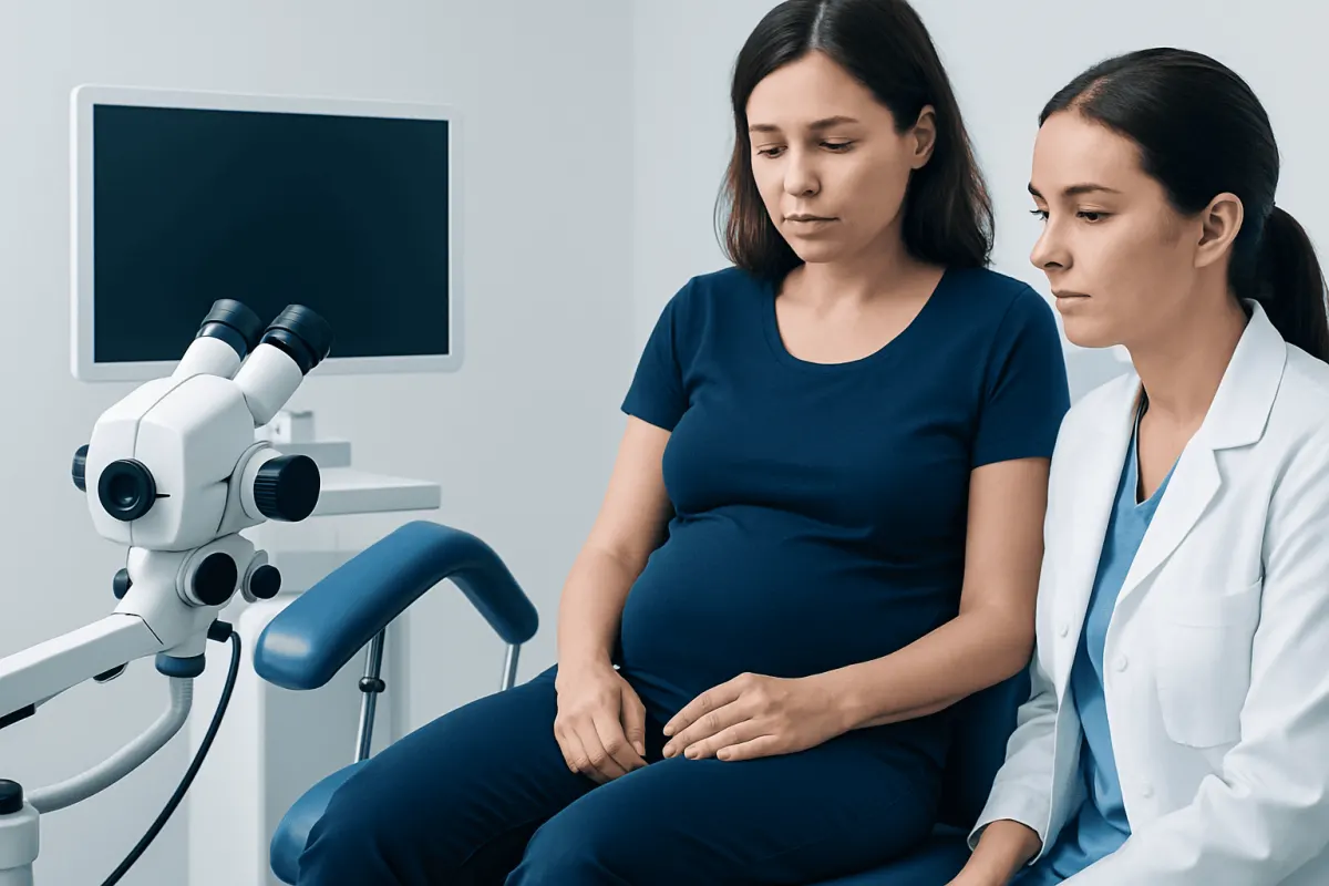
Diagnosis begins with a medical history and careful examination of the vulva and nearby groin lymph nodes. A gynecologist, dermatologist, or gynecologic oncologist may use magnification or a lighted instrument to inspect an area more closely. The examination is usually brief, and patients can ask questions or request support throughout the visit.
A biopsy is the only reliable way to determine whether a suspicious area is cancerous or precancerous. During a biopsy, a clinician removes a small sample of tissue, often using local anesthetic, and a pathologist examines it under a microscope. The biopsy also identifies the cancer type and other features that guide treatment planning.
If cancer is confirmed, imaging tests such as ultrasound, CT, MRI, or PET/CT may be recommended in selected cases to assess the size of the tumor and whether lymph nodes or other areas may be involved. Doctors use this information to determine the stage, which describes how far the cancer has spread. Staging helps the care team discuss appropriate treatment options and outlook.
Modern treatment approaches for vulvar cancer
Treatment is individualized according to the tumor’s type, location, size, depth, stage, and the person’s overall health and preferences. Care is often coordinated by a multidisciplinary team that may include a gynecologic oncologist, surgeon, radiation oncologist, medical oncologist, pathologist, specialist nurse, and supportive-care professionals.
Surgery is commonly used for localized vulvar cancer. The aim is to remove the cancer while preserving as much normal tissue and function as safely possible. Depending on the case, procedures may include removal of the lesion with a margin of surrounding tissue and assessment of nearby lymph nodes. Sentinel lymph node biopsy may be appropriate for some early cancers, helping avoid more extensive node surgery when it is not needed.
Radiotherapy may be used after surgery when there is a higher risk of cancer returning, when lymph nodes are involved, or when surgery alone is not suitable. Chemotherapy may be given with radiotherapy for some locally advanced cancers, and systemic treatments may be considered when disease has spread or returned. Reconstructive techniques, symptom management, sexual health support, and emotional care are also important parts of treatment planning.
Acıbadem Health Point’s multidisciplinary specialists and JCI-accredited hospitals diagnose and treat vulvar cancer for international patients, with treatment plans based on individual pathology and staging findings.
Outlook, follow-up, and living well after treatment
The outlook for vulvar cancer depends largely on the stage at diagnosis, the cancer type, lymph node involvement, and response to treatment. Cancers found while limited to the vulva generally have a more favorable outlook than cancers that have spread to lymph nodes or distant organs. Because each diagnosis is different, the treating team is best placed to explain an individual prognosis.
Follow-up appointments are important after treatment because they allow clinicians to check healing, manage long-term effects, and look for possible recurrence. Follow-up may include symptom review, examination of the vulva and groin, and further tests if a concern is found. Patients should report any new lump, persistent itch, bleeding, skin change, or pain between scheduled visits.
Recovery can involve physical and emotional adjustment. Depending on treatment, some people experience temporary or lasting changes in comfort, body image, urination, sexual function, or lymphatic swelling in the legs. Pelvic health physiotherapy, counseling, sexual health services, pain management, and support groups may help people regain confidence and quality of life.
Prevention and vulvar self-care
Not all vulvar cancers can be prevented, but several measures may lower risk. HPV vaccination helps protect against HPV types linked to several cancers, including some vulvar cancers. Vaccination recommendations vary by age and country, so a doctor or local public health service can advise on eligibility.
Avoiding smoking, attending recommended cervical screening, and seeking care for persistent vulvar symptoms are sensible preventive steps. People diagnosed with lichen sclerosus or vulvar precancer should follow their clinician’s treatment and surveillance plan, even if symptoms improve.
Gentle vulvar care can reduce irritation but cannot treat cancer. This may include avoiding fragranced washes and harsh products, wearing breathable underwear, and using prescribed treatments as directed. New or persistent symptoms should not be repeatedly treated with over-the-counter products without a medical review, particularly if there is an ulcer, growth, bleeding, or changing pigmented area.
When to seek medical care
Medical care should be arranged promptly for a vulvar lump, sore, ulcer, wart-like lesion, or mole-like mark that is new, enlarging, changing color, or not healing. It is also important to seek assessment for ongoing itching, burning, pain, unexplained bleeding, or discharge, especially when symptoms persist despite simple measures or treatment for a suspected infection.
Urgent assessment is appropriate if bleeding is heavy, pain is severe, there is fever or signs of significant infection, or a lesion is rapidly changing. In most cases, these symptoms have causes other than cancer, but timely examination can provide reassurance and ensure that appropriate treatment is not delayed.
People may feel embarrassed discussing vulvar symptoms, but clinicians are trained to assess these concerns respectfully and confidentially. Bringing a written list of symptoms, when they started, and any treatments already tried can make the appointment more useful.
Frequently asked questions
01Can vulvar cancer look like a pimple or ingrown hair?
A vulvar cancer lesion can sometimes resemble a pimple, cyst, wart, or ingrown hair, particularly early on. An ordinary pimple or irritated hair follicle usually improves within a short time, while a cancerous lesion may persist, enlarge, ulcerate, bleed, or repeatedly return. A clinician should examine any lump or sore that does not resolve.
02Is vulvar itching usually a sign of cancer?
No. Vulvar itching is common and is more often caused by irritation, infections, hormonal changes, or skin conditions than cancer. However, itching that is persistent, focused in one area, or accompanied by skin changes, a lump, bleeding, or a sore should be evaluated.
03Can a Pap test detect vulvar cancer?
A Pap test is designed to screen for abnormal cells in the cervix, not the vulva. It may identify cervical changes related to HPV, but it does not rule out vulvar cancer. A vulvar examination and biopsy of a suspicious area are needed for diagnosis.
04How quickly does vulvar cancer develop?
Vulvar cancer often develops gradually, and some precancerous changes can be present before invasive cancer develops. The timeline differs between individuals and cancer types. Any persistent lesion or symptom should be checked rather than monitored indefinitely at home.
05What does early vulvar cancer feel like?
Early vulvar cancer may cause no discomfort at all. When symptoms occur, they can include itching, burning, tenderness, pain, or a feeling of a small lump or rough patch. These sensations are not specific to cancer, so examination is important when they persist.
06Can vulvar cancer be cured?
Many vulvar cancers can be treated successfully, especially when they are diagnosed before they have spread. Treatment and outlook depend on the stage, tumor characteristics, and overall health of the individual. A gynecologic oncology team can explain the expected goals and likely outcomes of treatment in a specific case.
This article is for general information only and is not a substitute for professional medical advice. Please consult a qualified doctor about your individual situation.
Persistent digestive symptoms? Get evaluated in Turkey
JCI-accredited · board-certified surgeons · reply within 24h
Add us as a Preferred Source to see more of our trusted health content across Google Search, AI Overviews and Discover.