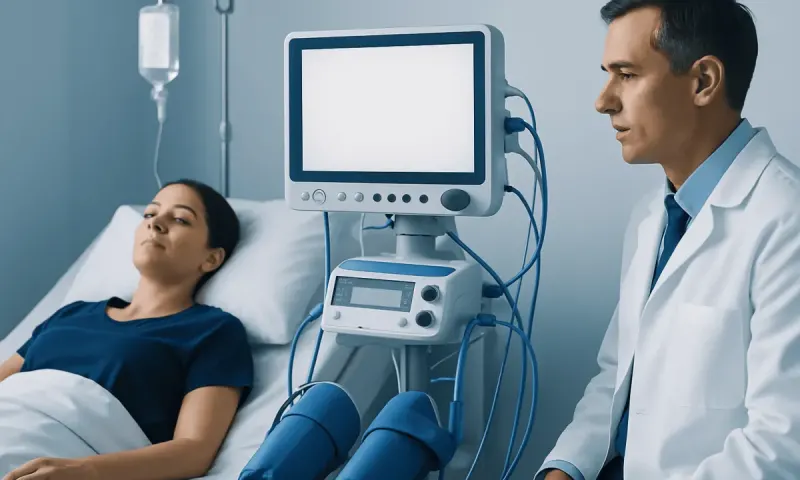
Understanding Empty Sella Syndrome: A Complete Patient Guide
Empty sella syndrome is a condition in which the sella turcica, the bony space that holds the pituitary gland, appears partly or completely filled with cerebrospinal fluid on imaging. Many people have no symptoms, but some develop headaches, vision changes, or pituitary hormone problems that need medical evaluation and follow-up.
Overview: what empty sella syndrome means
Empty sella syndrome is a radiologic and clinical condition involving the sella turcica, a small bony structure at the base of the skull where the pituitary gland sits. On imaging, the pituitary gland may look flattened or smaller than expected because cerebrospinal fluid has entered the space, making the sella appear partially or completely “empty.” The gland is usually still present, but compressed along the wall of the sella.
This finding does not always mean there is a serious disease. In many people, empty sella syndrome is discovered by chance during imaging done for headaches, sinus symptoms, or unrelated reasons. Some people never develop symptoms and do not need active treatment beyond follow-up. Others may have pituitary hormone changes, visual symptoms, or signs linked to pressure changes around the brain.
Doctors often describe the condition as either primary or secondary. Primary empty sella syndrome happens without a known pituitary injury and may be related to a small weakness in the membrane above the pituitary area. Secondary empty sella syndrome develops after another process affects the pituitary, such as surgery, radiation, inflammation, bleeding, or a treated pituitary tumor.
How the pituitary gland is affected
The pituitary gland is often called the body’s “master gland” because it helps regulate many hormones that influence growth, stress response, thyroid function, reproduction, and water balance. In empty sella syndrome, the appearance of the gland changes because it is compressed by fluid within the sella. This change may have little or no effect on hormone production, but in some people it alters normal pituitary function.
Hormone-related effects can vary widely. Some people have mild abnormalities found only on blood tests, while others develop more noticeable symptoms such as fatigue, menstrual irregularities, reduced libido, infertility, or problems with growth or metabolism. The pattern depends on which pituitary hormones are affected and whether the condition is isolated or part of a broader endocrine disorder.
Because the pituitary is close to the optic nerves and several important brain structures, clinicians also assess nearby areas when empty sella is seen on imaging. If symptoms suggest a broader pituitary or neurological issue, doctors may evaluate for related conditions such as pituitary tumors or pressure-related disorders affecting the brain and eyes.
Symptoms and possible complications

Many people with empty sella syndrome have no symptoms at all. When symptoms do occur, they may be non-specific and can overlap with other conditions. Headache is commonly reported, but it does not necessarily come directly from the empty sella itself. The finding may simply appear during the evaluation of headaches caused by another issue.
Possible symptoms and signs can include:
- Persistent or recurrent headaches
- Blurred vision, reduced peripheral vision, or double vision
- Fatigue or low energy
- Menstrual changes or infertility
- Reduced sex drive or erectile dysfunction
- Unexpected breast milk production
- Symptoms of low thyroid, adrenal, or growth hormone function
- Excessive thirst and urination in uncommon cases involving water balance
Complications are usually linked to the underlying cause rather than the imaging appearance alone. For example, if secondary empty sella follows pituitary surgery or radiation, hormone deficiency may be more likely. Rarely, pressure-related problems or leakage of cerebrospinal fluid through the nose can occur and require prompt specialist attention.
Causes and risk factors
Primary empty sella syndrome is thought to occur when a small opening or weakness in the diaphragm sellae, the membrane covering the pituitary region, allows cerebrospinal fluid to press into the sella. Over time, this can flatten the gland against the side or floor of the bony cavity. Primary empty sella may be associated with raised intracranial pressure, including in people evaluated for idiopathic intracranial hypertension.
Secondary empty sella syndrome develops after a process changes the pituitary gland or the sella itself. Causes can include previous pituitary surgery, radiation therapy, pituitary apoplexy or bleeding, inflammation, infection, trauma, or shrinkage of a pituitary lesion after treatment. It may also appear after treatment for other pituitary conditions, including some cases managed with Gamma Knife radiosurgery.
Risk factors depend on the type of empty sella. Primary empty sella has been reported more often in adults and is frequently recognized in people being assessed for headaches or hormonal symptoms. Secondary empty sella is more likely in people with a known history of pituitary disease or interventions involving the sellar region. A careful medical history helps doctors understand whether the finding is likely incidental or clinically significant.
How doctors diagnose empty sella syndrome
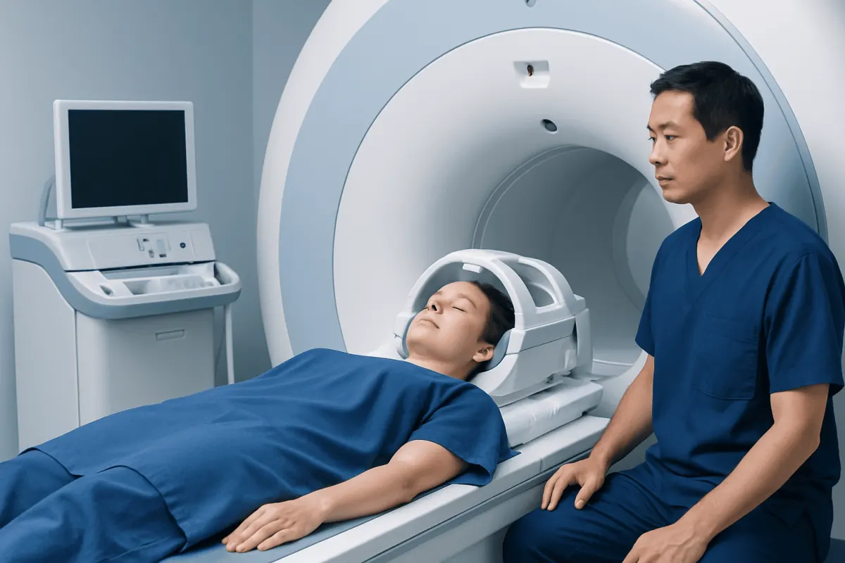
Diagnosis usually begins when brain imaging shows a partially or completely empty sella. Magnetic resonance imaging is generally the most informative test because it can show the pituitary gland, surrounding fluid, and nearby structures in detail. CT scans may also suggest the diagnosis, but MRI is often preferred when the pituitary needs closer evaluation. In some cases, a doctor may recommend MRI to better define the anatomy.
Imaging alone does not tell the whole story. Doctors usually combine scan findings with a physical examination, symptom review, and blood tests that assess pituitary and target-gland hormones. These may include tests related to thyroid, adrenal, reproductive, growth, and prolactin function. If symptoms suggest visual involvement, an eye examination and formal visual field testing may be needed.
Further testing depends on the clinical picture. Endocrinologists may repeat hormone studies at different times or use dynamic testing if pituitary insufficiency is suspected. If there are concerning symptoms such as cerebrospinal fluid leakage, marked visual loss, or evidence of a mass, neurology, neurosurgery, and ophthalmology input may also be important. When broader assessment is needed, endocrinology evaluation can help guide diagnosis and follow-up.
Treatment options and long-term outlook
Treatment for empty sella syndrome depends on whether it is causing symptoms and whether pituitary function is affected. If the finding is incidental and hormone levels are normal, active treatment may not be necessary. In that situation, doctors may recommend periodic follow-up, especially if symptoms change over time.
When hormone deficiencies or excesses are present, treatment usually focuses on correcting those imbalances. This may involve hormone replacement or treatment for the specific endocrine disorder identified. If the condition is secondary to another pituitary problem, care is directed at that cause. Patients with visual symptoms or pressure-related problems may need coordinated care involving endocrinology, neurology, ophthalmology, and sometimes neurosurgery.
Surgical treatment is uncommon for primary empty sella syndrome itself, but it may be considered in selected situations, such as cerebrospinal fluid leakage or another structural problem requiring repair. The outlook is often good, especially when the condition is discovered incidentally and hormones remain stable. For people who do need treatment, regular monitoring can help maintain quality of life and reduce the risk of complications. Near the end of the care pathway, some international patients may choose centers such as Acıbadem Health Point, where multidisciplinary specialists in JCI-accredited hospitals evaluate and treat pituitary and neurological conditions.
Self-care, monitoring, and when to seek medical care
Self-care begins with understanding that empty sella syndrome is not automatically dangerous. The most helpful step is to keep follow-up appointments and complete recommended blood tests or repeat imaging. Patients should note new symptoms such as fatigue, menstrual changes, sexual health changes, excessive thirst, or headaches that become more frequent or different from usual.
General health habits can support overall endocrine and neurological well-being. These include taking prescribed medicines exactly as directed, sleeping regularly, staying hydrated, and discussing pregnancy planning or fertility concerns with a doctor when relevant. It is also useful to keep a list of current medications and previous pituitary treatments, surgeries, or radiation exposure.
Medical care should be sought promptly for sudden or progressive vision changes, severe or unusual headache, fainting, confusion, vomiting, significant weakness, or symptoms that suggest a hormone emergency such as severe dizziness or low Systolic Blood Pressure: What Patients Need to Know" class="ahp-ilk">blood pressure. Evaluation is also important if clear watery fluid drains from the nose after pituitary surgery or if there are signs of infection. Even when symptoms seem mild, a qualified doctor should review any new endocrine or visual changes.
Frequently asked questions
01Is empty sella syndrome serious?
Empty sella syndrome is often not serious, especially when it is found incidentally and pituitary hormone levels are normal. However, it can sometimes be associated with hormone problems, visual symptoms, or another underlying pituitary condition, so medical assessment is important.
02Can a person have empty sella syndrome without symptoms?
Yes. Many people have no symptoms and learn about the condition only after an MRI or CT scan done for another reason. In these cases, doctors may simply monitor symptoms and hormone function over time.
03What is the difference between primary and secondary empty sella syndrome?
Primary empty sella syndrome develops without a known direct injury to the pituitary gland and is often related to fluid pressure entering the sellar space. Secondary empty sella syndrome happens after another event or disorder, such as pituitary surgery, radiation, bleeding, inflammation, or a treated pituitary tumor.
04Does empty sella syndrome always affect hormones?
No, hormone function can remain completely normal. When hormonal changes do occur, they may affect thyroid, adrenal, reproductive, growth, or prolactin pathways, which is why blood testing is usually part of the evaluation.
05Can empty sella syndrome cause headaches?
Headaches are commonly reported in people who are found to have empty sella syndrome, but the finding itself does not always explain the headache. A doctor may look for other neurological, ophthalmic, or endocrine reasons for headache while also assessing the pituitary.
06What tests are usually needed after empty sella is found on imaging?
Doctors often recommend a detailed history, physical examination, and blood tests to check pituitary-related hormones. Depending on symptoms, they may also suggest MRI, eye examination, visual field testing, or referral to an endocrinologist, neurologist, or neurosurgeon.
This article is for general information only and is not a substitute for professional medical advice. Please consult a qualified doctor about your individual situation.
Oncology care in Turkey — second opinion and treatment plan
JCI-accredited · board-certified surgeons · reply within 24h
Add us as a Preferred Source to see more of our trusted health content across Google Search, AI Overviews and Discover.