Tumor PET Scan: Cancer Diagnosis, Staging, and Next Steps

A tumor PET scan is an imaging test that highlights areas of increased cellular activity, helping clinicians assess suspected or known cancer. It is usually interpreted together with medical history, examination findings, CT or MRI images, laboratory tests, and sometimes a biopsy.
How a tumor PET scan helps evaluate cancer
A tumor PET scan is a type of nuclear medicine imaging that helps doctors identify tissues with unusual metabolic activity. Cancer cells often use glucose differently from many normal cells, so they may absorb more of a small amount of radioactive tracer given before the scan. The resulting images can show areas that may need further evaluation.
In cancer care, the most common examination is a PET/CT scan. It combines PET images, which show metabolic activity, with CT images, which show body structures in detail. This combined approach can help locate an active area more precisely and determine whether it corresponds to a lymph node, organ, bone, or another tissue.
A tumor PET scan is not a stand-alone cancer test. Some cancers do not take up the usual tracer strongly, and noncancerous processes can also look active. For this reason, radiologists and cancer specialists interpret the result in the context of other imaging, symptoms, blood tests, and pathology findings when available.
The scan may be used before treatment, during treatment, or after treatment. Depending on the cancer type and clinical question, it can support diagnosis, guide the best site for a biopsy, assess the extent of disease, evaluate response to therapy, or investigate a possible recurrence.
What happens before, during, and after a PET scan
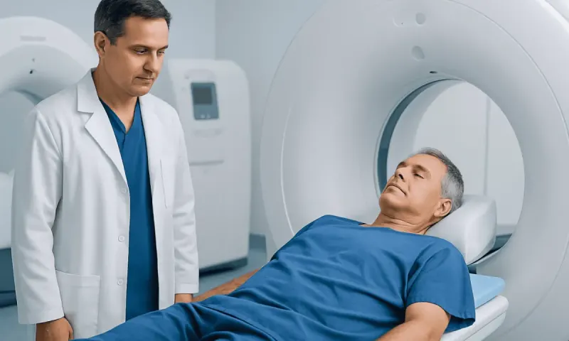
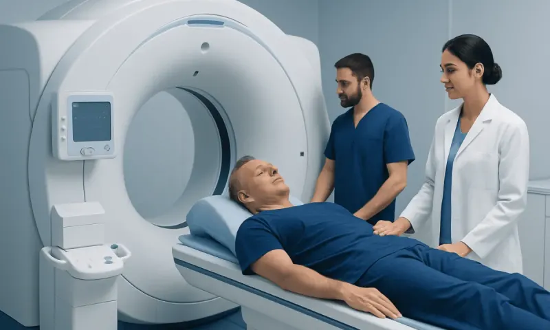
For many cancer PET scans, a glucose-like tracer called fluorodeoxyglucose (FDG) is injected into a vein. The person then rests quietly for a period while the tracer circulates and is taken up by tissues. The imaging itself is painless. During the scan, the person lies on a table that moves slowly through the scanner.
Preparation instructions vary, but patients are commonly asked to avoid eating for several hours before an FDG PET scan and to drink water unless told otherwise. Physical exertion may need to be limited beforehand because active muscles can take up tracer. People should tell the imaging team about diabetes, pregnancy, breastfeeding, medicines, recent surgery, infection, radiation therapy, or chemotherapy, as these can affect preparation or interpretation.
The appointment may take several hours in total because it includes preparation, tracer uptake time, and scanning. The tracer leaves the body naturally, mainly through urine, over a short period. The imaging department may recommend drinking fluids afterward and following practical instructions about close contact with infants or pregnant people for the rest of the day.
Most people can return to usual activities after the scan unless they have been given sedation or specific instructions for another procedure. A radiologist reviews the images and sends a report to the referring clinician, who discusses what the findings mean in the individual clinical setting.
Can you tell what stage cancer is from a PET scan?

A PET scan can contribute important information to cancer staging, but it usually cannot determine the stage by itself. Staging describes the size and location of the primary tumor, whether nearby lymph nodes are involved, and whether cancer has spread to distant parts of the body. PET/CT can be particularly useful for looking for metabolically active lymph nodes or distant lesions that may not be evident on other tests.
Its role differs by cancer type. PET imaging is often helpful in several lymphomas and in cancers such as lung cancer, head and neck cancer, melanoma, and some colorectal cancers when specific clinical questions arise. In other cancers, CT, MRI, ultrasound, bone scans, specialized PET tracers, surgery, or pathology may be more informative.
A staging decision is made by the cancer care team using all available evidence. A suspicious PET finding may need confirmation with additional imaging or a biopsy before it changes the diagnosis or treatment plan. For information about comprehensive cancer care, patients should discuss which staging tests are appropriate for their specific tumor type.
When cancer has already been diagnosed, PET results may help clinicians select a treatment approach, such as surgery, radiation therapy, systemic treatment, or a combination. The final plan depends on the cancer type, stage, molecular features where relevant, overall health, and personal treatment goals.
How accurate is a PET scan in diagnosing cancer?
The accuracy of a PET scan in diagnosing cancer depends on the tumor type, size, location, tracer used, and reason for testing. PET can be highly informative when a cancer is known to be metabolically active and the scan is used for an appropriate clinical question. However, it cannot reliably distinguish cancer from every noncancerous cause of increased activity.
False-positive findings can occur with infection, inflammation, autoimmune conditions, recent surgery, healing injuries, and effects of radiation or other treatments. Normal tissues such as the brain, heart, kidneys, bladder, and sometimes the bowel may also show tracer uptake. A bright area on a PET image therefore does not automatically mean cancer.
False-negative results can also occur. Very small lesions may be below the scan’s resolution, and some slow-growing or low-metabolism tumors may show little tracer uptake. Blood sugar levels, timing of imaging, and technical factors can influence FDG PET imaging as well.
For these reasons, a biopsy is often needed to confirm a new or uncertain diagnosis. PET may help identify the most suitable area to sample, especially when there is more than one abnormality. The scan is best understood as one part of a careful diagnostic process rather than a definitive answer on its own.
What is an alternative to a PET scan for cancer?
There is no single replacement for a PET scan because each imaging test provides different information. The best alternative depends on the suspected cancer, the body area being examined, and whether the goal is diagnosis, staging, treatment planning, or follow-up. A doctor may recommend CT, MRI, ultrasound, X-ray imaging, or a bone scan instead of, or in addition to, PET.
CT is commonly used to assess the size and location of tumors and to look for spread in the chest, abdomen, and pelvis. MRI provides detailed images of soft tissues and is especially valuable for the brain, spinal cord, liver, pelvis, and certain bone or muscle concerns. Ultrasound can assess some organs and lymph nodes and can guide needle biopsies.
In many situations, tissue sampling remains the most direct way to establish whether a suspicious area is cancerous and to identify its type. Biopsy procedures can also provide molecular and other laboratory information that imaging alone cannot provide. For selected cancers, specialized blood tests, endoscopy, or surgical assessment may be part of the evaluation.
Alternative imaging is not necessarily less advanced or less useful than PET. It may simply answer a different question. The care team considers the expected benefit of each test, prior imaging results, radiation exposure where relevant, kidney function, contrast allergies, and the person’s individual circumstances.
Is it serious if you need a PET scan?
Needing a PET scan does not by itself mean that a person has cancer or that a condition is advanced. Doctors order PET imaging when they need more information about an abnormal symptom, a finding on another scan, a known cancer, or possible treatment response. It is a tool for reducing uncertainty and making decisions based on a more complete picture.
For example, a PET scan may be requested after a CT scan shows an area that needs clarification, when a biopsy has confirmed cancer and staging is needed, or when clinicians are checking whether treatment is working. It may also be used to assess a change that could be related to inflammation, infection, scar tissue, or cancer recurrence.
Waiting for imaging and results can feel stressful. Patients can ask why the scan was requested, what body area will be examined, whether preparation is needed, and when results are likely to be available. It can be helpful to bring a list of questions to the follow-up appointment.
A result that requires more testing does not necessarily indicate a serious outcome. Additional testing is often the safest way to confirm or rule out an explanation before treatment decisions are made.
How PET findings support modern treatment approaches
Modern Skin Cancer Treatment: Procedure, Recovery and Results" class="ahp-ilk">cancer treatment is increasingly individualized. PET/CT can help the multidisciplinary team understand where active disease is located and whether it appears limited to one area or involves multiple sites. This information can support decisions about surgery, radiation therapy, drug treatment, or careful surveillance.
In some cancers, a PET scan performed after treatment begins can show whether metabolic activity is decreasing. This may complement physical examinations, blood tests, CT or MRI scans, and symptom review. The timing of follow-up PET imaging matters, since inflammation from treatment can temporarily increase uptake and make interpretation more complex.
For a localized tumor, PET findings may help radiation specialists define areas that require attention during radiation therapy planning. For cancers that need whole-body treatment, imaging can also be part of the assessment before or during systemic cancer treatment. The exact approach is always based on the cancer’s biology and the person’s health needs, not on a scan result alone.
At Acıbadem Health Point, multidisciplinary specialists in JCI-accredited hospitals can evaluate PET findings alongside pathology and other imaging when caring for international patients. A coordinated review can help patients understand the purpose of each test and the options being considered.
When to seek medical care
A person should arrange medical assessment for persistent or unexplained symptoms such as a new lump, ongoing unexplained weight loss, coughing up blood, continuing changes in bowel or bladder habits, unusual bleeding, persistent pain, or worsening fatigue. These symptoms can have many causes, and most do not automatically mean cancer, but timely evaluation is important.
Anyone who has been advised to have a PET scan should contact the referring clinician or imaging center if they are pregnant, may be pregnant, are breastfeeding, have diabetes, or have recently had a significant infection, surgery, or cancer treatment. These details help the team plan the scan safely and interpret it appropriately.
After a PET scan, patients should seek urgent medical help for severe symptoms such as trouble breathing, chest pain, sudden weakness, confusion, or signs of a serious allergic reaction. Such symptoms are uncommon and may not be related to the scan, but they should not be ignored.
For nonurgent questions about a report, the most appropriate next step is usually a follow-up discussion with the clinician who ordered the test. They can explain whether an abnormality is likely to need monitoring, another scan, specialist referral, or biopsy.
Frequently asked questions
01What does a tumor PET scan show?
A tumor PET scan shows patterns of tracer uptake that may reflect increased cellular activity. It can help identify areas that need further assessment, including possible tumor sites, involved lymph nodes, or sites of cancer spread. It cannot confirm cancer without considering other clinical information.
02Can you tell what stage cancer is from a PET scan?
A PET scan can provide useful staging information, particularly about possible lymph node involvement or distant spread. However, cancer stage is determined using the full set of findings, which may include CT, MRI, pathology, surgery, and other tests. Suspicious PET findings sometimes need biopsy confirmation.
03What is an alternative to a PET scan for cancer?
Depending on the clinical question, alternatives include CT, MRI, ultrasound, bone scanning, endoscopy, or biopsy. These tests are not interchangeable in every situation because they reveal different features of a suspected cancer. A clinician chooses the test most likely to answer the specific question.
04Is it serious if you need a PET scan?
Not necessarily. A PET scan may be ordered to clarify an abnormality found on another test, to stage a known cancer, or to check treatment response. The need for the scan reflects a need for more information rather than a diagnosis or prediction by itself.
05How accurate is a PET scan in diagnosing cancer?
Accuracy varies by cancer type, tumor size, location, and the tracer used. PET scans can be very helpful, but inflammation, infection, and healing tissue can sometimes look similar to cancer, while some cancers may not show strongly. Doctors often use PET results together with other tests and may recommend a biopsy for confirmation.
06Does a PET scan hurt or have risks?
The injection may feel similar to a routine blood test, while the scan itself is painless. PET imaging uses a small amount of radioactive tracer, and the radiation exposure is considered when the test is recommended. The imaging team can explain individual considerations, including pregnancy, breastfeeding, diabetes, and contrast use if a CT component requires it.
This article is for general information only and is not a substitute for professional medical advice. Please consult a qualified doctor about your individual situation.
Oncology care in Turkey — second opinion and treatment plan
JCI-accredited · board-certified surgeons · reply within 24h
Add us as a Preferred Source to see more of our trusted health content across Google Search, AI Overviews and Discover.