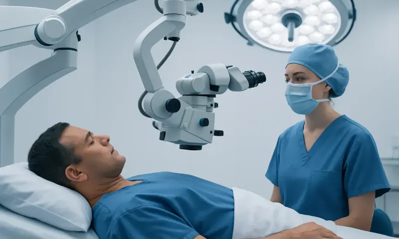
MRI Vs CT Scan for Cancer: Which Test Fits?
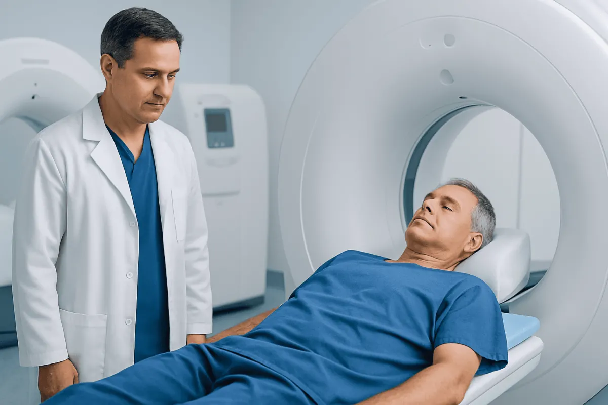
For cancer imaging, neither MRI nor CT is universally better. CT is often the first choice when speed, lung detail, bone assessment, or broad body staging is needed, while MRI is especially valuable for detailed views of soft tissues such as the brain, spinal cord, liver, pelvis, and prostate.
MRI vs CT Scan Cancer: Side-by-Side Comparison
When comparing MRI vs CT scan cancer imaging, the practical question is not which test is better overall, but which one can answer the clinical question most clearly. Both scans can help find a possible tumour, show its size and location, evaluate whether cancer may have spread, and guide treatment planning. However, they produce images in different ways and have different strengths.
CT, or computed tomography, uses a series of X-ray images to create detailed cross-sectional pictures of the body. MRI, or magnetic resonance imaging, uses a powerful magnet and radio waves to create highly detailed images, particularly of soft tissues. A clinician may request one scan first and the other later if additional detail is needed.
- CT: usually faster; highly useful for the lungs, chest, abdomen, pelvis, bones, bleeding, and urgent situations.
- MRI: provides stronger soft-tissue contrast; especially useful for the brain, spinal cord, nerves, liver, pelvic organs, muscles, and bone marrow.
- Radiation: CT uses ionizing radiation; MRI does not use ionizing radiation.
- Scan experience: CT is generally brief and less enclosed; MRI takes longer and can be noisy or challenging for people with severe claustrophobia.
- Confirmation: neither test alone always proves cancer. Biopsy and pathology may be needed for a definite diagnosis.
Results should always be interpreted alongside a person’s symptoms, physical examination, laboratory tests, and medical history. A scan may detect an abnormality that is benign, or it may miss disease that is very small or difficult to distinguish from normal tissue.
How Clinicians Choose Between MRI and CT
Clinicians choose cancer imaging based on the organ being assessed and the reason for the scan. They may be looking for a primary tumour, checking whether symptoms could be related to cancer, determining whether a known cancer has spread, assessing response to treatment, or planning surgery, radiotherapy, or biopsy.
CT is frequently chosen when a rapid overview of the chest, abdomen, and pelvis is needed. It is particularly effective for evaluating the lungs and can identify enlarged lymph nodes, some liver lesions, bone changes, fluid collections, and complications such as obstruction. CT also plays an important role in staging many cancers because it can image several body regions during one appointment.
MRI is selected when precise soft-tissue definition may change care. It can show the relationship between a tumour and nearby nerves, blood vessels, organs, or the spinal cord. MRI may also help distinguish some types of tissue and assess local tumour extent before treatment. For example, it is often part of the evaluation of brain, prostate, rectal, cervical, uterine, liver, and certain musculoskeletal tumours.
Safety and practicality matter as well. MRI may not be suitable for some people with specific implanted devices or metal fragments, although many modern implants are MRI-compatible. CT contrast and MRI contrast have different considerations, so the imaging team may ask about kidney disease, previous contrast reactions, medications, pregnancy, and implanted devices before the appointment.
Can MRI Detect Cancer Better Than CT Scan?
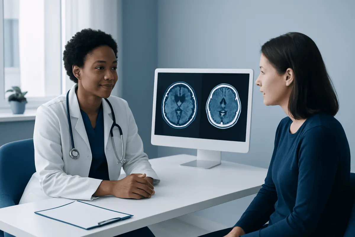
MRI can detect cancer better than CT in certain body areas, especially where soft-tissue detail is essential. It is often more informative for suspected abnormalities in the brain, spinal cord, prostate, female pelvic organs, rectum, liver, bone marrow, and muscles. Its detailed images can help clinicians assess the local size and boundaries of a tumour.
However, MRI is not automatically more sensitive or more accurate for every cancer. CT is usually better for viewing the lungs because air-filled lung tissue is well assessed with CT, and it is commonly preferred for rapid chest imaging. CT can also be more practical when a person is acutely unwell, cannot lie still for a longer examination, or needs a broad assessment of multiple body regions.
The comparison also depends on scan protocol and contrast use. A dedicated liver MRI, for instance, may characterize a liver lesion more clearly after an indeterminate CT result. Conversely, a chest CT may be more appropriate than MRI when evaluating a suspected lung mass or small lung nodules. Specialists may use both studies at different stages because each contributes distinct information.
Even the best imaging test has limits. A suspicious result does not necessarily mean cancer, and a normal result does not completely exclude cancer when symptoms or clinical concern persist. Follow-up imaging, endoscopy, blood tests, or a tissue biopsy may be recommended depending on the findings.
Which Cancers Cannot Be Detected by MRI?
There is no single group of cancers that MRI can never detect. Rather, MRI may be less suitable or less sensitive for certain cancers, particularly when lesions are extremely small, located in moving organs, hidden by air, or require a different imaging method to be seen reliably. Scan quality, the use of contrast, and the expertise of the radiology team also influence detection.
MRI is generally not the preferred first test for many lung cancers because small lung nodules are usually better demonstrated on CT. MRI may also be less practical for finding some cancers of the bowel lining, where colonoscopy directly examines the colon and allows tissue sampling. Very early skin cancers are usually diagnosed through clinical skin examination and biopsy rather than MRI.
Some cancers are difficult to identify on any imaging scan in their earliest stages. Small tumours may not create a visible mass, and diffuse or microscopic cancer cells cannot always be seen. Certain blood cancers, such as leukemia, are primarily diagnosed with blood testing and bone marrow examination, although imaging can be useful in selected situations.
If imaging does not explain ongoing symptoms, a clinician may recommend a different test rather than assuming cancer has been ruled out. The next step may include repeat imaging, targeted ultrasound, endoscopy, laboratory testing, specialist review, or biopsy.
Which Scan Is Most Accurate for Cancer?
No single scan is the most accurate for all cancers. Accuracy depends on the type of cancer, its location, whether the question is detection or staging, and whether clinicians need to identify the primary tumour, lymph node involvement, distant spread, or treatment response.
For example, CT is central to assessing many cancers of the chest, abdomen, and pelvis, while MRI is often preferred for detailed assessment of the brain, spine, pelvis, prostate, and certain liver lesions. Ultrasound may be useful for the thyroid, breasts, abdomen, pelvis, and guiding biopsies. Mammography remains important for breast cancer screening, and PET/CT can be useful in selected cancers to evaluate metabolically active disease.
Pathology is usually the standard method for confirming cancer. During a biopsy, a clinician removes cells or tissue from an abnormal area for examination in a laboratory. Imaging helps identify the safest and most informative biopsy site and shows the extent of disease, but it cannot always determine the exact cancer type.
A multidisciplinary team may combine radiology, pathology, oncology, surgery, and other specialist input to reach an accurate diagnosis and treatment plan. This approach is especially helpful when scan findings are unclear or when more than one imaging method appears appropriate.
What Cancers Are Best Seen on MRI?
MRI is especially valuable for cancers that arise in, spread to, or closely involve soft tissues. It is commonly used to assess brain tumours and possible spread to the brain, because it provides detailed views of brain structures, swelling, and the relationship of a lesion to surrounding tissue. It is also a key test for many spinal tumours and suspected spinal cord compression.
In the pelvis, MRI is often used for prostate cancer evaluation, local staging of rectal cancer, and assessment of cervical, uterine, ovarian, bladder, and other pelvic abnormalities when greater detail is needed. In the abdomen, specialized MRI can help characterize liver lesions and may be used for pancreatic, kidney, or adrenal findings in selected circumstances.
MRI is also useful for sarcomas and other tumours involving muscles, tendons, bones, and soft tissues because it can define tumour extent and its relationship to nearby structures. In breast care, breast MRI may be recommended for selected people, such as those at high risk or those with unclear findings on other breast imaging, but it does not replace routine screening approaches for everyone.
When a scan identifies a suspicious lesion, the next steps depend on its appearance and location. The care team may advise comparison with prior scans, a repeat study after an interval, a targeted biopsy, or referral to a specialist. Imaging decisions should be individualized rather than based solely on a scan type’s general reputation.
What to Do Before, During, and After Each Scan
Before either test, patients should tell the imaging team about pregnancy or possible pregnancy, kidney problems, previous reactions to contrast, asthma or severe allergies, diabetes medicines, and all implants or devices. For MRI, it is particularly important to mention pacemakers, cochlear implants, aneurysm clips, neurostimulators, insulin pumps, metal fragments, and prior surgery. The team will determine whether MRI is safe and whether special precautions are needed.
Some CT and MRI examinations use contrast material to make blood vessels, organs, inflammation, and tumours easier to see. Contrast may be given through a vein or, for some examinations, by mouth or another route. Patients may receive preparation instructions, including whether to avoid food for a specified time beforehand; these instructions should be followed exactly.
During a CT scan, the person lies on a moving table that passes through a ring-shaped scanner. The examination is typically short. During an MRI scan, the person lies still on a table that moves into a longer scanner; the machine makes repetitive loud sounds, and ear protection is provided. If anxiety, pain, or difficulty lying flat may be a concern, patients should discuss this beforehand.
After the scan, a radiologist reviews the images and sends a report to the referring clinician. Patients should ask when and how results will be explained, what the findings mean in their specific situation, and whether further tests are needed. For international patients, Acıbadem Health Point’s multidisciplinary specialists and JCI-accredited hospitals can coordinate diagnostic imaging and cancer care where appropriate.
When to Seek Medical Care
A person should arrange medical assessment for persistent or unexplained symptoms that could need investigation, such as a new lump, unexplained bleeding, ongoing change in bowel or bladder habits, persistent cough, coughing up blood, unexplained weight loss, worsening pain, difficulty swallowing, or unusual fatigue. These symptoms are common and often have causes other than cancer, but timely assessment can help clarify the reason.
Urgent medical care is appropriate for severe or rapidly worsening symptoms, including sudden weakness or numbness, new seizures, significant shortness of breath, severe chest pain, confusion, severe headache with neurological symptoms, or loss of bowel or bladder control with back pain. These symptoms may have several possible causes and should not be self-diagnosed.
People with a previous cancer diagnosis should contact their care team if they develop new symptoms, have concerns about treatment side effects, or are unsure about a planned scan. The treating clinician can decide whether CT, MRI, or another investigation is appropriate and explain the expected benefits and limitations.
Screening scans should not be arranged solely because of worry about cancer without medical advice. Recommended screening tests depend on age, sex, family history, genetic risk, smoking history, and local guidance. A qualified clinician can help determine whether screening, diagnostic imaging, or another form of assessment is suitable.
Frequently asked questions
01Is MRI safer than CT for cancer imaging?
MRI does not use ionizing radiation, while CT uses X-rays. However, safety is not determined by radiation alone: MRI may not be appropriate for everyone with certain implants or metal fragments, and both tests may involve contrast material. The most suitable scan is the one that provides the needed information with appropriate safety precautions.
02Can a CT scan show cancer that an MRI misses?
Yes. CT can be more useful for some findings, especially small lung nodules and certain changes in the chest or bones. MRI and CT reveal different tissue features, so one scan may clarify an abnormality that is not well characterized on the other.
03Can MRI show whether a tumour is cancerous?
MRI can show features that make a lesion appear more or less suspicious, but it cannot always confirm whether it is cancer. In many cases, a biopsy is needed to examine cells or tissue under a microscope and establish a diagnosis.
04Why might a doctor order both an MRI and a CT scan?
The scans may answer different questions. CT can provide a quick overview of the chest, abdomen, pelvis, lungs, or bones, while MRI can add detailed information about a tumour's local extent and nearby soft tissues. Using both can support more accurate staging and treatment planning.
05Does contrast make MRI or CT better for cancer detection?
Contrast can improve the visibility of many tumours, blood vessels, and areas of inflammation by making differences between tissues clearer. Whether contrast is needed depends on the body area and clinical question. The imaging team considers kidney function, allergy history, and other safety factors before administering it.
06How long does it take to get MRI or CT cancer scan results?
Timing varies by facility, scan complexity, and whether the images require specialist comparison with previous studies. A radiologist reviews the images and sends a report to the referring clinician, who explains what the findings mean and discusses any next steps. Patients can ask at the appointment when they should expect results.
This article is for general information only and is not a substitute for professional medical advice. Please consult a qualified doctor about your individual situation.
Joint and spine care in Turkey — expert assessment & treatment
JCI-accredited · board-certified surgeons · reply within 24h
Add us as a Preferred Source to see more of our trusted health content across Google Search, AI Overviews and Discover.