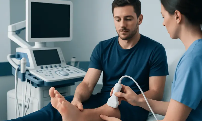
Subchondral Edema Beneath Cartilage: Causes and Care

Subchondral edema describes an area of increased fluid signal in bone just below a joint’s cartilage, most often identified on an MRI scan. It is not a diagnosis by itself; it is an imaging finding that can occur after injury, joint overload, arthritis, inflammation, or less commonly a problem with blood supply to bone.
What Is Subchondral Edema?
Subchondral edema is a term used to describe fluid-related changes in the bone immediately beneath the smooth cartilage that covers the ends of bones in a joint. It is usually found on magnetic resonance imaging (MRI), where it appears as an area of altered signal within the bone marrow. Clinicians may also call it subchondral bone marrow edema or a bone marrow edema pattern.
It is important to understand that subchondral edema is a finding, not a final diagnosis. The image shows that the bone has responded to stress, injury, inflammation, or another process. A clinician interprets it alongside a person’s symptoms, physical examination, medical history, and other MRI features to identify the likely cause.
This finding can occur in many joints, including the knee, hip, ankle, foot, shoulder, and wrist. Its significance varies widely. A small area after a minor injury may settle with time and activity modification, while more extensive changes can signal an underlying cartilage, bone, inflammatory, or circulation-related condition that needs targeted care.
How It May Feel and Where It Occurs
Subchondral edema itself does not always cause symptoms. When it is associated with an active joint problem, the most common complaint is pain near the affected joint. The pain may be deep, aching, or sharp with movement, and it can become more noticeable during walking, running, climbing stairs, lifting, or other load-bearing activities.
Other possible symptoms include stiffness after rest, reduced range of motion, tenderness around the joint, swelling, or a sense that the joint cannot tolerate usual activity. In weight-bearing joints such as the hip, knee, ankle, and foot, pain may lead to limping or avoiding pressure on the limb.
The severity of symptoms does not always match the size of the MRI finding. Some people have substantial MRI changes with modest discomfort, while others experience significant pain from a smaller area. The location, the surrounding cartilage and soft tissues, and the underlying cause all influence how a person feels.
- Knee: pain with stairs, squatting, prolonged standing, or twisting.
- Hip: groin, buttock, or thigh pain, often worse with weight-bearing.
- Ankle or foot: pain after running, jumping, or a change in training load.
- Shoulder: pain during lifting, reaching, or overhead activities.
Why Subchondral Edema Develops
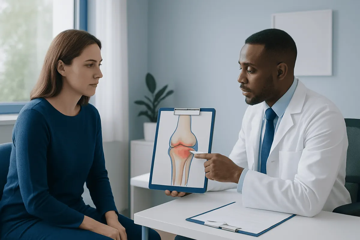
Subchondral edema can develop when the bone beneath cartilage is exposed to more stress than it can comfortably absorb. An acute injury, such as a fall, twist, impact, or sports-related trauma, may cause a bone bruise or tiny structural injuries within the bone. Repetitive loading can have a similar effect, particularly when activity increases quickly or recovery time is limited.
Joint degeneration is another common context. In osteoarthritis, cartilage becomes less effective at distributing force across the joint, and the underlying bone may react with marrow edema changes. Meniscus tears, ligament injuries, cartilage defects, and altered joint alignment can also concentrate stress in one area of subchondral bone.
Inflammatory arthritis, infection, crystal-related joint inflammation, and conditions affecting bone metabolism may sometimes be associated with bone marrow edema. In the hip and other joints, clinicians also consider whether reduced blood supply to bone, known as osteonecrosis, could be contributing. This is different from routine overuse injury and requires careful assessment.
Risk factors depend on the cause but may include previous joint injury or surgery, high-impact or repetitive exercise, sudden training changes, excess mechanical load, poor joint alignment, osteoporosis, inflammatory disease, and use of certain medicines. Having risk factors does not mean subchondral edema will occur, but it can help guide evaluation.
How Doctors Interpret MRI Findings
MRI is the most sensitive imaging test for identifying subchondral edema because it can detect changes in bone marrow and surrounding soft tissues. X-rays may be normal early on, although they can be useful for identifying arthritis, fractures, alignment changes, or later structural changes in bone. CT scans may be considered when detailed assessment of bone structure is needed.
An MRI report may describe the location and extent of the edema, as well as associated findings such as cartilage thinning, a meniscal or ligament injury, fracture line, joint fluid, cyst-like changes, or collapse of the bone surface. These details help distinguish a temporary stress response from a condition that may need closer follow-up.
Diagnosis begins with the person rather than the scan. A doctor will ask about the timing and pattern of pain, injury, sport or work demands, prior joint problems, medicines, and general health. Examination may assess walking, joint movement, tenderness, stability, swelling, strength, and signs of inflammation.
Blood tests are not required for every person. They may be used when symptoms or examination raise concern for infection, inflammatory arthritis, metabolic bone disease, or another systemic cause. Prompt and accurate interpretation is particularly important if there is severe weight-bearing pain, a possible fracture, or concern for osteonecrosis.
Treatment Options and Recovery
Treatment is directed at the underlying reason for the subchondral edema. For an injury- or overload-related finding, temporary reduction of painful impact and weight-bearing activity may allow the bone and joint to recover. A clinician may recommend modifying exercise, using supportive footwear, a brace, or, in selected cases, crutches or another device to reduce pressure on the joint.
Physical therapy can support recovery by improving joint movement, muscle strength, balance, and movement patterns. The program should be individualized: pushing through sharp or escalating pain can prolong symptoms, while complete inactivity for long periods can weaken muscles and reduce function. Lower-impact activities may sometimes be appropriate during recovery, depending on the involved joint and diagnosis.
Pain relief may include non-drug measures such as ice or heat when suitable, as well as medicines recommended by a healthcare professional. Anti-inflammatory medicines are not appropriate for everyone, especially people with certain stomach, kidney, heart, bleeding, or medication-related risks. Injections, procedures, or surgery are considered only for selected underlying conditions, such as significant structural injury, advanced joint disease, or bone collapse.
Recovery time varies from weeks to months and depends on the joint, cause, size of the affected area, and whether cartilage or bone structure is involved. Follow-up imaging is not always necessary, but may be recommended when pain persists, symptoms worsen, or the original findings suggest a risk of structural change. Care for related osteoarthritis or a significant joint injury may be part of the overall plan.
Self-Care and Protecting the Joint
Until a clinician advises otherwise, it is generally sensible to avoid activities that reliably provoke significant pain in the affected joint. For lower-limb symptoms, this may mean pausing running, jumping, deep squatting, or long walks on uneven ground. Gradual return to activity is usually safer than rapidly resuming previous training levels.
Supportive habits can reduce avoidable strain on joints. These include maintaining a healthy body weight where appropriate, wearing footwear suited to the activity, warming up before exercise, and increasing training volume or intensity gradually. Strengthening the muscles around a joint can improve control and load distribution, ideally with professional guidance when pain is ongoing.
People should not assume that every MRI finding needs aggressive treatment, nor should they ignore persistent pain because an injury seems minor. Continuing high-impact activity despite worsening pain may aggravate some underlying problems. A healthcare professional can help determine which activities are safe and when return to sport or work is appropriate.
Nutrition that supports overall bone and muscle health, adequate sleep, and avoiding tobacco are also sensible components of recovery. They do not replace diagnosis or treatment for a structural joint problem, but they can contribute to general musculoskeletal health.
When to Seek Medical Care
A person should arrange medical assessment for joint pain that persists beyond a short period of rest, recurs with normal activity, limits walking or daily tasks, or follows a significant injury. Evaluation is also appropriate if there is ongoing swelling, reduced movement, a limp, or pain that continues to worsen rather than improve.
More urgent medical care is needed after a major fall or impact, if a person cannot bear weight, if the joint looks deformed, or if severe pain develops suddenly. Fever, feeling unwell, redness and warmth around a swollen joint, or pain with signs of infection should be assessed promptly.
People with known inflammatory arthritis, osteoporosis, immune suppression, or risks for osteonecrosis should not delay discussing new, persistent joint pain with a doctor. Early assessment can help identify the cause and protect joint function.
At Acıbadem Health Point, multidisciplinary specialists in JCI-accredited hospitals can assess joint symptoms and coordinate imaging, rehabilitation, and orthopedic care for international patients when needed.
Frequently asked questions
01Is subchondral edema serious?
Subchondral edema can range from a temporary response to injury or overload to a sign of a more significant joint problem. Its importance depends on the location, symptoms, MRI features, and underlying cause. A clinician can explain what it means in the individual situation.
02Can subchondral edema heal on its own?
Some cases, especially those related to a minor injury or temporary overuse, improve with appropriate reduction in joint loading and a gradual return to activity. Healing may take weeks or months. Persistent or worsening symptoms should be reassessed because treatment depends on the cause.
03Does subchondral edema mean arthritis?
No. It may occur with osteoarthritis, but it can also follow trauma, repetitive stress, cartilage injury, inflammation, or other conditions. MRI findings and clinical assessment are needed to determine whether arthritis is present.
04Will subchondral edema show on an X-ray?
Subchondral edema is usually best seen on MRI. An X-ray may appear normal, especially early, because it shows bone structure rather than bone marrow fluid changes. X-rays can still help identify fractures, arthritis, alignment issues, or other structural changes.
05Should exercise be avoided with subchondral edema?
The appropriate level of activity depends on the joint and the cause. Activities that cause sharp, increasing, or lingering pain should generally be reduced until a clinician provides guidance. Rehabilitation often includes a gradual, individualized return to movement rather than complete long-term rest.
06What is the difference between bone marrow edema and subchondral edema?
Bone marrow edema is a broad imaging term for fluid-related changes within bone marrow. Subchondral edema refers specifically to these changes in bone just beneath the cartilage surface of a joint. Both are MRI descriptions and require clinical context to determine the cause.
This article is for general information only and is not a substitute for professional medical advice. Please consult a qualified doctor about your individual situation.
Joint and spine care in Turkey — expert assessment & treatment
JCI-accredited · board-certified surgeons · reply within 24h
Add us as a Preferred Source to see more of our trusted health content across Google Search, AI Overviews and Discover.