Spinal Stenosis Diagnosis: Exams and MRI Scans
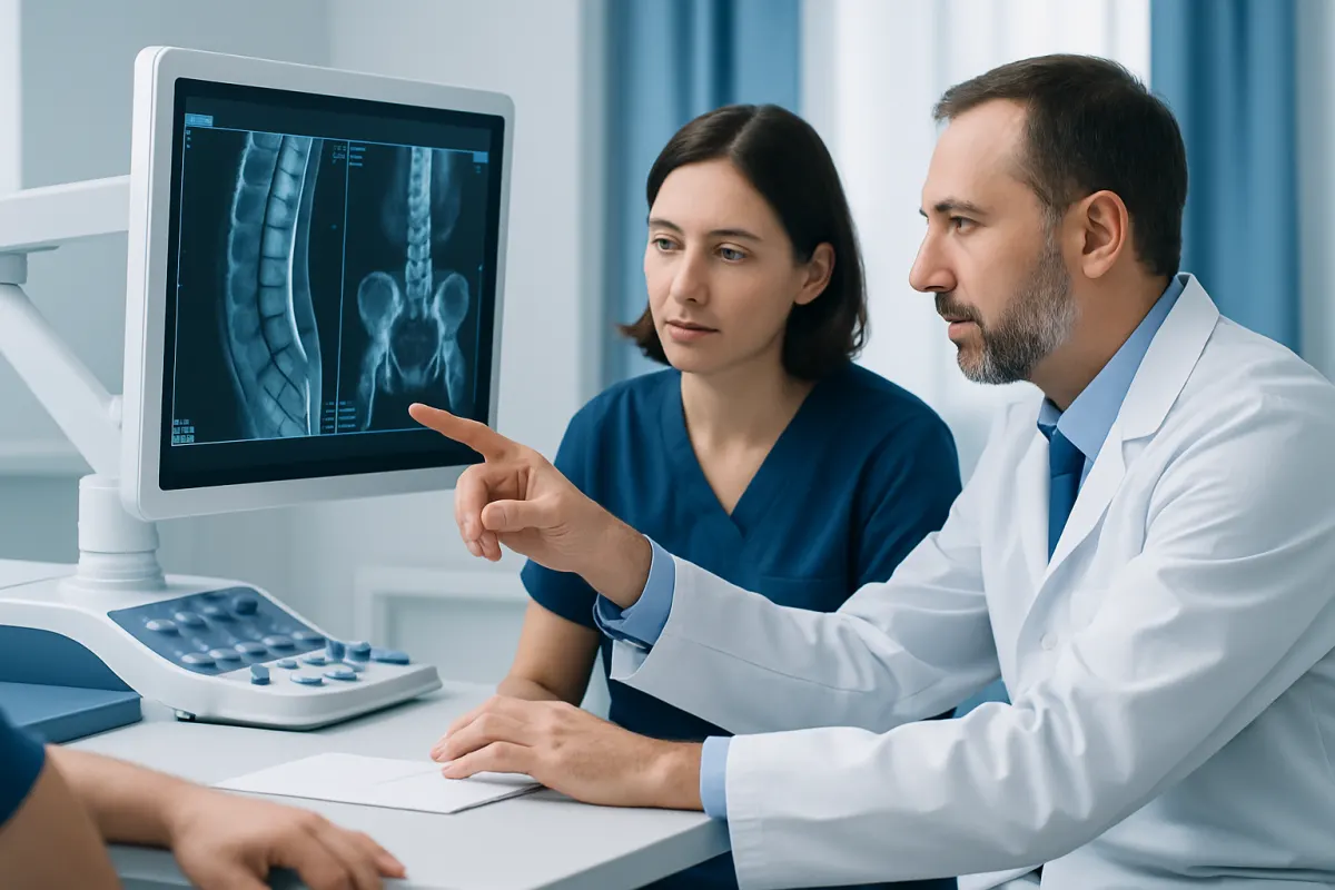
Spinal stenosis diagnosis usually starts with a discussion of symptoms and a physical and neurological examination, followed by imaging when needed. MRI is often the most useful test because it shows the spinal canal, nerves, discs and surrounding soft tissues in detail.
Overview: How Spinal Stenosis Is Diagnosed
Spinal stenosis diagnosis is based on a person’s symptoms, medical history, physical examination and, when appropriate, imaging tests. Doctors look for evidence that narrowing in the spinal canal or nerve openings is affecting nerves and causing symptoms such as leg pain while walking, numbness, tingling or weakness.
Spinal stenosis means that spaces in the spine have become narrower. This may occur in the lower back (lumbar spine), neck (cervical spine) or, less commonly, the middle back. However, an image showing narrowing alone does not confirm that it is the cause of symptoms; many people have age-related spinal changes without pain or nerve problems.
The diagnostic process is therefore designed to connect the clinical picture with the anatomy seen on scans. It also helps exclude other causes of leg, back, neck or arm symptoms, such as circulation problems, hip disease, peripheral neuropathy or another nerve condition.
Symptoms and History Doctors Review
During the appointment, the clinician will ask when symptoms began, where they are felt, what brings them on and what eases them. Lumbar spinal stenosis commonly causes pain, heaviness, cramping, tingling or numbness in one or both buttocks or legs during standing or walking. Many people feel better when sitting down or leaning forward, such as over a shopping trolley.
In cervical spinal stenosis, symptoms may include neck pain, tingling or weakness in the hands or arms, reduced hand coordination, balance difficulties or changes in walking. These features can suggest that the spinal cord, rather than only an individual nerve root, may be affected.
The doctor will also review prior injuries, spine surgery, inflammatory conditions, cancer history, infections, medications and family history. Information about daily activity, work, falls and walking distance can help clarify how symptoms are affecting function and whether further assessment is needed.
- Does pain travel into an arm or leg, or remain in the back or neck?
- Are symptoms worse with walking, standing, extension or certain positions?
- Is there numbness, weakness, clumsiness or difficulty with balance?
- Have bladder or bowel habits changed, or is there numbness around the groin or buttocks?
Physical and Neurological Examination

A physical examination helps determine whether symptoms follow a pattern consistent with nerve compression. The clinician may observe posture and gait, ask the person to walk on heels and toes, and assess how movements of the back or neck affect symptoms. They may also examine the hips, knees, pulses in the legs and other areas that can produce similar symptoms.
A neurological examination commonly includes testing muscle strength, reflexes, sensation and coordination. For example, reduced sensation in a particular part of the leg or arm, weakness in a specific muscle group, or altered reflexes may point to involvement of a particular spinal nerve.
In suspected cervical stenosis, the examination may include tests for signs of spinal cord involvement, as well as balance and hand-function assessment. Examination findings are important, but a normal examination does not always rule out stenosis, particularly if symptoms only occur after prolonged walking or standing.
Imaging and Other Tests
Imaging is selected according to symptoms, examination findings and the clinical question. It is not always required immediately for uncomplicated back pain, but it is often appropriate when symptoms persist, affect function, suggest nerve involvement or raise concern for a more serious cause.
MRI is generally the preferred imaging test for spinal stenosis because it provides detailed views of the spinal canal, spinal cord, nerve roots, discs, ligaments and other soft tissues. It can show where narrowing occurs and whether nerves or the spinal cord appear compressed. In many settings, timely MRI access can help clinicians clarify the diagnosis and plan next steps when symptoms warrant imaging.
X-rays show bone alignment, arthritis-related changes, curvature and possible vertebral slippage. Flexion-extension X-rays may sometimes be used to look for abnormal movement or instability. CT scanning offers detailed bone images and may be used when MRI is unsuitable or when more information about bony narrowing is required. A CT myelogram, involving contrast dye in the spinal fluid space, is reserved for selected situations.
Electromyography and nerve conduction studies may occasionally be recommended if the diagnosis remains uncertain. These tests assess nerve and muscle function and can help distinguish a spinal nerve problem from peripheral neuropathy or another nerve disorder. Blood tests are not used to diagnose stenosis directly, but may help investigate alternative explanations for symptoms.
What the Diagnostic Process May Involve
For most people, the process begins with a consultation and examination. If imaging is indicated, the clinician will request the most appropriate scan and explain what information it is expected to provide. The results are then reviewed alongside symptoms and examination findings rather than considered in isolation.
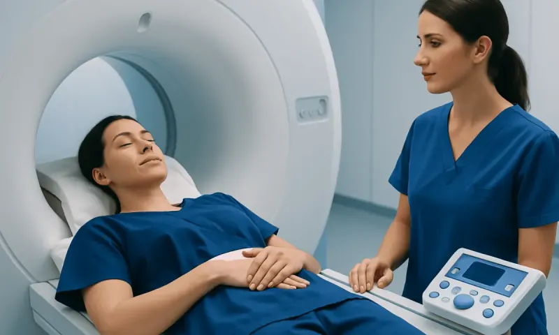
An MRI scan is painless and does not use ionising radiation. The person lies still on a moving table while images are taken inside the scanner; the examination commonly takes between 20 and 60 minutes, depending on the area being studied and the protocol used. The machine can be noisy, and staff provide communication and hearing protection. People who have claustrophobia, implanted devices, metal fragments or possible pregnancy should tell the imaging team beforehand.
Contrast material is not needed for most routine spinal stenosis MRI examinations. It may be considered in specific circumstances, such as prior surgery, suspected infection, inflammation or tumour. After imaging, the clinician explains whether the findings match the symptoms and discusses options, which may range from observation and conservative care to referral for specialist evaluation.
For an overview of the condition and its management, see spinal stenosis. If diagnosis identifies symptomatic narrowing that needs specialist care, spinal stenosis treatment options can be discussed with the treating team.
Benefits, Limits and Possible Risks of Testing
The main benefit of diagnostic testing is greater clarity. Imaging can locate narrowing, identify possible causes such as disc changes, enlarged joints or vertebral slippage, and help doctors decide whether non-surgical care, rehabilitation, injections or surgical consultation may be appropriate. It can also identify findings that require more prompt attention.
Testing has limitations. Degenerative changes are common with age, and their severity on a scan may not match a person’s symptoms. A scan may show narrowing in someone who has no symptoms, while another person may have significant symptoms with relatively modest visible narrowing. This is why decisions should be individualized and based on the whole assessment.
MRI is generally very safe, although it may not be suitable for everyone with certain implanted devices or metal in the body. CT and X-rays involve radiation, so they are used thoughtfully. CT myelography is more invasive and carries uncommon risks such as headache, contrast reaction, bleeding or infection. The care team will explain why a test is recommended and address questions before it is performed.
When to Seek Medical Care
People should arrange a medical assessment for persistent back or neck symptoms that spread into the limbs, interfere with walking, cause numbness or weakness, or do not improve as expected. Earlier review is sensible if symptoms are progressively limiting daily activities, balance, hand coordination or sleep.
Urgent medical assessment is needed for new or worsening weakness, difficulty walking that develops quickly, loss of bladder or bowel control, inability to urinate, or numbness in the groin, genital or inner buttock area. These symptoms can have several causes, but they need prompt evaluation to check for significant nerve compression.
Acıbadem Health Point’s multidisciplinary spine specialists and JCI-accredited hospitals assess and treat spinal conditions for international patients. A qualified clinician can help determine whether symptoms suggest spinal stenosis and which tests are appropriate for the individual.
Frequently asked questions
01Can spinal stenosis be diagnosed without an MRI?
A doctor may suspect spinal stenosis from the symptom pattern and neurological examination. MRI is often used to confirm the location and extent of narrowing, but it may not be necessary in every case. X-rays, CT or other assessments may be more appropriate for some individuals.
02What does an MRI show in spinal stenosis?
MRI can show narrowing of the central spinal canal or the openings where nerves leave the spine. It also shows discs, ligaments, nerve roots and the spinal cord, helping clinicians assess whether these structures may be compressed. The findings need to be matched with the person’s symptoms and examination.
03Does severe narrowing on a scan always mean surgery is needed?
No. Scan findings alone do not determine treatment, because some people have substantial narrowing with few or no symptoms. Decisions are usually based on symptom severity, functional limitations, neurological findings and the response to non-surgical care.
04What is the difference between spinal stenosis and a slipped disc?
Spinal stenosis describes narrowing of spaces in the spine that can affect nerves. A disc herniation, sometimes called a slipped disc, is one possible contributor to narrowing or nerve irritation. Stenosis can also result from joint enlargement, thickened ligaments, vertebral slippage or other age-related changes.
05Are X-rays enough to diagnose spinal stenosis?
X-rays can show alignment, arthritis, bone changes and vertebral slippage, but they do not show nerves and soft tissues as clearly as MRI. They may be useful as part of the evaluation, especially when instability or structural alignment is being assessed. Additional imaging may be needed if nerve compression is suspected.
06How quickly should a person seek help for leg pain when walking?
Leg pain or heaviness that repeatedly occurs with walking and improves with sitting should be discussed with a doctor, especially if it is limiting activity or worsening. The clinician can assess whether the pattern may relate to spinal stenosis, circulation or another condition. New weakness, bladder or bowel changes, or saddle-area numbness need urgent assessment.
This article is for general information only and is not a substitute for professional medical advice. Please consult a qualified doctor about your individual situation.
Joint and spine care in Turkey — expert assessment & treatment
JCI-accredited · board-certified surgeons · reply within 24h
Add us as a Preferred Source to see more of our trusted health content across Google Search, AI Overviews and Discover.