Spinal Stenosis Causes: Changes That Narrow the Spine
Spinal stenosis develops when spaces within the spine become narrower and place pressure on the spinal cord or nearby nerves. Most cases are linked to gradual, age-related changes such as arthritis and disc degeneration, although some people are born with a naturally narrow spinal canal.
Overview: What Causes Spinal Stenosis?
Spinal stenosis causes are usually gradual changes that narrow the passageways in the spine where the spinal cord and nerves travel. These changes commonly include Pain Relief, and Mobility Care" class="ahp-ilk">osteoarthritis, bulging or herniated discs, thickened ligaments and bony overgrowths. As the available space becomes smaller, nerves may become irritated or compressed.
Spinal stenosis is most often found in the lower back, called lumbar spinal stenosis, or the neck, called cervical spinal stenosis. Narrowing may also affect the openings where individual spinal nerves leave the spine, known as foraminal stenosis. Some people have narrowing visible on imaging but no symptoms, while others develop pain, numbness, weakness or problems with balance and walking.
The condition is not always preventable, particularly when it results from normal ageing or inherited anatomy. However, understanding the contributing factors can help people recognise symptoms early, protect spinal health and discuss appropriate assessment with a qualified clinician.
How the Spine Narrows Over Time
The spine is made of bones called vertebrae, shock-absorbing discs, small joints, ligaments and nerves. The spinal canal is the central tunnel that contains the spinal cord in the neck and upper back, and the bundle of lower spinal nerves in the lumbar region. There are also side openings through which nerve roots exit toward the arms, chest or legs.
With time, discs can lose water content and height. This may reduce the space between vertebrae and shift more stress onto the facet joints at the back of the spine. In response, the joints may enlarge because of arthritis, and small bone spurs may develop. Ligaments that support the spine can also become thicker or stiffer. Together, these changes can reduce room around the nerves.
A disc bulge or disc herniation can further crowd the canal or nerve exit opening. In some cases, one vertebra slips slightly forward over another, a condition called spondylolisthesis, which can add to narrowing. Symptoms depend not only on the degree of narrowing but also on which nerve structures are affected and how well a person tolerates pressure on them.
Common Causes and Risk Factors

Age-related degeneration is the leading contributor to spinal stenosis. It is more common later in adulthood because joint arthritis, disc changes and ligament thickening tend to accumulate over years. However, age alone does not determine whether someone will have symptoms or need treatment.
Other factors can make spinal narrowing more likely or cause it at a younger age:
- A naturally narrow spinal canal: Some people are born with less space around the spinal cord or nerves.
- Osteoarthritis: Degeneration of the facet joints may lead to joint enlargement and bone spur formation.
- Disc degeneration or herniation: A bulging or displaced disc can press into the canal or nerve opening.
- Spondylolisthesis and spinal curvature: Vertebral slippage, scoliosis or other alignment changes can alter spinal space.
- Past injury or surgery: Fractures, healing changes, scar tissue or instability following injury may contribute in some people.
- Less common bone or inflammatory conditions: Disorders such as Paget disease, ankylosing spondylitis or abnormal bone growth may occasionally narrow the canal.
Occupational strain, repeated heavy lifting and smoking may contribute to disc and joint degeneration, but they are not direct explanations for every case. A clinician will consider the whole history, examination and imaging findings rather than assuming a single cause.
Symptoms and Patterns to Recognise
Lumbar spinal stenosis often causes symptoms in the buttocks, thighs, calves or feet. People may notice aching, tingling, numbness, heaviness or weakness when standing or walking. Symptoms may ease when sitting down or leaning forward, such as over a shopping trolley or bicycle handlebars, because these positions can temporarily increase space for the nerves.
Cervical spinal stenosis can affect the neck, shoulders, arms and hands. It may cause neck pain, tingling, numbness or weakness in an arm or hand. If pressure affects the spinal cord itself, symptoms can include reduced hand coordination, difficulty with buttons or handwriting, unsteadiness, altered walking and stiffness in the legs. These signs need timely medical assessment.
Symptoms may develop slowly and fluctuate. Not everyone experiences pain, and the severity of symptoms does not always match the appearance of an MRI or CT scan. Similar leg symptoms can also arise from circulation problems, hip disease, peripheral neuropathy or other conditions, which is why an accurate diagnosis is important.
Diagnosis: Identifying the Source of Nerve Pressure
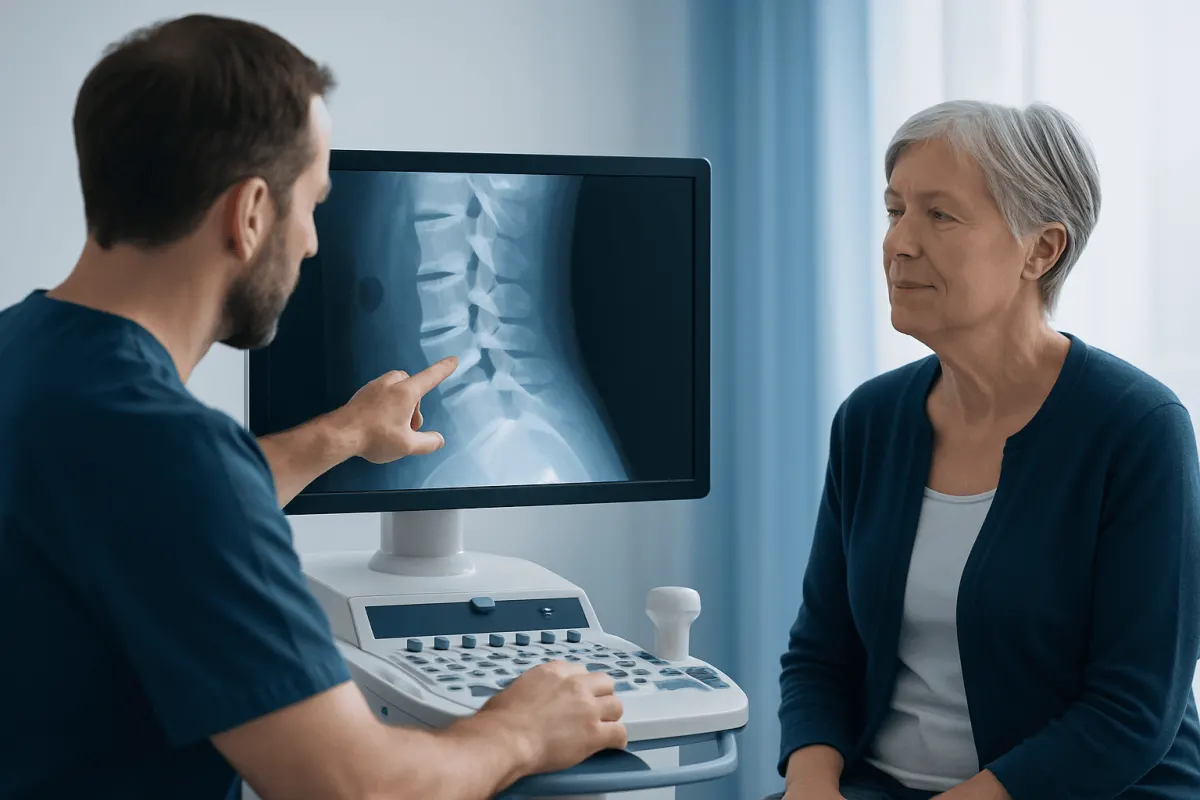
Diagnosis begins with a discussion of symptoms, daily activities, previous injuries, medical conditions and treatments. A clinician will examine posture, walking, spinal movement, muscle strength, reflexes, sensation and balance. The pattern of symptoms can help distinguish narrowing in the spine from other causes of pain or numbness.
Imaging is often used when symptoms persist, affect function or suggest nerve compression. X-rays can show alignment changes, arthritis or vertebral slippage. MRI is particularly useful for viewing discs, ligaments, nerves and the spinal cord. CT scanning or CT myelography may be considered when MRI is unsuitable or when more detail about bone anatomy is needed.
Imaging findings are interpreted alongside symptoms and examination results. Many adults have age-related changes on scans without troublesome symptoms, so treatment decisions should focus on the person rather than the scan alone. More detailed information on assessment and management is available through spinal stenosis care.
Treatment Options and When a Procedure May Help
Treatment is tailored to the location of stenosis, symptom severity, neurological findings and personal goals. For mild to moderate symptoms, clinicians may recommend activity modification, physiotherapy, exercises to improve strength and mobility, and pain-relief strategies when appropriate. Some people benefit from guided injections to reduce inflammation around irritated nerves, although these do not remove the underlying narrowing.
Surgery may be considered when symptoms remain limiting despite non-surgical treatment, when there is progressive weakness or when spinal cord compression is affecting walking, balance or hand function. The main aim is to create more room for the nerves. A decompression procedure, often called laminectomy, removes part of the bone or thickened tissue contributing to pressure. If there is significant instability or vertebral slippage, fusion may sometimes be performed at the same time.
Suitable candidates are assessed individually. The surgeon considers imaging, nerve findings, overall health, bone quality, previous spinal operations and whether symptoms are likely to come from the narrowed area. A spinal procedure does not reverse all age-related changes, and it may not resolve symptoms caused by another condition, so shared decision-making is essential. For a fuller explanation of operative options, patients can review spinal stenosis treatment.
When surgery is chosen, preparation commonly includes pre-operative medical assessment and planning with the surgical team. During the operation, the surgeon reaches the affected spinal level, removes or reshapes structures narrowing the nerve space, and adds stabilisation only if needed. Hospital stay and recovery differ according to the procedure and a person’s health. Walking often starts soon after surgery, while return to work, driving and fuller activity may take weeks to months. Potential benefits include improved walking tolerance and reduced nerve symptoms; risks include infection, bleeding, blood clots, spinal fluid leak, nerve injury, persistent symptoms and a possible need for further treatment.
Prevention and Everyday Self-care
Not all spinal stenosis can be prevented, especially when inherited anatomy or normal ageing plays a role. Still, regular physical activity can support muscle strength, flexibility, balance and overall function. Low-impact options such as walking, swimming, cycling and clinician-guided strengthening may be more comfortable for people whose symptoms increase with standing upright.
Maintaining a weight that is appropriate for the individual, avoiding tobacco and using safe lifting techniques may help reduce strain on the spine and support general musculoskeletal health. A physiotherapist can adapt an exercise plan to symptoms and mobility. Exercises should not cause rapidly worsening pain, new weakness or spreading numbness.
Simple practical adjustments can also be useful, such as pacing activities, taking seated breaks, using supportive footwear and improving fall safety at home if balance is affected. Self-care measures should complement, not replace, professional assessment when neurological symptoms are present.
When to Seek Medical Care
A doctor should assess back or neck symptoms that persist, limit walking or daily activities, or are accompanied by tingling, numbness or weakness in an arm or leg. Early evaluation is especially helpful when symptoms are gradually worsening or when a person notices declining hand coordination, balance or endurance.
Urgent medical care is needed for new loss of bladder or bowel control, numbness around the groin or inner thighs, sudden or rapidly increasing limb weakness, or major difficulty walking. These symptoms can indicate significant nerve compression and require prompt assessment.
Acıbadem Health Point’s multidisciplinary spine specialists and JCI-accredited hospitals diagnose and treat spinal conditions for international patients. A clinician can help determine whether symptoms are related to spinal stenosis and discuss the safest next steps for the individual.
Frequently asked questions
01What is the most common cause of spinal stenosis?
The most common cause is age-related degeneration of the spine. Arthritis of the small spinal joints, loss of disc height, bone spur formation and thickened ligaments can gradually narrow the spaces around nerves.
02Can spinal stenosis occur in younger adults?
Yes. Although it is more common with ageing, younger adults may develop stenosis because they were born with a narrow spinal canal, have a disc herniation, experienced an injury or have certain spinal alignment or bone conditions. A clinician can identify the likely contributing factors.
03Does a bulging disc always mean spinal stenosis?
No. Disc bulges are common and may not cause symptoms or significant narrowing. Spinal stenosis is diagnosed by considering whether the spinal canal or nerve openings are narrowed and whether that finding matches a person’s symptoms and examination.
04Why does lumbar spinal stenosis often feel better when sitting?
Sitting or bending forward can slightly open the spaces in the lower spine, reducing pressure on irritated nerves. This is why some people find walking uphill or leaning forward more comfortable than standing upright for long periods.
05Can exercise make spinal stenosis worse?
Appropriate exercise is often helpful for strength, mobility and function, but the programme should be matched to the person’s symptoms. New weakness, worsening numbness or severe pain during activity should be discussed with a clinician or physiotherapist.
06When is surgery considered for spinal stenosis?
Surgery may be considered when non-surgical care has not adequately improved disabling symptoms, or when there is progressive weakness or spinal cord compression. The decision depends on symptoms, examination findings, imaging, general health and the person’s preferences.
This article is for general information only and is not a substitute for professional medical advice. Please consult a qualified doctor about your individual situation.
Joint and spine care in Turkey — expert assessment & treatment
JCI-accredited · board-certified surgeons · reply within 24h
Add us as a Preferred Source to see more of our trusted health content across Google Search, AI Overviews and Discover.