Skin Cancer Rash: Signs, Diagnosis, and Treatment
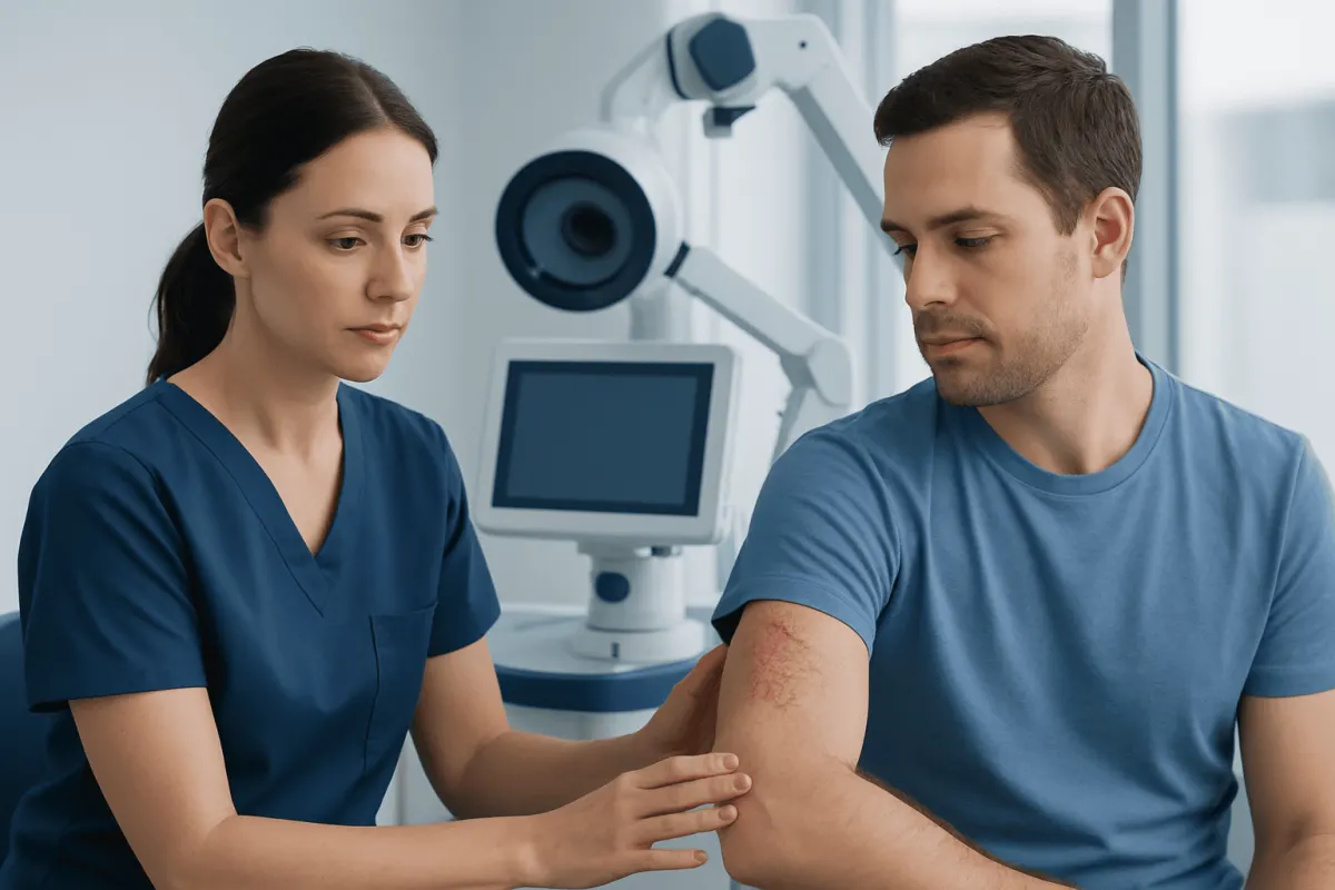
A skin cancer rash may look like a scaly, crusted, red, itchy, bleeding, or non-healing area of skin, but many harmless conditions can look similar. Any spot or rash that persists, changes, or repeatedly returns should be examined by a qualified clinician.
Overview: Can a Rash Be Skin Cancer?
A skin cancer rash is not a single, specific rash. It describes a skin change that may resemble eczema, psoriasis, an allergic reaction, a sore, or a patch of dry skin but is actually caused by cancerous or precancerous cells. The area may remain in one place, slowly enlarge, crust over, itch, bleed, or fail to heal despite basic skin care.
Most rashes are not skin cancer. Irritant dermatitis, fungal infections, eczema, psoriasis, insect bites, and medication reactions are much more common explanations. However, a persistent or unusual patch deserves clinical assessment because visual appearance alone cannot reliably distinguish a benign rash from skin cancer.
The most common skin cancers are basal cell carcinoma and squamous cell carcinoma. Melanoma is less common but can be more likely to spread if not detected early. Some uncommon cancers, including cutaneous T-cell lymphoma, may also begin with rash-like patches. A dermatologist or other qualified clinician can assess the pattern, examine the whole skin surface when appropriate, and arrange testing if needed.
How a Skin Cancer Rash May Appear
Skin cancer may develop on sun-exposed areas such as the face, ears, scalp, neck, chest, shoulders, forearms, hands, and lower legs. It can also occur in less exposed areas, including the soles, nails, genital region, and areas between the toes. People of every skin tone can develop skin cancer, although signs may be less obvious in darker skin.
A rash-like skin cancer may appear as a rough, scaly, pink, red, brown, purple, or darker-than-surrounding patch. It may be flat or raised, tender or painless. Squamous cell carcinoma may look like a persistent scaly plaque or a firm, crusted bump. Basal cell carcinoma may present as a pearly or translucent bump, a pink patch, or a sore that repeatedly heals and reopens.
Features that should prompt attention include a spot that changes in size, shape, color, thickness, or sensation; bleeding without a clear injury; persistent crusting; a sore that does not heal; or a patch that does not respond as expected to treatment for eczema or another common rash. Itching can occur, but its presence or absence does not determine whether a lesion is cancerous.
- Persistent: present for weeks and not improving.
- Progressive: slowly enlarging, thickening, or becoming more raised.
- Fragile: bleeding, ulcerating, or forming a recurring scab.
- Different: unlike other spots on the person’s skin, sometimes called an “ugly duckling” lesion.
What Does Stage 1 Skin Cancer Look Like?
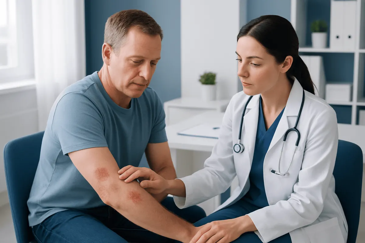
Stage 1 skin cancer usually refers to an early invasive cancer that is small and has not spread to nearby lymph nodes or distant organs. It cannot be identified with certainty by looking at the skin alone. Staging depends on the cancer type, its measured size and depth, features seen under a microscope, and sometimes imaging or lymph node assessment.
For many early non-melanoma skin cancers, the lesion may appear as a small localized scaly patch, persistent sore, shiny bump, or crusted area. An early melanoma may be a new or changing pigmented spot, although some melanomas are pink, red, skin-colored, or darker rather than brown or black. The ABCDE approach can be helpful for pigmented lesions: asymmetry, irregular border, color variation, diameter that is increasing, and evolution or change.
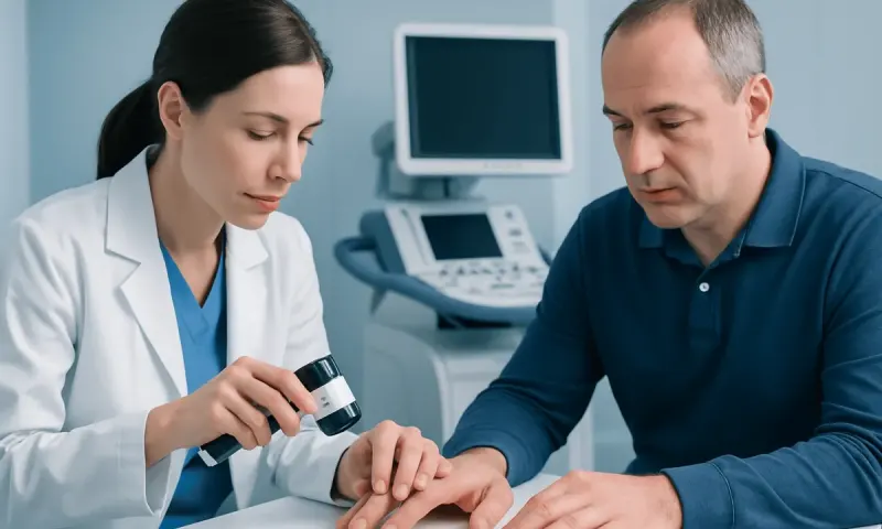
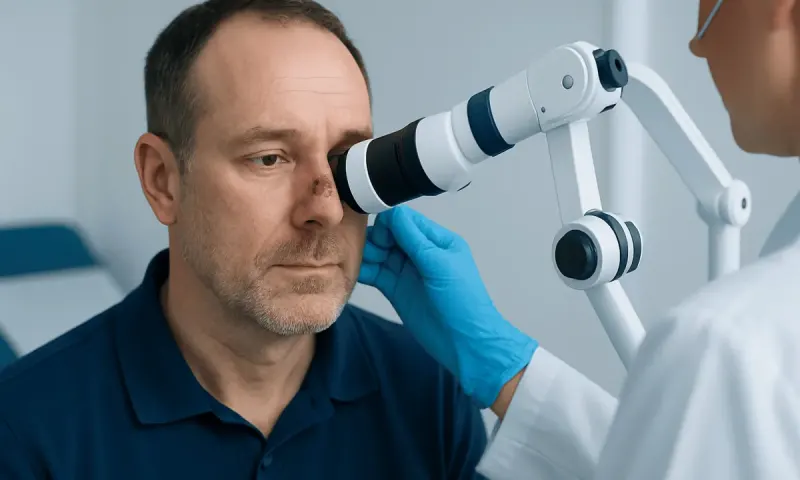
Importantly, a small lesion is not automatically harmless, and a larger lesion is not automatically cancer. A clinician may use dermoscopy, a handheld magnifying device with light, to inspect skin structures that are not visible to the unaided eye. A biopsy is the definitive way to confirm the diagnosis and provide information used for staging.
Causes and Factors That Increase Risk
Skin cancer develops when genetic changes build up in skin cells. Ultraviolet radiation from sunlight and tanning devices is a major preventable cause. Repeated sun exposure over years is associated with many basal and squamous cell carcinomas, while intense intermittent sun exposure and sunburns can contribute to melanoma risk.
Risk can also be influenced by fair or easily burning skin, a history of sunburn, many moles, atypical moles, previous skin cancer, a family history of melanoma, older age, outdoor work or recreation, radiation exposure, and immune suppression. Medicines or health conditions that suppress the immune system may increase the likelihood of certain skin cancers and can change how closely a person needs to be monitored.
Skin cancer can still occur without obvious risk factors. It may develop in people with darker skin and in areas that receive little sun. Rather than trying to decide risk based on one factor, it is more useful to notice new, changing, non-healing, or unexplained skin changes and seek medical advice when they persist.
Diagnosis: Why a Biopsy Matters
Diagnosis begins with a medical history and careful examination of the affected area. The clinician may ask when the rash began, whether it is changing, whether it bleeds or hurts, prior sun exposure, personal or family history of skin cancer, and whether creams or other treatments have helped. A full-body skin examination may be recommended depending on the finding and the person’s risk profile.
If skin cancer is suspected, the clinician usually performs or arranges a biopsy. This involves removing a small sample or, in some cases, the entire lesion under local anesthetic. A pathologist examines the tissue under a microscope to determine whether cancer is present and, if so, its exact type and features.
Not every rash needs a biopsy. When a diagnosis is uncertain, a clinician may first consider other causes, use dermoscopy, or recommend a short period of treatment and review. A rash that does not improve as expected, however, should be reassessed rather than repeatedly treated without a clear diagnosis. Confirmed skin cancers may then be staged to guide the most suitable plan.
What Are the Treatment Options for a Skin Cancer Rash?
Treatment is directed at the diagnosed cancer, not the rash-like appearance alone. The best option depends on the cancer type, size, depth, location, stage, previous treatment, the person’s general health, and cosmetic or functional considerations. For many localized skin cancers, surgery to remove the cancer with an appropriate margin of normal tissue is the main treatment.
Specialized techniques may be used for cancers on high-risk or cosmetically sensitive areas, such as the face, ears, hands, or genital region. Other options can include curettage and electrodessication, cryotherapy for selected precancerous lesions, topical medicines for certain superficial cancers, photodynamic therapy, or radiation therapy when surgery is not appropriate. A clinician will explain expected benefits, side effects, wound care, and follow-up needs.
For melanoma and higher-risk squamous cell carcinoma, treatment may include wider surgery and, in selected cases, assessment of nearby lymph nodes. Advanced skin cancers may be treated with systemic therapies such as immunotherapy, targeted therapy, or other anticancer medicines. These decisions are made by a specialist team. Information about skin cancer can help patients understand the broader range of diagnoses and care pathways.
Acıbadem Health Point’s multidisciplinary specialists and JCI-accredited hospitals assess and treat skin cancer for international patients, with care plans based on the confirmed diagnosis and individual clinical needs.
When to Seek Medical Care
A person should arrange a medical appointment for a rash or skin lesion that lasts longer than about two weeks without clear improvement, especially if it is growing, bleeding, crusting, painful, or repeatedly breaking down. Prompt assessment is also sensible for a new or changing mole, a dark streak under a nail, or a lesion on the lip, ear, scalp, genital skin, palm, or sole.
More urgent review is appropriate if a lesion is rapidly enlarging, persistently bleeding, becoming ulcerated, or accompanied by a new swollen lymph node. These symptoms do not necessarily mean cancer, but timely examination can clarify the cause and prevent delays in care.
It is helpful to take a clear photograph with the date, note any changes, and avoid picking at the area before the appointment. Over-the-counter steroid creams may reduce inflammation from benign rashes but should not be relied upon for a persistent, unexplained lesion. A clinician can advise whether a dermatology referral or biopsy is needed.
What is the 2 week rule for skin cancer? The “2 week rule” is a practical reminder, not a diagnostic test: a skin spot, sore, or rash that has not healed or clearly improved after around two weeks should be checked by a healthcare professional. Some benign skin problems also last longer than two weeks, while some cancers change slowly, so any suspicious or evolving lesion should be assessed even if the timing does not fit this rule exactly.
How to Deal With a Skin Cancer Diagnosis
A skin Prostate Cancer Diagnosis: Tests and Next Steps" class="ahp-ilk">cancer diagnosis can feel unexpected, but it is reasonable to take time to understand the pathology result and proposed treatment. Patients may wish to ask what type of skin cancer it is, whether it has been staged, what treatment is recommended, whether there are alternatives, what the scar or recovery may involve, and how often follow-up skin checks are advised.
Bringing a family member or trusted friend to appointments, writing down questions, and requesting a copy of the pathology report can make discussions easier. Emotional support is also important. Speaking with the clinical team, a counselor, or a cancer support organization may help a person manage uncertainty while decisions are being made.
After treatment, regular self-examination and follow-up visits help identify new skin cancers or recurrence early. Sun protection remains important: seek shade when practical, wear protective clothing and a broad-brimmed hat, use broad-spectrum sunscreen as part of a wider sun-safety routine, and avoid indoor tanning. These measures reduce additional ultraviolet damage but do not replace professional skin assessment for a suspicious rash.
Frequently asked questions
01Can skin cancer look like eczema or a dry rash?
Yes. Some skin cancers, particularly certain squamous cell carcinomas and cutaneous lymphomas, can resemble eczema, psoriasis, or a dry scaly patch. A rash that remains in the same place, fails to improve with appropriate treatment, or changes over time should be assessed by a clinician.
02Is an itchy rash usually skin cancer?
Itching is common in many harmless skin conditions and does not by itself mean skin cancer. However, a spot that itches and also grows, bleeds, crusts, changes color, or does not heal should be checked.
03Can a doctor diagnose a skin cancer rash just by looking at it?
A clinician can often identify suspicious features during examination, sometimes with dermoscopy. However, a biopsy is usually needed to confirm whether cancer is present and to identify its type accurately.
04Can skin cancer appear suddenly?
Some skin cancers become noticeable over a relatively short period, while others grow slowly over months or years. A new spot that looks different from other marks on the skin, or any existing spot that changes, is worth medical review.
05Does a skin cancer rash go away with steroid cream?
A steroid cream may reduce redness or itching around some benign rashes, but it does not treat skin cancer. Temporary improvement does not rule out cancer, particularly if the patch persists or returns after treatment is stopped.
06What happens after a skin cancer biopsy?
The sample is examined by a pathologist, and the result often identifies the exact type of lesion and whether further treatment is needed. The treating clinician will discuss the result, treatment choices, wound care, and an appropriate follow-up plan.
This article is for general information only and is not a substitute for professional medical advice. Please consult a qualified doctor about your individual situation.
Hair transplant in Turkey — costs, graft counts & a free assessment
JCI-accredited · board-certified surgeons · reply within 24h
Add us as a Preferred Source to see more of our trusted health content across Google Search, AI Overviews and Discover.