Sciatica Diagnosis: Exams, Imaging, and Next Steps
Sciatica diagnosis begins with a clinician listening carefully to the pattern of leg pain and performing a focused back, strength, sensation and reflex examination. Scans and nerve tests are not always needed, but they can help identify the cause when symptoms are severe, persistent, unusual or associated with warning signs.
Overview: how sciatica is diagnosed
Sciatica diagnosis is usually made by combining a person’s symptoms with a focused physical and neurological examination. A clinician looks for pain that travels from the lower back or buttock into one leg, together with findings that suggest a spinal nerve root is irritated or compressed. Imaging is often unnecessary in the first weeks unless there are warning signs or the result is likely to change treatment.
The term sciatica describes nerve-related leg pain rather than one single disease. A common cause is a lumbar disc herniation, but spinal narrowing, age-related changes, injury and, less commonly, other conditions can produce similar symptoms. The aim of assessment is to identify the likely pain source, understand how it affects movement and daily life, and rule out conditions needing prompt care.
Many people improve with time and non-surgical care. A careful diagnosis helps clinicians avoid tests that are unlikely to help while arranging timely imaging or specialist review when it is appropriate. For an overview of the condition and its management, see sciatica.
The first appointment: symptoms, history and examination
A clinician will ask when the pain began, where it travels, and whether it is sharp, burning, tingling or numb. Pain that extends below the knee and follows a narrow path down the back or side of the leg can support sciatica, although symptoms vary. They will also ask whether coughing, sneezing, sitting, standing or walking changes the pain.
The medical history includes recent injuries, work or activity demands, previous back episodes, fever, Weight Loss: Possible Causes and Medical Evaluation" class="ahp-ilk">unexplained weight loss, cancer history, immune suppression, osteoporosis and use of medicines that affect bleeding. These questions are important because they help identify less common causes of back and leg pain.
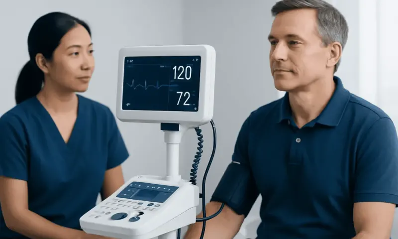
The examination usually includes observing posture and walking, checking lower-back movement and gently assessing the hips. A clinician tests leg and foot strength, skin sensation and tendon reflexes. Differences between sides can indicate which nerve root may be affected and whether there is significant nerve dysfunction.
A commonly used manoeuvre is the straight-leg raise. While the person lies down, the clinician slowly raises the straight leg; reproduction of familiar shooting leg pain at a particular point may suggest nerve-root irritation. This test is interpreted alongside the full history and examination, as tight muscles or hip conditions may also cause discomfort.
Tests used in sciatica diagnosis

There is no single blood test that confirms sciatica. In people with a typical symptom pattern and no concerning findings, routine blood tests are often not needed. Blood tests may be requested if infection, inflammation, cancer or another systemic illness is considered possible based on symptoms or medical history.
Imaging is selected to answer a specific clinical question. MRI is the main scan used when detailed pictures of discs, nerves, the spinal canal and nearby soft tissues are needed. It can show a disc herniation or spinal narrowing that matches the person’s symptoms and examination. MRI findings must be interpreted carefully, because disc bulges and other age-related changes are also common in people without pain.
CT scanning can provide detailed images of bone and may be used when MRI is not suitable or is unavailable. A CT myelogram, in which contrast dye is placed around the spinal nerves before CT imaging, is reserved for selected situations. X-rays can show fractures, alignment changes or some degenerative bone changes, but they do not directly show discs or nerve compression.
Electromyography and nerve conduction studies, often called EMG/NCS, measure electrical activity in muscles and nerves. They may be useful when the diagnosis is uncertain, when symptoms may come from a peripheral nerve problem rather than the spine, or when clinicians need further information about nerve injury. These studies are not routinely required for every person with suspected sciatica.
When imaging is appropriate and what to expect
For uncomplicated recent sciatica, early MRI often does not improve outcomes and may reveal incidental changes that are not responsible for symptoms. Imaging is more likely to be recommended if pain remains troublesome despite an appropriate period of conservative care, symptoms are progressing, there is marked weakness, or a spinal injection or surgery is being considered.
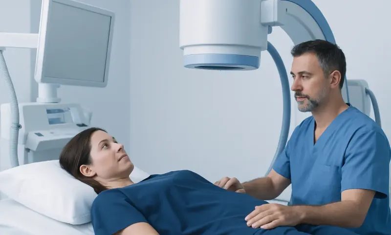
MRI uses a strong magnetic field and radio waves, not ionising radiation. Before the scan, staff check for implanted devices, metal fragments, pregnancy considerations and severe claustrophobia. The person lies still on a moving table while it passes through the scanner. The scan is painless, but the machine is noisy; ear protection is normally provided. Contrast material is not routinely required for standard sciatica assessment, though it may be used in selected circumstances.
CT is faster and can be helpful in certain clinical settings, but it uses X-rays. The most suitable test depends on the suspected cause, urgency, medical history and local availability. Faster access to MRI may be valuable when imaging is clinically indicated, but a scan should remain part of a full assessment rather than a replacement for it.
A radiologist interprets the images, and the treating clinician explains whether the findings fit the pain pattern and examination. An imaging abnormality alone does not automatically mean a procedure is necessary. Decisions are based on symptoms, function, neurological findings, scan results and the person’s preferences.
How results guide treatment decisions
Once sciatica diagnosis is established, care is tailored to the likely cause and severity. Most people are initially advised to remain gently active as tolerated, avoid prolonged bed rest, and use appropriate pain-relief and rehabilitation strategies recommended by a clinician. Physiotherapy may support gradual return to usual movement, strength and confidence.
If symptoms continue or substantially limit function, a specialist may discuss targeted options such as image-guided spinal injections in carefully selected cases. These procedures may reduce inflammation-related pain for some people, but their expected benefits, limitations and risks should be discussed individually.
Surgery is generally considered when there is persistent disabling leg pain with a matching structural cause on imaging after non-surgical treatment, or when there is significant or worsening neurological deficit. A common operation for disc-related nerve compression is removal of the part of the disc pressing on the nerve. Information on sciatica treatment options can help patients understand the broader care pathway.
The benefit of a procedure is greatest when the symptoms, examination and scan all point to the same pain source. Risks depend on the treatment and may include infection, bleeding, nerve injury, incomplete symptom relief or recurrence. A spine specialist can explain the likely recovery timeline: recovery after diagnostic testing is immediate, while rehabilitation after injections or surgery varies according to the procedure and individual health needs.
Preparation, follow-up and self-care while awaiting assessment
Before an appointment, it can help to note when symptoms started, where pain travels, what makes it better or worse, and whether numbness or weakness is present. Bringing a list of medicines, relevant medical history and previous scan reports can make the consultation more efficient. It is also useful to describe how symptoms affect sleep, walking, work and daily activities.
While awaiting assessment, gentle movement and position changes are generally preferable to prolonged inactivity. Activities should be paced to avoid major flare-ups, and heavy lifting or movements that sharply worsen leg pain may need to be limited temporarily. A clinician or physiotherapist can advise on safe activity adjustments for the individual.
People should avoid interpreting scan reports without clinical guidance. Terms such as “disc bulge” or “degeneration” are common and do not always explain pain. The key question is whether a finding corresponds with the person’s symptoms and neurological examination.
For international patients who need coordinated assessment, Acıbadem Health Point’s multidisciplinary specialists and JCI-accredited hospitals diagnose and treat spinal conditions, with imaging and relevant specialty consultation arranged according to clinical need.
When to seek medical care
Medical assessment is appropriate for leg pain that is severe, lasts beyond a short period, repeatedly returns, interferes with walking or sleep, or is accompanied by numbness or weakness. An earlier appointment is especially important if foot weakness, frequent tripping or difficulty standing on the toes or heels develops.
Urgent medical care is needed for new difficulty controlling urine or bowel movements, inability to pass urine, numbness around the genitals, buttocks or inner thighs, or rapidly worsening weakness in one or both legs. These symptoms can rarely signal serious compression of the lower spinal nerves and should not be managed at home.
Prompt evaluation is also advisable when back and leg pain follows a significant injury or occurs with fever, feeling unwell, unexplained weight loss, a history of cancer, or factors that increase the risk of infection or fracture. Most sciatica is not caused by these conditions, but checking for them is an important part of safe diagnosis.
Frequently asked questions
01Can sciatica be diagnosed without an MRI?
Yes. Many cases can be diagnosed from the symptom pattern and a neurological examination. MRI is usually reserved for persistent, severe, progressive or atypical symptoms, or when the result may guide an injection or surgery.
02What does a neurological examination for sciatica involve?
The clinician checks how a person walks, moves and balances, then assesses leg strength, sensation and reflexes. They may also perform a straight-leg raise or related test to see whether leg movement reproduces typical nerve pain.
03Will an X-ray show sciatica?
An X-ray cannot directly show the sciatic nerve or most disc herniations. It may help assess bone alignment, fractures or certain structural changes, while MRI is usually more informative for discs and nerve compression.
04How long does an MRI for sciatica take?
The scanning time varies by facility and the body area being assessed, but a lumbar spine MRI commonly takes less than an hour. The person needs to lie still, and the examination itself is painless.
05Do all disc bulges cause sciatica?
No. Disc bulges are common on imaging, including in people with no back or leg symptoms. A clinician considers whether a disc finding is in the expected location and matches the pain pattern and examination findings.
06When is sciatica an emergency?
Sciatica requires urgent assessment if there is new loss of bladder or bowel control, inability to urinate, numbness around the groin or buttocks, or rapidly worsening leg weakness. These signs may indicate serious nerve compression and need immediate medical attention.
This article is for general information only and is not a substitute for professional medical advice. Please consult a qualified doctor about your individual situation.
Medical weight-loss options in Turkey — costs & eligibility
JCI-accredited · board-certified surgeons · reply within 24h
Add us as a Preferred Source to see more of our trusted health content across Google Search, AI Overviews and Discover.