Posterior Disc Bulge: Symptoms, Causes, And Care
A posterior disc bulge occurs when a spinal disc extends beyond its usual boundary toward the back of the spinal canal. Many posterior disc bulges cause no symptoms, while others may contribute to back or neck pain, tingling, numbness, or weakness if nearby nerve structures are irritated.
Posterior Disc Bulge: What It Means
A posterior disc bulge means that part of an intervertebral disc extends outward toward the back of the spine. Discs are flexible cushions between the vertebral bones. Their outer ring, called the annulus fibrosus, surrounds a softer center and helps distribute load during movement.
The word posterior describes direction: the bulge points toward the spinal canal, where the spinal cord and nerve roots travel. A bulge may be broad-based and involve a larger portion of the disc edge, rather than being a small focal area. It can occur in the neck, mid-back, or lower back, although the lower back and neck are common locations.
Importantly, a scan finding does not always explain a person’s symptoms. Disc bulges are frequently seen in people without back pain or neck pain, especially as they get older. Clinicians therefore consider the scan together with the location of symptoms, neurological examination, medical history, and day-to-day function.
Bulge, Protrusion, Herniation and Degeneration: Important Differences
Terms used in spine imaging can sound concerning, but they describe anatomy rather than predict a person’s future. A disc bulge generally refers to disc material extending beyond the usual disc margin over a broad area. A protrusion is a more focal displacement. A herniation is an umbrella term that may include protrusion or extrusion, depending on how disc material extends through the outer disc fibers.
Disc degeneration refers to gradual age- and load-related changes in the disc, such as loss of water content and flexibility. These changes can make a bulge more likely, but they are not necessarily a disease requiring treatment. Many people remain active and comfortable despite degenerative findings on imaging.
Whether a posterior disc bulge is clinically significant depends largely on whether it narrows the space for a nerve root or, less commonly, the spinal cord. A report may also mention spinal canal narrowing or foraminal narrowing, which means reduced space where nerves travel. These terms should be discussed with a qualified clinician in the context of the individual’s symptoms.
Possible Symptoms and How They Vary
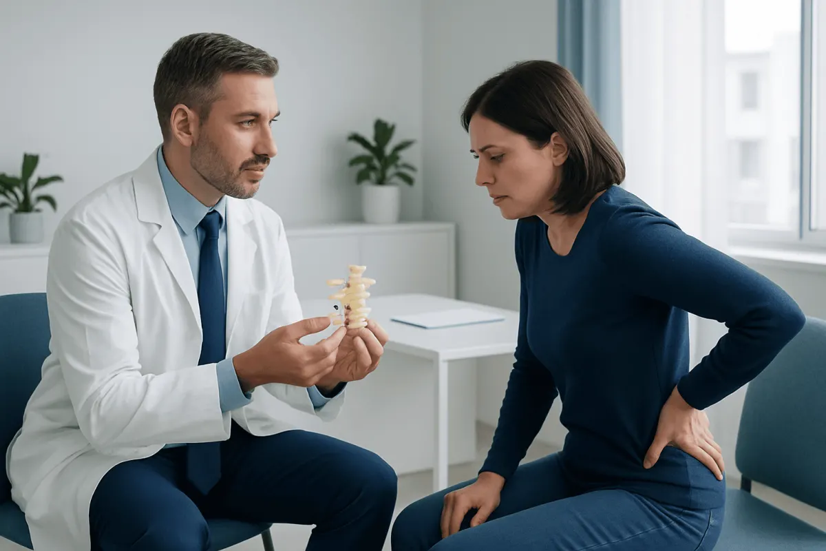
A posterior disc bulge may cause no symptoms at all. When it is related to symptoms, the most common concern is local neck or back pain. Pain may fluctuate with posture, prolonged sitting, lifting, bending, coughing, or certain movements, although no single movement pattern can diagnose a disc problem.
If a bulge irritates or compresses a nerve root, symptoms may travel along that nerve’s pathway. In the lower back, this can include pain, pins and needles, numbness, or weakness in the buttock, leg, foot, or toes. This pattern is often called sciatica and may be related to sciatica, although several conditions can produce similar symptoms.
In the neck, nerve irritation can cause pain that spreads to the shoulder, arm, or hand, with tingling or altered sensation. More significant compression in the cervical spine can occasionally affect hand coordination, balance, or walking. These symptoms deserve timely medical review because they may indicate involvement beyond a single nerve root.
- Symptoms may be mild, intermittent, or absent.
- The intensity of pain does not always match the size of a bulge on MRI.
- Weakness, progressive numbness, or loss of coordination needs clinical assessment.
Why Posterior Disc Bulges Develop
Most posterior disc bulges arise through a combination of normal aging, repeated physical loading, and individual susceptibility. Over time, discs can lose some water content and elasticity. The outer disc fibers may gradually weaken, allowing the disc contour to extend outward under everyday pressure.
Genetics may influence how early or how rapidly disc changes develop. Work or sport involving repetitive bending, twisting, heavy lifting, vibration exposure, or long periods in one posture can add strain, but a single activity is rarely the only explanation. A sudden injury can also contribute to a disc injury, particularly if it occurs during forceful lifting or an accident.
Smoking, low levels of physical activity, excess body weight, and reduced trunk strength may be associated with poorer spinal health or more persistent symptoms. These factors are not reasons for self-blame. They are potential areas where gradual, supported changes may improve overall health, resilience, and recovery.
How Doctors Assess a Posterior Disc Bulge
Assessment usually begins with a history and physical examination. A clinician asks about the onset and pattern of pain, activities that affect it, previous injuries, medical conditions, and any symptoms such as tingling, numbness, weakness, balance changes, or bowel and bladder changes. They may assess movement, reflexes, sensation, muscle power, walking, and nerve-tension signs.
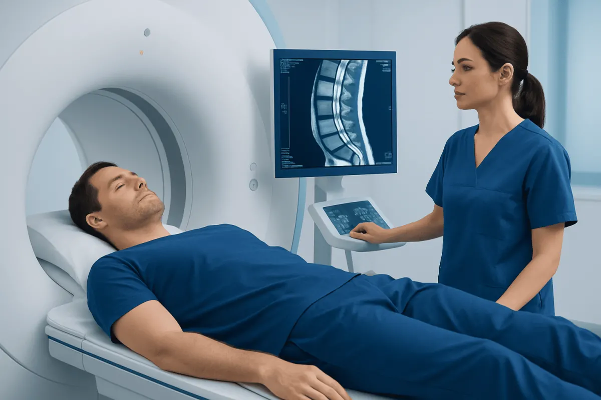
Imaging is not always needed immediately for uncomplicated back or neck pain. When symptoms are persistent, severe, associated with neurological findings, or suggest another condition, magnetic resonance imaging (MRI) may provide detailed pictures of discs, nerves, and other soft tissues. X-rays may help assess bones and alignment but do not show discs in the same detail. CT scans can be useful in selected situations.
An MRI can identify a posterior disc bulge, but it cannot by itself determine whether that bulge is painful. The most useful interpretation is one that matches imaging findings to the person’s symptoms and examination. This helps avoid treating an incidental finding and supports an individualized care plan.
Treatment Options: Usually Starting Without Surgery
For most people, initial care is non-surgical. Relative rest from movements that clearly aggravate symptoms may be helpful briefly, but extended bed rest is generally discouraged. Maintaining gentle movement and returning gradually to usual activities can support recovery, provided symptoms are not worsening.
A clinician may recommend short-term pain-relief options based on the person’s medical history, other medicines, and potential risks. Physical therapy can help restore movement, build trunk and hip strength, improve confidence with activity, and teach safer ways to lift, sit, and move. A personalized physical therapy and rehabilitation program can be particularly useful when pain limits daily life or keeps returning.
If nerve pain remains significant despite conservative care, a specialist may discuss options such as image-guided spinal injections for carefully selected patients. These do not correct every disc finding, but may reduce inflammation or pain enough to help a person participate in rehabilitation.
Surgery is considered when there is persistent, disabling nerve-related pain that has not improved with appropriate non-surgical treatment, or when there is clinically important weakness or nerve compression. Procedures are chosen according to the spinal level and anatomy; for example, selected lumbar cases may be assessed for lumbar disc herniation surgery. The purpose is usually to relieve pressure on a nerve, not simply to remove an imaging finding.
Self-Care, Activity and Prevention
There is no guaranteed way to prevent disc changes, as aging and genetics play a role. However, regular physical activity is one of the most practical ways to support spinal function. Walking, swimming, cycling, and strength exercises can be adapted to individual comfort and fitness. A physiotherapist or doctor can advise on appropriate exercise when symptoms are acute or recurrent.
Helpful habits include changing position regularly, using sound lifting technique, avoiding sudden increases in training loads, and pacing demanding tasks. For lifting, keeping an object close to the body and avoiding combined twisting and heavy lifting can reduce strain. Ergonomic adjustments at work may be useful, but frequent movement is usually more important than finding one perfect posture.
Stopping smoking, getting adequate sleep, managing stress, and working toward a sustainable weight where appropriate may support general musculoskeletal health. Pain can be influenced by sleep, mood, workload, and fear of movement as well as tissue changes. A supportive, gradual plan is often more effective than trying to avoid all activity.
When to Seek Medical Care
Medical assessment is appropriate for new or persistent neck or back pain that is not improving, pain that travels into an arm or leg, or symptoms that interfere with sleep, work, walking, or everyday activities. A clinician can identify whether symptoms may be related to a disc, a joint, a muscle, or another health condition and can recommend suitable next steps.
Urgent assessment is needed for new or worsening weakness, difficulty walking, loss of hand coordination, numbness around the genitals or inner thighs, or new difficulty controlling bladder or bowel function. These symptoms are uncommon but may signal significant nerve compression and should not be managed at home.
Prompt review is also sensible after major trauma or when spinal pain occurs with fever, Weight Loss: Possible Causes and Medical Evaluation" class="ahp-ilk">unexplained weight loss, a history of cancer, immune suppression, or other serious health concerns. Acıbadem Health Point’s multidisciplinary specialists and JCI-accredited hospitals evaluate and treat spinal conditions for international patients, with care plans based on clinical findings and individual needs.
Frequently asked questions
01Is a posterior disc bulge serious?
A posterior disc bulge is not automatically serious. Many are found incidentally and do not cause pain or nerve problems. Its importance depends on symptoms, the physical examination, and whether nearby nerves or the spinal cord are affected.
02Can a posterior disc bulge heal on its own?
Symptoms related to a disc bulge often improve over time with conservative care and gradual activity. The disc may not return exactly to its earlier shape on imaging, but an imaging change does not need to disappear for a person to feel better and function well. Recovery varies, so persistent or worsening symptoms should be assessed.
03Does a posterior disc bulge always require surgery?
No. Most people are treated without surgery using education, activity modification, exercise-based rehabilitation, and appropriate pain management. Surgery is generally reserved for selected cases with persistent disabling nerve symptoms or significant neurological deficits.
04What is the difference between a posterior disc bulge and a herniated disc?
A bulge usually describes a broader extension of disc tissue beyond its normal boundary. A herniated disc is a broader term for disc displacement and may involve a focal protrusion or extrusion. The terminology is less important than whether the finding matches symptoms and affects nerve structures.
05Can exercise make a posterior disc bulge worse?
Appropriate exercise is usually helpful rather than harmful, especially when increased gradually. During a painful flare, some movements may temporarily aggravate symptoms and should be adjusted. A clinician or physiotherapist can recommend exercises that fit the person’s symptoms and level of function.
06When should someone go to emergency care for a disc bulge?
Emergency assessment is needed for new bladder or bowel control problems, numbness in the groin or saddle area, or rapidly developing leg weakness. Urgent care is also appropriate for severe symptoms after major trauma or for significant difficulty walking or using the hands. These signs require prompt evaluation to rule out serious nerve compression.
This article is for general information only and is not a substitute for professional medical advice. Please consult a qualified doctor about your individual situation.
Joint and spine care in Turkey — expert assessment & treatment
JCI-accredited · board-certified surgeons · reply within 24h
Add us as a Preferred Source to see more of our trusted health content across Google Search, AI Overviews and Discover.