Renal Ultrasound: What It Shows and How to Prepare

A renal ultrasound is a common imaging test used to look at the kidneys and often the bladder without radiation. It can help doctors check for kidney stones, cysts, swelling, urinary blockage, and other structural changes, and preparation is usually simple.
Overview
A renal ultrasound, also called kidney ultrasound or renal ultrasonography, is an imaging test that uses high-frequency sound waves to create pictures of the kidneys. In many cases, the bladder is also examined during the same appointment because the kidneys and urinary tract work together. The test is widely used because it is noninvasive, does not use ionizing radiation, and can often be performed quickly.
Doctors may request a renal ultrasound to investigate urinary symptoms, flank pain, repeated urinary infections, blood in the urine, reduced kidney function, or suspected urinary blockage. It is also used to follow known kidney conditions over time. The test focuses mainly on structure, meaning it helps show the size, shape, and appearance of the kidneys and nearby urinary system.
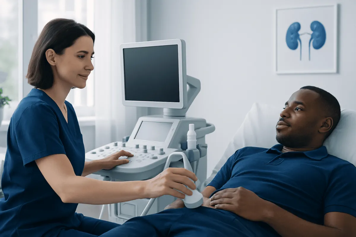
For many patients, this scan is reassuring because it is painless and straightforward. A gel is placed on the skin, and a handheld probe is moved over the abdomen and side to collect images. If a bladder view is needed, the patient may be asked to come with a comfortably full bladder or drink water before the exam.
What a Renal Ultrasound Can Show

A renal ultrasound can help identify a range of structural kidney and urinary tract changes. It may show whether the kidneys are normal in size, whether one kidney is smaller than expected, or whether the collecting system is enlarged because urine is not draining properly. This is useful when a doctor suspects obstruction, back pressure, or swelling of the kidney.
The scan may also reveal kidney cysts, some kidney masses, anatomical differences present from birth, and signs that suggest chronic kidney damage. In some patients, it can help detect or support the diagnosis of kidney stones, especially when stones are causing blockage or when the kidney appears swollen. If stones are a concern, further evaluation may sometimes include assessment for nephrolithiasis or additional imaging.
When the bladder is scanned too, the test can provide extra information about urine retention, bladder wall changes, and whether the bladder empties completely. In men, this can be useful when lower urinary tract symptoms suggest an enlarged prostate or another cause of poor urine flow. Ultrasound findings are interpreted together with symptoms, examination, and laboratory tests.
- Kidney size and shape
- Cysts and fluid-filled areas
- Swelling from urinary blockage
- Some kidney stones
- Bladder filling and emptying
- Structural abnormalities of the urinary tract
Why It Is Done
Doctors order renal ultrasound for many reasons. Common indications include side or back pain near the kidneys, blood in the urine, recurrent urinary tract infections, suspected kidney stones, abnormal kidney blood tests, and poor urine flow. It is also useful in people with Kidney Disease: Stages, Blood Tests, and Slowing Progression" class="ahp-ilk">chronic kidney disease, known cysts, or inherited kidney conditions that need monitoring over time.
In men, ultrasound may be part of the workup for urinary frequency, urgency, weak stream, or incomplete bladder emptying. Although the test does not diagnose every cause of these symptoms, it can reveal whether the kidneys are affected by back pressure from obstruction or whether the bladder is retaining urine. In the setting of infection, it may help identify complications or structural reasons for repeated episodes, such as those explored in urinary infection evaluation.
Renal ultrasound is also used when a safer imaging option is preferred, such as during pregnancy or when avoiding radiation is important. In people with a family history of cystic kidney disease, it may help check for enlarged kidneys or multiple cysts, findings that can be relevant to polycystic kidney disease care.
How to Prepare for a Renal Ultrasound
Preparation for a renal ultrasound is usually simple, but instructions can vary depending on whether the kidneys alone are being scanned or whether the bladder is included. Many imaging centers ask patients to drink several glasses of water before the appointment and avoid urinating so that the bladder is comfortably full. A full bladder can improve the view of the bladder itself and the lower urinary tract.
Patients should follow the specific guidance given by their doctor or imaging department. In some cases, no fasting is needed. In others, especially if a broader abdominal ultrasound is planned at the same visit, eating restrictions may be advised. Wearing loose, comfortable clothing can make the appointment easier, and patients may be asked to expose the abdomen and sides.
It is helpful to bring a list of medications and any previous imaging results if available. Most regular medicines can be taken as usual unless a doctor says otherwise. If there are concerns about discomfort from holding urine, patients should mention this in advance so the team can advise on the best timing for drinking water.
What Happens During the Test
During the scan, the patient usually lies on an examination table on the back or side. A technician or radiologist applies a clear gel to the skin and moves a small device called a transducer over the area. The transducer sends sound waves into the body and receives echoes that are turned into images on a screen.
The patient may be asked to hold the breath briefly, roll to one side, or change position so the kidneys can be seen from different angles. If the bladder is being assessed, images may be taken before and after urination to see how much urine remains. The test is generally painless, though mild pressure from the probe or discomfort from a full bladder is possible.
A typical renal ultrasound takes about 20 to 45 minutes, depending on what needs to be examined. Afterward, patients can usually return to normal activities right away. In some centers, this may be part of a wider renal ultrasonography evaluation within a kidney or urology workup.
Understanding the Results
The ultrasound report describes how the kidneys and bladder look. It may comment on kidney size, symmetry, cysts, masses, stones, swelling of the collecting system, bladder wall appearance, and post-void residual urine if the bladder was checked after urination. A normal result can be very helpful, but it does not always rule out all kidney or urinary tract problems.
Ultrasound is excellent for many structural questions, but it does not directly measure how well the kidneys filter blood. Kidney function is assessed with blood tests and sometimes urine tests. Small stones, early tumors, and some causes of pain or blood in the urine may not be fully explained by ultrasound alone, so a doctor may recommend other studies if symptoms continue.
Depending on the findings, the next step may include blood and urine analysis, specialist review, or more detailed imaging such as CT or MRI for urology. In selected cases, tissue sampling with renal biopsy may be considered, but this is only done when clinically appropriate and after careful evaluation.
Limitations, Safety, and Self-care After the Scan
Renal ultrasound is considered very safe because it uses sound waves rather than radiation. There are no known recovery restrictions after a routine scan, and patients can usually eat, drink, work, and drive as normal afterward. The gel is wiped off at the end, and no sedation is usually required.
Like all tests, ultrasound has limits. Image quality can be affected by body shape, bowel gas, or difficulty holding a full bladder. It may not show every stone or every abnormality, and it is less detailed than CT for some conditions. This does not mean the test is ineffective; rather, it is one part of a broader assessment.
After the scan, patients should continue any care plan already discussed with their doctor, especially if they have high blood pressure, diabetes, recurrent urinary infections, or known kidney disease. Drinking enough fluids, taking medicines as prescribed, and attending follow-up appointments are practical ways to support kidney health while awaiting or reviewing results.
When to See a Doctor
A person should speak with a doctor if they have symptoms that suggest a kidney or urinary tract problem, such as blood in the urine, pain in the side or back, fever with urinary symptoms, swelling, changes in urination, or repeated urinary infections. These symptoms do not always mean serious disease, but they do deserve proper assessment. Early evaluation can help identify whether imaging, urine testing, or specialist care is needed.
Urgent medical attention is especially important if pain is severe, urine output drops significantly, fever is high, or symptoms are accompanied by vomiting, dehydration, or confusion. These situations may suggest infection, obstruction, or another problem needing prompt treatment. Men with a suddenly painful inability to pass urine also need urgent care.
For international patients seeking coordinated kidney and urology assessment, Acıbadem Health Point’s multidisciplinary specialists and JCI-accredited hospitals diagnose and treat urinary and kidney conditions using modern imaging and follow-up care. The exact tests recommended depend on the patient’s symptoms, medical history, and examination findings.
Frequently asked questions
01Is a renal ultrasound the same as a kidney ultrasound?
Yes. The terms renal ultrasound and kidney ultrasound are commonly used to describe the same test. In many cases, the bladder is also examined during the same appointment.
02Does a renal ultrasound hurt?
A renal ultrasound is usually painless. Patients may feel mild pressure from the probe or some discomfort if they need to keep the bladder full, but the scan itself does not normally cause pain.
03Do patients need to fast before a renal ultrasound?
Not always. Many renal ultrasounds do not require fasting, but some centers give different instructions if the bladder or other abdominal organs are being scanned too. It is best to follow the preparation advice from the imaging department.
04Can a renal ultrasound show kidney stones?
It can show some kidney stones, especially if they are causing swelling or blockage. However, very small stones or stones in certain locations may be harder to detect, so other imaging may sometimes be needed.
05Can a renal ultrasound diagnose kidney disease?
It can help identify structural changes linked to kidney disease, such as small kidneys, cysts, or obstruction. But it does not measure kidney function directly, so doctors usually combine it with blood tests, urine tests, and clinical assessment.
06How long does a renal ultrasound take?
Most renal ultrasounds take about 20 to 45 minutes. The exact time depends on whether the kidneys alone are scanned or whether the bladder and post-urination images are also included.
This article is for general information only and is not a substitute for professional medical advice. Please consult a qualified doctor about your individual situation.
Explore treatments in Turkey — costs, top hospitals & a free quote
JCI-accredited · board-certified surgeons · reply within 24h
Add us as a Preferred Source to see more of our trusted health content across Google Search, AI Overviews and Discover.