Prostate Cancer PET Scan: Normal vs Abnormal Results
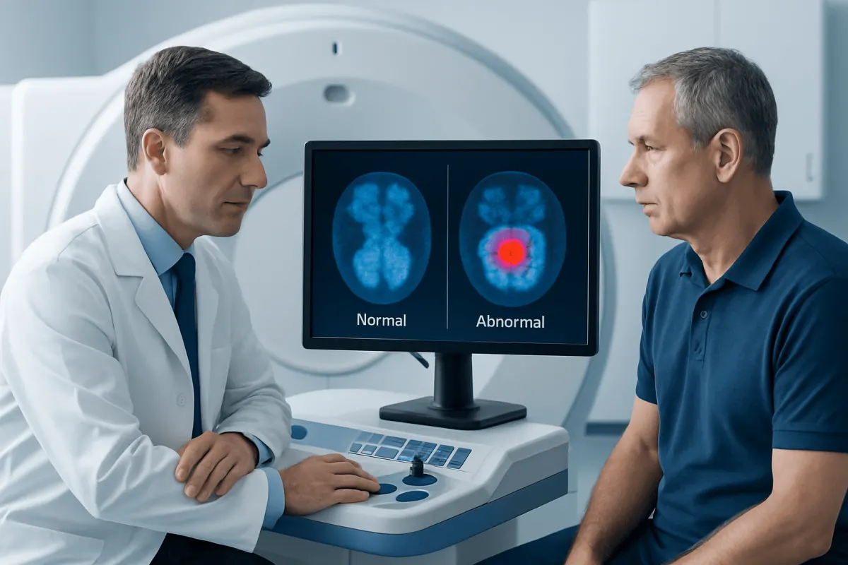
A normal prostate cancer PET scan generally means there is no suspicious tracer uptake outside expected areas, while an abnormal scan shows uptake that may represent prostate cancer, recurrence, spread, or a non-cancer cause. PET findings must be interpreted alongside PSA results, biopsy grade, prior treatment, symptoms, and other imaging.
Overview: Normal vs Abnormal Prostate Cancer PET Scan
In a prostate cancer normal vs abnormal PET scan comparison, a normal result means the scan has not identified suspicious tracer uptake in locations where prostate cancer would be expected to be present or spread. An abnormal result means there is a focus of uptake that requires clinical interpretation; it may represent prostate cancer, but it can also reflect normal body activity, inflammation, healing, or another benign condition.
Most modern prostate cancer PET scans use a prostate-specific membrane antigen (PSMA) tracer. PSMA is commonly present at higher levels on prostate cancer cells, allowing the tracer to highlight possible disease in the prostate, lymph nodes, bones, or other organs. The scan is often used for staging a newly diagnosed cancer with a meaningful risk of spread or for finding cancer that may have returned after treatment.
A PET scan result is not read in isolation. The care team considers the person’s PSA level and trend, biopsy findings such as Gleason Grade Group, previous surgery or radiation, symptoms, and CT or MRI images obtained with the PET scan. This combined approach helps avoid overinterpreting a single bright area on an image.
| Feature | Normal or expected PET appearance | Abnormal or suspicious PET appearance |
|---|---|---|
| Tracer uptake | Expected activity in normal organs that process or eliminate the tracer | Focal uptake in an unexpected location or with a pattern concerning for cancer |
| Prostate bed after treatment | No convincing focal uptake suggesting remaining or recurrent tumor | Localized uptake that may suggest recurrence, depending on timing and anatomy |
| Lymph nodes | No suspicious nodes identified | Uptake in pelvic or more distant lymph nodes that may indicate spread |
| Bones | No suspicious focal skeletal uptake | Focal bone uptake that needs correlation with CT, MRI, symptoms, and sometimes follow-up |
| Meaning | No disease seen on this scan; microscopic disease may still be below detection limits | Possible cancer location, not always a final diagnosis on its own |
How Clinicians Tell Normal and Abnormal Findings Apart
Radiologists evaluate the location, intensity, shape, and pattern of tracer uptake. They also review the accompanying CT or MRI images to see whether there is a corresponding enlarged lymph node, bone change, mass, scar, fracture, or other structural explanation. A small focus of uptake may be important in a person with a rising PSA after prostate removal, whereas the same finding may be less specific in another situation.
Some uptake is expected. Depending on the tracer used, normal activity may be seen in the salivary and tear glands, liver, spleen, kidneys, urinary tract, bladder, bowel, and certain ganglia near the spine. Because tracer passes into urine, activity in the bladder or ureters can sometimes obscure nearby areas in the pelvis.
Not every abnormal-looking spot is prostate cancer. Benign bone disease, recent fractures, infection, inflammation, reactive lymph nodes, and some non-prostate tumors can occasionally show PSMA uptake. Conversely, a negative scan does not exclude very small deposits of cancer or tumor cells that do not take up enough tracer to be visible. When uncertainty remains, the team may compare prior imaging, arrange targeted MRI or CT, repeat imaging later, or recommend a biopsy when the result would change management.
For broader context on diagnosis and staging, patients can review prostate cancer information. The most useful report is one that describes where uptake is seen, how suspicious it appears, whether there is a matching structural finding, and how the result affects the likely stage.
What to Do After a Normal or Abnormal Result

After a normal PET scan, the next step depends on why the scan was ordered. In a newly diagnosed cancer, a negative scan may support disease that appears confined to the prostate, but treatment decisions still depend on PSA, MRI, biopsy Grade Group, overall health, and personal preferences. In people previously treated for prostate cancer, a negative scan with a rising PSA may lead to close monitoring, additional imaging at a later time, or discussion of treatment based on the full clinical picture.
After an abnormal PET scan, the clinician first determines whether the finding is likely to be prostate cancer and whether it changes the stage or treatment plan. A suspicious finding limited to the prostate or nearby pelvic nodes may be approached differently from disease seen in distant lymph nodes, bone, or organs. Further tests are not always needed, but they may be useful when a lesion is unusual or when confirmation would alter treatment.
Management can include active surveillance for selected localized cancers, surgery, radiation therapy, hormone therapy, systemic medicines, or carefully selected targeted approaches. For localized or locally advanced disease, prostate Skin Cancer Treatment: Procedure, Recovery and Results" class="ahp-ilk">cancer treatment options are individualized according to cancer risk and extent. A PET result helps guide care; it does not determine treatment without the rest of the assessment.
Patients benefit from asking for a clear explanation of the report: what was found, how certain the interpretation is, whether cancer appears localized or metastatic, and whether any follow-up is recommended. Keeping copies of the imaging report and prior PSA values can make comparison over time easier.
What Gleason Score Requires a PET Scan?
There is no single Gleason score that automatically requires a PET scan for every person. Clinicians generally consider PSMA PET/CT for newly diagnosed prostate cancer that is unfavorable intermediate risk, high risk, or very high risk, especially when the result could change the treatment plan. Biopsy Grade Group 3 or higher, which commonly includes Gleason 4+3=7 or above, may prompt discussion of staging imaging when other risk features are present.
PSA level, clinical stage, the amount of cancer in biopsy samples, and imaging findings are considered alongside the Gleason score. For example, a person with Gleason 3+4=7 disease may or may not need a PET scan depending on PSA, examination findings, MRI results, and the expected risk of spread. A person with Gleason 4+3=7, Gleason 8, or Gleason 9 to 10 disease is more likely to be considered for advanced staging imaging.
For low-risk prostate cancer, including many Grade Group 1 cancers with low PSA, PET scanning is often unlikely to add useful information because the chance of detectable spread is low. The treating urologist or oncologist can explain whether PET imaging is appropriate before curative treatment or at the time of biochemical recurrence.
Which Prostate Cancer Test Is the Most Accurate?
No single test is the most accurate for every prostate cancer question. A prostate biopsy is the standard method for confirming whether cancer is present and identifying its microscopic grade. However, biopsies sample only selected areas of the prostate, so multiparametric MRI can help identify suspicious regions and guide a more targeted biopsy.
PSA testing is valuable for detecting a possible problem and monitoring change over time, but PSA alone cannot diagnose prostate cancer. It can rise for non-cancer reasons, including benign prostate enlargement, inflammation, infection, urinary retention, and recent prostate manipulation. Digital rectal examination also provides important information but cannot confirm cancer.
For staging and recurrence, PSMA PET/CT is among the most sensitive imaging tools available for finding prostate cancer outside the prostate, particularly at relatively low PSA levels after treatment. It is not a replacement for a biopsy when tissue confirmation is needed. The best diagnostic strategy combines PSA assessment, MRI where appropriate, targeted and systematic biopsy, pathology review, and PET imaging when indicated.
How Accurate Is a PET Scan for Prostate Cancer?
The accuracy of a PET scan for prostate cancer depends on the tracer used, the reason for the scan, PSA level, cancer biology, size of any lesions, and whether the disease is newly diagnosed or suspected to have returned. PSMA PET/CT is generally more sensitive than conventional CT and bone scanning for detecting many sites of prostate cancer, especially small lymph node or bone metastases that are not yet structurally obvious.
Even so, PET scans have limits. Very small cancer deposits may not be detected, and some prostate cancers express little PSMA, making them harder to see. A scan can also have false-positive findings, meaning uptake appears suspicious but is due to a non-cancer cause. For these reasons, a report may use terms such as indeterminate, equivocal, or recommend correlation with another scan or clinical follow-up.
Accuracy is also different for different clinical questions. A negative scan is reassuring but does not prove that no cancer cells are present. A positive scan can identify a likely location of disease and guide treatment planning, but confirmation may be considered if the finding is unexpected or would lead to a major treatment decision.
What Is the 2 Week Rule for Prostate Cancer?
The “2 week rule” is not a universal medical rule that describes how quickly prostate cancer grows or how long a person can safely wait for treatment. In some healthcare systems, it refers to an urgent referral pathway designed to help people with symptoms or test findings that could indicate cancer be assessed by a specialist within about two weeks.
Whether an urgent appointment is needed depends on the individual situation. A markedly abnormal PSA result, an abnormal rectal examination, blood in the urine, unexplained bone pain, unintentional weight loss, or symptoms suggesting urinary blockage should be reviewed promptly. However, many prostate cancers develop slowly, and a short period needed to complete PSA review, MRI, biopsy, pathology, and staging tests is often appropriate and helps ensure treatment decisions are well informed.
People should not assume that a two-week timeline applies to every test, diagnosis, or treatment. The safest approach is to follow the timetable recommended by the clinician and to contact the care team if symptoms worsen, test results are unclear, or an appointment is delayed beyond the advised timeframe.
When to Seek Medical Care
Medical review is appropriate for persistent urinary symptoms, such as difficulty starting urination, weak flow, needing to urinate often at night, pain or burning with urination, or visible blood in urine or semen. These symptoms are common and are often caused by non-cancer conditions, but they should be assessed rather than self-diagnosed.
People with a known prostate cancer diagnosis should contact their care team if they develop new or worsening bone pain, unexplained weakness or numbness, loss of bladder or bowel control, significant urinary obstruction, or a rapid decline in general health. Sudden inability to pass urine, severe back pain with leg weakness, or loss of bladder or bowel control requires urgent medical attention.
Anyone receiving a PET scan should arrange a follow-up discussion with the clinician who ordered it. Acıbadem Health Point’s multidisciplinary specialists and JCI-accredited hospitals diagnose and treat prostate cancer for international patients, with care plans based on pathology, imaging, and individual clinical needs.
Frequently asked questions
01Does a normal PSMA PET scan mean there is no prostate cancer?
No. A normal PSMA PET scan means there is no cancer detected at the resolution and sensitivity of that scan. Cancer may still be confined to the prostate, present in microscopic amounts, or have low PSMA expression. Biopsy, PSA, MRI, and clinical findings remain important.
02Can an abnormal prostate PET scan be benign?
Yes. Inflammation, infection, fractures, degenerative bone changes, benign lymph nodes, and normal tracer distribution can sometimes produce uptake. The radiologist evaluates the pattern and compares it with CT or MRI images before determining how suspicious a finding is.
03What does uptake in a pelvic lymph node mean on a PSMA PET scan?
Pelvic lymph node uptake may indicate that prostate cancer has spread to a nearby lymph node, particularly when the appearance and clinical context are consistent. It is not always definitive, as some benign processes can cause uptake. The finding may affect staging and decisions about surgery, radiation fields, or systemic treatment.
04Why can the bladder make a prostate PET scan difficult to interpret?
Many PET tracers are removed from the body through urine, creating strong normal activity in the kidneys, ureters, and bladder. This activity can obscure small areas near the prostate bed or pelvic lymph nodes. Imaging protocols and review of CT or MRI images help reduce this limitation.
05Is a PSMA PET scan painful?
The injection of the tracer is similar to a routine intravenous injection. During imaging, the person needs to lie still on the scanner table, which may be uncomfortable for some people but is not usually painful. The imaging center provides preparation instructions based on the specific tracer and scan protocol.
06Can PET scan results change prostate cancer treatment?
Yes. If a scan identifies cancer outside the prostate or shows a likely site of recurrence, it may change the planned treatment or the area targeted by radiation. Decisions should be made with the full clinical picture, because PET findings alone do not always establish a final diagnosis.
This article is for general information only and is not a substitute for professional medical advice. Please consult a qualified doctor about your individual situation.
Oncology care in Turkey — second opinion and treatment plan
JCI-accredited · board-certified surgeons · reply within 24h
Add us as a Preferred Source to see more of our trusted health content across Google Search, AI Overviews and Discover.