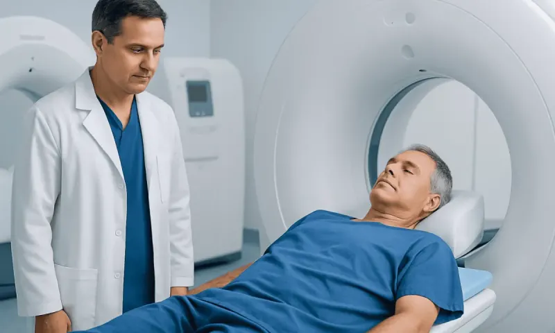
PET Scan Versus MRI for Cancer: Which Is Used?
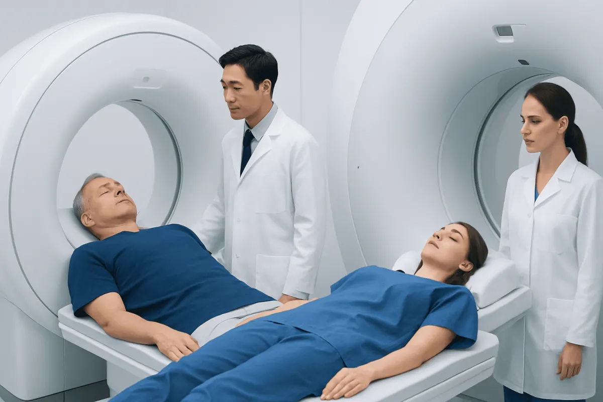
PET scans and MRI scans provide different but complementary information in cancer care. A PET scan helps identify areas of increased metabolic activity, while MRI provides highly detailed images of soft tissues and body structures; the most suitable scan depends on the suspected cancer, body area, and purpose of testing.
PET Scan Versus MRI for Cancer: Side-by-Side Comparison
When considering a PET scan versus MRI for cancer, the key difference is what each test measures. Positron emission tomography (PET) shows how tissues are functioning, often by detecting how much of a small amount of radioactive tracer is taken up by cells. Magnetic resonance imaging (MRI) uses a powerful magnet and radio waves to create detailed pictures of organs, tissues, and structures.
Neither examination is automatically “better.” PET may be particularly helpful when a clinician needs to assess whether cancer is active or whether it may have spread. MRI may be preferred when precise anatomical detail is needed, such as defining a tumor’s relationship to nerves, blood vessels, muscles, or nearby organs. In many situations, their findings are interpreted together.
| Feature | PET scan | MRI scan |
|---|---|---|
| Main information | Cellular activity and tracer uptake | Detailed anatomy and tissue characteristics |
| Common cancer uses | Staging, checking treatment response, looking for recurrence or spread | Characterizing a local tumor and assessing soft tissues |
| Radiation | Uses a radioactive tracer; PET is often combined with CT | Does not use ionizing radiation |
| Strengths | Can show active disease throughout much of the body | Excellent contrast for many soft tissues |
| Limitations | Inflammation and infection can also show uptake; some cancers have low uptake | May not distinguish active cancer from scar tissue as clearly in some settings |
| Typical duration | Includes tracer uptake time and scanning time | Often takes longer than a standard CT examination |
Results should always be considered alongside a person’s symptoms, medical history, physical examination, and prior scans. If a suspicious area is found, a biopsy may still be needed to confirm whether it is cancer and identify its exact type.
How Clinicians Tell PET and MRI Apart
Clinicians select imaging based on the question that needs answering. If there is a known cancer, they may ask whether it has spread to lymph nodes, bone, or other organs, whether treatment is reducing active disease, or whether a new finding may represent recurrence. A PET scan, often performed as PET-CT, can be valuable for these whole-body questions.
MRI is commonly selected when the location and boundaries of a finding matter most. It can provide excellent detail in the brain, spinal cord, pelvis, liver, breast, muscles, joints, and many other soft-tissue areas. It is also used to help plan surgery, radiation therapy, or targeted biopsy by showing the location of abnormal tissue more precisely.
The choice is also influenced by the suspected cancer type. Some tumors tend to take up the most commonly used PET tracer strongly, while others may not. MRI protocols can likewise be adapted for particular organs, sometimes with contrast material, to improve the ability to distinguish normal from abnormal tissue.
For some people, practical considerations also matter. MRI can be challenging for those with certain implanted devices, severe claustrophobia, or difficulty lying still. PET may require dietary preparation depending on the tracer and medical indication. The imaging team reviews safety, kidney function when contrast is considered, allergies, medications, pregnancy status, and other relevant details before the scan.
Which Is More Accurate for Detecting Cancer, an MRI or a PET Scan?
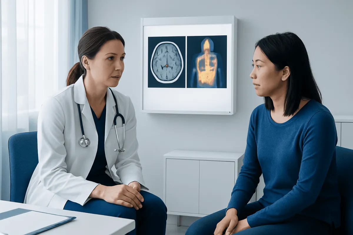
Accuracy depends on the cancer being assessed, its size, its location, and the purpose of the test. PET can be more informative when the goal is to identify metabolically active cancer in multiple parts of the body or to evaluate whether a treated cancer remains active. MRI can be more informative when the goal is to detect or define an abnormality within soft tissues with high anatomical precision.
For example, MRI is often a central imaging test for suspected tumors of the brain, spinal cord, prostate, uterus, ovaries, rectum, and breast. PET may play an important role in staging several cancers, assessing certain lymphomas, evaluating many lung cancer cases, and investigating possible recurrence when other tests are unclear. The exact use varies by cancer type and national clinical guidance.
A PET result can be positive for reasons other than cancer. Infection, inflammation, healing after surgery, and some benign conditions can increase tracer uptake. Conversely, a small tumor, a slow-growing tumor, or a tumor with low tracer uptake may not be clearly visible on PET. MRI findings can also be indeterminate, particularly if inflammation, bleeding, treatment-related changes, or benign growths resemble cancer.
For this reason, clinicians do not usually make a cancer diagnosis from one scan alone. They may compare previous imaging, order a complementary study, perform blood or laboratory testing, or recommend a biopsy. Cancer evaluation and diagnosis generally involves combining several sources of evidence rather than relying on one test result.
What Can an MRI Show That a PET Scan Cannot?
MRI can show exceptionally detailed structural information. It can help clinicians see the exact size, shape, margins, and internal features of a lesion, as well as its relationship to nearby organs, nerves, blood vessels, bone marrow, and soft tissues. This detail is particularly useful when treatment planning requires a clear map of the area involved.
In the brain and spinal cord, MRI is often especially important because it can show edema, pressure on nerve tissue, bleeding, fluid changes, and detailed anatomy that may not be adequately defined by PET. MRI can also identify features such as tissue composition and blood flow patterns through specialized sequences, which may help characterize a finding.
MRI is useful for evaluating local disease in areas where normal PET tracer uptake can complicate interpretation, including the brain, urinary tract, and some pelvic organs. In certain situations, MRI can show small abnormalities that have not caused enough metabolic change to stand out on PET imaging.
However, MRI does not always show whether a structural abnormality is active cancer. After treatment, for instance, a mass or area of scarring may remain visible even when there are no viable cancer cells. PET may then provide useful complementary information about whether the area is metabolically active, although its result must still be interpreted cautiously.
Which Scan Is Most Accurate for Cancer?
There is no single scan that is most accurate for all cancers. The best examination is the one that matches the clinical question and the suspected or confirmed cancer type. In many cancer pathways, clinicians use more than one imaging method because each contributes different information.
For an initial suspicious symptom or finding, the first test may be an ultrasound, X-ray, CT, mammogram, or MRI rather than PET. Once a diagnosis is established, PET-CT, MRI, CT, bone scanning, or other specialized imaging may be used to determine the extent of disease and guide treatment planning. A biopsy is commonly required before Skin Cancer Treatment: Procedure, Recovery and Results" class="ahp-ilk">cancer treatment begins, unless a doctor explains why it is not appropriate or safe.
Imaging can also be repeated during or after treatment. The scan type is chosen according to the cancer and treatment received. MRI can be important for following local changes, while PET can sometimes help evaluate metabolic response. Timing matters because treatment-related inflammation can affect scan appearance.
People should ask their clinician what question the scan is intended to answer and whether another test may be needed afterward. A radiologist and the treating specialist interpret the images in the context of the whole clinical picture, rather than treating a scan result as a diagnosis by itself.
Can a PET Scan Pick Up All Cancers?
No. A PET scan cannot pick up all cancers, and it is not a general cancer screening test for people without symptoms or a specific clinical reason for imaging. The most widely used PET tracer highlights glucose use, which is often increased in cancer cells but can also be increased in normal tissues and non-cancerous inflammation.
Some slow-growing cancers and some tumor types may show little or no uptake with the standard tracer. Very small tumors can also fall below the scan’s detectable resolution. In addition, areas with naturally high tracer activity, such as the brain, heart, kidneys, and bladder, may be more difficult to assess with standard PET imaging.
Different PET tracers may be used in selected circumstances because they target particular biological features. These specialized studies are ordered only when appropriate for the suspected cancer and clinical question. They do not remove the need for other imaging or tissue sampling when confirmation is required.
A negative PET scan can be reassuring in the right setting, but it does not always rule out cancer. Persistent concerning symptoms, an abnormal examination, or a suspicious result on another test should be discussed with a qualified doctor even if PET findings are negative.
What to Do Before and After Each Scan
Preparation differs between PET and MRI. For many PET scans, patients are asked to follow instructions about eating, drinking, exercise, and diabetes medication before the appointment because blood glucose levels can affect the tracer’s distribution. They should tell the imaging department about diabetes, pregnancy or breastfeeding, current medicines, recent infections, and recent surgery or treatment.
Before an MRI, patients should report any implanted medical device, metal fragment injury, surgical clips, cochlear implant, medication pump, or kidney problem. Many implants are MRI-compatible under specific conditions, but the radiology team must confirm this in advance. MRI contrast may be recommended in some cases, and the team will explain its purpose and relevant safety considerations.
After either examination, most people can return to usual activities unless they receive sedation or are given different instructions. The images are reviewed by a radiologist, often together with prior scans. The referring clinician then explains what the findings mean, whether they are conclusive, and what follow-up is appropriate.
For international patients, Acıbadem Health Point’s multidisciplinary specialists and JCI-accredited hospitals can coordinate cancer imaging, pathology review, and treatment planning when these are clinically needed.
When to Seek Medical Care
A person should arrange medical assessment for new, persistent, or worsening symptoms that concern them, particularly unexplained lumps, ongoing bleeding, unexplained weight loss, persistent pain, a lasting change in bowel or bladder habits, difficulty swallowing, or a cough that does not improve. These symptoms are common and often have non-cancerous causes, but a clinician can determine whether testing is needed.
Urgent medical care is appropriate for severe or rapidly worsening symptoms, such as trouble breathing, sudden weakness or numbness, new seizures, coughing or vomiting blood, severe headache with neurological changes, or uncontrolled pain. Emergency services should be contacted when symptoms are sudden, severe, or potentially life-threatening.
People who have already had cancer should follow their planned surveillance schedule and contact their oncology team about new symptoms rather than waiting for a routine scan. If imaging has been recommended, patients can ask why it is needed, whether contrast or a tracer will be used, how to prepare, and when results are expected.
Frequently asked questions
01Is PET-CT the same as a PET scan?
PET is frequently performed together with CT in one appointment, called PET-CT. PET provides metabolic information, while CT provides anatomical detail and helps localize areas of tracer uptake. The term “PET scan” is often used informally to refer to PET-CT.
02Can MRI detect cancer without contrast?
MRI can detect many abnormalities without contrast, and some MRI sequences are designed to provide useful information without it. Contrast may be recommended when it can improve visualization or help characterize a lesion. The decision depends on the body area, clinical question, and the person’s health history.
03Can PET scans give false-positive results?
Yes. Increased tracer uptake can occur with infection, inflammation, healing tissue, and some non-cancerous conditions. A positive PET finding may therefore need comparison with other imaging or confirmation by biopsy.
04Are PET and MRI scans painful?
Both scans are generally painless, although remaining still can be uncomfortable for some people. MRI can be noisy and enclosed, while PET involves an injection of tracer and a period of waiting before imaging. The radiology team can discuss comfort measures before the appointment.
05Why might a doctor order both PET and MRI?
A doctor may order both tests because they provide complementary information. MRI can define the location and anatomy of a lesion in detail, while PET can indicate whether tissue is metabolically active and whether there are concerning areas elsewhere in the body.
06Does a normal MRI or PET scan mean there is no cancer?
A normal scan lowers concern in many situations but does not exclude every cancer. Some cancers are too small, slow-growing, or not well detected by that particular technique. Doctors interpret normal imaging alongside symptoms, examination findings, and any other relevant tests.
This article is for general information only and is not a substitute for professional medical advice. Please consult a qualified doctor about your individual situation.
Oncology care in Turkey — second opinion and treatment plan
JCI-accredited · board-certified surgeons · reply within 24h
Add us as a Preferred Source to see more of our trusted health content across Google Search, AI Overviews and Discover.